

 PDF
PDF ePub
ePub Citation
 Print
Print


INTRODUCTION
Traditional elliptical excisions with length:width ratios of ≥3:1 are the preferred method for cutaneous annular lesion extirpation1,2. These excisions require scar lengths three times the diameter of the lesion so dog ears can be prevented.
Nowadays, patients have high expectations when it comes to minimization of postoperative scarring after dermatologic surgery. Many surgical options other than elliptical excision have been introduced for reducing scarring.
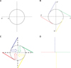
We introduce a new flap design that produces a short, X-shaped scar (O-X flap). The new flap design, which leads to reduced scar length in annular defects, consists of four rotation flaps arranged in open and closed configuration3. This design results in a scar as large as the diameter of the lesion. The surgical procedure is as follows. Draw imaginary vertical and horizontal axes at 90 degrees to each other, considering a relaxed skin tension line and adjacent structures. Make one point on the axis at a radius distance from the lesion (Fig. 1A). Draw a tangent line from the point to the circle. Repeat three more triangles at 90 degrees, creating a vane-like structure (Fig. 1B). Excise four straight lines and four tangent lines including the margin of the lesion. Rotate each flap into the center of the circle, situated in a cruciform manner. Suture each point of two rotated flaps together at the center of the circle. To prevent tip necrosis, place the tips of the sutures of two rotated flaps slightly away from each other. Rotate the others as above (Fig. 1C). Close the sides together, creating an X-shaped closure line (Fig. 1D). The flap design and clinical applications are described in subsequent cases. Acceptable cosmetic results were obtained.
CASE REPORT
Case 1
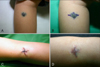
A 24-year-old woman presented with an asymptomatic, solitary, 1.5×1.5 cm sized annular congenital melanocytic nevus on the right forearm. The lesion consisted of a centrally located, slightly elevated, black papule with a surrounding bluish patch (Fig. 2A). The quadruple fan flap was designed as described above (Fig. 2B), and the lesion was excised along the marking lines. This design made it easier to excise the peripheral bluish patch without lengthening the scar. We then applied a lazy S-plasty design on each arm of the flap, considering the cylindrical structure of the forearm. We rotated the excised flaps to the center, sutured the flap points together around the center, and closed the sides together (Fig. 2C). Follow-up imaging done forty days later showed that the scar length of the quadruple fan flap was much shorter than that of the elliptical excision (Fig. 2D).
Case 2
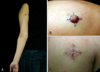
A 27-year-old woman presented with a 1.6×1.2 cm sized soft nodule on the right arm (Fig. 3A). She also had café-au-lait macules and axillary freckles and had been previously diagnosed with neurofibromatosis. We created the quadruple fan flap while saving some parts of the skin above the neurofibroma, in the interest of making the scar smaller (Fig. 3B). The neurofibroma was excised along the marking lines, leaving some parts of the skin above the lesion. Each flap was rotated into the center, and the suture tips of two rotated flaps were placed slightly away from each other to prevent tip necrosis. Follow-up imaging after eight days showed that the scar length was almost the same as the diameter of the lesion and that the flap tips were placed slightly away from each other (Fig. 3C).
DISCUSSION
We used the quadruple fan flap to reduce scar length and achieve results superior to those seen with elliptical excisions. Our method consists of four fan-like rotation flaps arranged in open and closed configuration3. This design results in a scar that is the same size as the lesion.
Elliptical excision has been the most commonly used surgical method for treating annular defects. But, if a lesion is excised in an elliptical fashion, the long axis needs to be three or four times the diameter of the lesion. If the ellipse is too short, or one side of the ellipse is shorter than the other, a dog ear is formed at the end of the repaired wound. In the interest of overcoming these disadvantages, various surgical techniques have been developed, including rotation flaps and advancement flaps that address lesion size, location, and orientation, and adjacent soft tissue elasticity. For example, the O-Z flap commonly used to treat annular defects offers the advantage of lowering closing tension. It can be performed when sufficient tissue is available, without distorting surrounding structures4. As with traditional rotation flaps, O-Z flap movement produces paired dog-ear redundancies near the flap's pivot points. Triple rotation flaps, especially those used in scalp reconstruction, offer the advantage of permitting distribution of tension over the surrounding tissue away from the suture lines5. However, this results in relatively large incision lines.
This new flap has some significant advantages. It does not require the 1:3 or 1:4 length ratio normally required in elliptical excisions6. The final length of each limb was actually approximately the same as the diameter of the defect. We were also able to apply a lazy S-plasty design7 to cylindrical areas like the arms and legs, and two limbs of the flap were directed along the relaxed skin tension line. This is useful for lesions located in areas with complex skin tension lines. Both the patients and the physicians were cosmetically satisfied with the scar lines. There were no complications, including tissue necrosis.
We suggest that the quadruple fan flap (O-X flap) is a good option for treating annular skin defects, because it shortens the scar line, preserves normal tissue, and offers a cosmetically favorable outcome.
 XML Download