

 PDF
PDF ePub
ePub Citation
 Print
Print


INTRODUCTION
Dermatitis neglecta (unwashed dermatosis) was first described by Poskitt et al1 in 1995. They reported 3 cases of pigmented hyperkeratotic plaques on various sites, and these plaques were the result of avoiding washing the affected areas. All of the lesions were rapidly resolved with normal washing by gently wiping with an alcohol swab.
We herein report on a case of dermatitis neglecta on the umbilicus, and this resolved after cleaning the lesions with H2O2 and a saline gauze swab during the skin biopsy. This is the first reported case to provide the histologic characteristics of dermatitis neglecta.
CASE REPORT
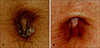
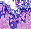
A 29-year-old man presented with an asymptomatic thick scaly plaque on the umbilicus (Fig. 1A). The lesion was incidentally found by the patient himself the day before he visited the clinic. He reported no history of any preceding eruption, injury to the skin or any family history of skin diseases. On the physical examination, there was a hyperkeratotic plague on the umbilicus with silvery scales. During skin biopsy, gentle swabbing with H2O2 and saline gauze was done. Histopathologic examination showed orthokeratotic hyperkeratosis and anastomosing rete ridges (Fig. 2). He was not given any other medications and the patient revisited our clinic 10 days after the skin biopsy. The hyperkeratotic lesion was cleared and he reported that the swabbing with H2O2 and saline gauze during skin biopsy made the lesion disappear (Fig. 1B).
DISCUSSION
Poskitt et al1 first described dermatitis neglecta (unwashed dermatosis) in 1995. They reported 3 cases of pigmented hyperkeratotic plaques on various sites, and these plaques were the result of avoiding washing the affected areas. Ruiz-Maldonado et al2 added two more cases of dermatitis neglecta in 1999, and the lesions resembled verrucous nevi around the areolas and pubic areas. All of the lesions were rapidly resolved by normal washing with gentle wiping from an alcohol swab. For our patient, the lesion was on the umbilicus and it resembled psoriasis or nevoid acanthosis nigricans localized to the umbilicus, and this type of case has rarely been reported3. On histologic examination, the epidermis showed orthokeratotic hyperkeratosis with anastomosing rete ridges, and a mild perivascular lymphocytic infiltration was noted in the dermis. Psoriasis could be promptly ruled out by its histopathologic features, but nevoid acanthosis nigricans possesses some common histological features with dermatitis neglecta; hyperkeratosis, slight acanthosis, undulating with dermal papillomatosis in the epidermis and a mild perivascular lymphocytic infiltration in the superficial dermis. However, nevoid acanthosis nigricans progresses for a certain period and then it remains stable, and so it requires treatment with various modalities; topical application of keratolytic agents, podophyllin, calcipotriol or retinoids, cryotherapy, dermabrasion, laser, excision, etc. In our case, gentle swabbing with H2O2 and saline gauze during skin biopsy treated the lesion, and this quick resolution of the lesion is a characteristic feature of dermatitis neglecta.
Dermatitis neglecta might occur in various circumstances. Poskitt et al1 stated that dermatitis neglecta is a result of willful and subconscious self-neglect, which is different from dermatitis artefacta that the lesions are not produced or aggravated by the patient's own actions. Dermatitis artefacta is classified as an impulsive-aggressive type of obsessive-compulsive disorder by Stein and Hollander4. This loss of self-control could be associated with guilt or this may occur during a period of a hysterical trance during which time the ordinary personality has no conscious memory5. Thus, the concept of dermatitis artefacta was not fulfilled for the previously reported cases or the present instance, since the lesions in these instances arose through acts of omission rather than commission1. Dermatitis neglecta may occur in areas of unwashed hyperaesthetic skin, for example, in patients suffering from trigeminal neuralgia or in those patients whose mobility is compromised by stroke or severe arthritis, and so they are unable to wash adequately2. In our case, patient had inadequate bathing habits so that he probably didn't give much attention to cleaning the umbilical area.
In most cases, dermatitis neglecta is probably due to accumulation of sebum, sweat, corneocytes and bacteria, and these all form a compact, adherent crust of dirt. Recognizing its existence and cause are important to avoid unnecessary biopsies and potentially aggressive therapeutic measures2. However, dermatitis neglecta might be overlooked and underdiagnosed.
We herein report on a case of dermatitis neglecta on the umbilicus. This case is the first report to describe the histologic characteristics of dermatitis neglecta.
 XML Download