

 PDF
PDF ePub
ePub Citation
 Print
Print


INTRODUCTION
Lichen planus pigmentosus-inversus (LPP-inversus) is an extremely rare variant of lichen planus (LP), and only a few cases have been reported12. We have already seen one patient with LPP-inversus, and that case has been published3. Recently, we saw two more cases of LPP-inversus. We report the details of those cases and offer suggestions concerning their possible origin.
CASE REPORT
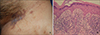
The first patient was a 49-year-old woman who presented complaining of violaceous reticulated patches and scattered rice grain-sized macules localized to the left inguinal area for several months (Fig. 1A). She had no subjective symptoms, such as pruritus or pain. She had not come into contact with any chemicals, animals, or plants, nor had she been using any medications that could prompt an allergic response. Her medical and family history were non-contributory. A skin biopsy from a violaceous patch revealed irregular acanthosis, vacuolar alteration of the basal layer, and marked band-like dermal lymphocytic infiltration with pigment incontinence (Fig. 1B). These histological features suggested the presence of classic LP. Thereafter, the lesions slowly flattened and changed color to brown. Although we could not examine the flattened lesions histologically, we hypothesized that lesions of classic LP located only in intertriginous areas may have changed into LPP sometime later.
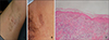
The second patient was a 25-year-old woman who complained of multiple brownish macules scattered on both axillae for one year (Fig. 2A). Recently, a solitary pigmented atrophic patch was also found on the left inner thigh (Fig. 2B). She was not symptomatic. The size of the lesions increased gradually. There were also some tiny papules around the lesions in the axilla. According to the patient, some of the papules had flattened into macular components. A skin biopsy was performed on the
axillary and inner thigh lesions. The papular lesions in the axilla showed histological features consistent with classic LP (Fig. 2C). Thinning of the epidermis and pigmentary incontinence were prominent features of the thigh lesions. These features found in the thigh lesions were consistent with LPP.
DISCUSSION
LPP, a disease of unknown etiology, manifests as hyperpigmented, dark brown, occasionally pruritic macules and/or papules. The course of the disease is characterized by exacerbations and remissions. It is known to be more chronic than classical LP is4.
With regard to the coexistence of classic LP in a number of LPP patients and the histopathological resemblance between these two disorders, many authors have suggested that LPP is a variant of LP5. However, classical LP shows a predilection for the wrist, thigh, ankle, and the dorsum of the hand, and to the best of our knowledge, there have been no reports of classical LP being confined to intertriginous areas. The two current cases and the case detailed in our previous report3 confirm classic LP lesions confined to skin folds, which developed LPP features over time through epidermal flattening.
Although verification of similar cases is needed in order to confirm our hypotheses, we suggest that LPP-inversus may originate from LP of flexural areas. Furthermore, classic LP can be located in the flexural area only, so we suggest that a new term, 'LP-inversus', be used to designate such an entity.
 XML Download