

 PDF
PDF ePub
ePub Citation
 Print
Print


INTRODUCTION
Steatocystoma simplex, first described as a distinct entity by Brownstein1 in 1982, is an extremely rare benign adnexal tumor. The individual lesion of steatocystoma simplex is usually identical with that of steatocystoma multiplex, both clinically and histologically, but is characterized by solitary, non-heritable growth in adulthood1. Steatocystoma simplex occurs predominantly on the face, but the incidence of steatocystoma simplex on the scalp is extremely low. Only three cases of steatocystoma simplex on the scalp have been reported worldwide123. We report an additional case of steatocystoma simplex which was developed on the right parietal scalp.
CASE REPORT
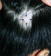
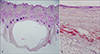
A 49-year-old man presented to our outpatient clinic with an asymptomatic papule on the right parietal scalp which had been present for about 1 year. The lesion had slowly enlarged a few months ago. The physical examination revealed a skin-colored, deep-seated, soft cystic mass on his right parietal scalp, 0.9 cm in diameter (Fig. 1). There was no remarkable family history of similar lesions. Clinically, the lesion appeared to be a trichilemmal cyst, and was completely excised. The histological examination revealed an undulating, configured cystic mass that was empty. The cyst was located in the dermis and the cystic walls were thin and lined by stratified squamous epithelium that lacked a stratum granulosum (Fig. 2A). The cyst contained eosinophilic hyalinized cuticle in the lumen and flattened sebaceous gland lobules close to the cystic wall (Fig. 2B). There have been no signs of recurrence or development of other cysts during the 4 month follow-up.
DISCUSSION
Steatocystoma simplex is thought to originate from a nevoid malformation of the pilosebaceous duct junction and a non-heritable benign adnexal tumor4. Brownstein1 first reported 30 cases of steatocystoma simplex as distinct clinical entity compared with steatocystoma multiplex as a counterpart disease in 1982.
The individual lesion of steatocystoma simplex is identical with that of steatocystoma multiplex clinically and histologically. The lesion of steatocystoma is usually asymptomatic, with a normal skin color and no inflammatory component. Steatocystoma simplex is a soft, movable intracutaneous cyst with a distinct boundary. To distinguish steatocystoma simplex from steatocystoma multiplex, it is important to make sure that the lesion is solitary and occurs in adulthood with a non-heritable pattern1.
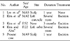
Steatocystoma simplex can occur on various parts of the body, but most commonly on the face and the chest. The upper limbs and axillae are also relatively common sites of occurrence. Rarely, steatocystoma simplex can occur on relatively uncommon sites, such as the back, leg, oral cavity, perineum, and scalp156. Four cases of steatocytoma simplx have been reported and only one case has been reported to involve the scalp in the Korean medical literature (Table 1)3478. Steatocytoma simplx involving the scalp is extremely rare and only two cases have been reported in foreign medical journals by Brownstein1 and Nakamura et al2. This case report on steatocystoma simplex occurring on an uncommon site, the scalp, is considered to be a very rare medical case.
On gross examination, the cyst contained oily, yellowish fluid. The histological examination revealed a cystic mass surrounded by stratified squamous epithelium with a saw-tooth appearance that lacked a stratum granulosum. In the innermost area of the cyst, a prominent, acellular, homogenous, eosinophilic, and compact horny layer was seen. Large, flattened sebaceous glands were within or near the cyst wall. If there is a hyaline cuticle in the cystic wall, it can be shown to be a steatocystoma simplex, even in the absence of sebaceous glands14.
The differential diagnosis of steatocystoma simplex includes steatocystoma multiplex, a dermoid cyst, cystic sebaceous hyperplasia, and hidrocystoma. Steatocystoma multiplex is differentiated by multiple lesions, usually over the trunk, and inherited in an autosomal dominant pattern. A dermoid cyst, cystic sebaceous hyperplasia, and hidrocystoma are distinguished by histologic characteristics14.
Above all, the treatment of choice is simple excision with an intact cyst wall to reduce the risk of recurrence. Also, aspiration, cryosurgery, electrocautery, and carbon dioxide laser therapy are included as possible treatment options of steatocystoma simplex35. There have been no reported case of recurrent steatocystoma simplex after excision9.
 XML Download