

 PDF
PDF ePub
ePub Citation
 Print
Print


INTRODUCTION
Juvenile xanthogranuloma (JXG) is a benign cutaneous histiocytic proliferation, and was first described by Helwig and Hackney in 19541. The pathophysiology of JXG is not well understood, although it is thought to originate from a histiocytic granulomatous reaction23.
The cells of JXG originate from the monocytemacrophage lineage, which can differentiate in diverse directions2. JXG normally occurs in infancy or early childhood45, and clinically usually manifests as yellowish or red-brown, firm papules, or nodules6. Lesions usually present on the head, neck, and trunk, however JXG involving a finger is rare4; only six cases of JXG of the fingers have been reported in the English literature.
CASE REPORT
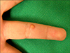
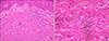
A 4-year-old girl presented with a papule of several months duration on the ventral aspect of the right fourth finger MCP joint (Fig. 1). The lesion was a firm, dome-shaped, yellowish, 0.4×0.4cm sized papule. There was no remarkable past or family history. On physical examination, there was no limitation of joint motion and no abnormal findings other than the cutaneous lesion. A 4 mm punch biopsy specimen of the lesion showed dense intradermal histiocytic infiltrates, some of which contained foamy cells, Touton giant cells, and foreign body giant cells (Fig. 2). Scattered lymphocytes and eosinophils were also presented. Histopathological findings were consistent with a diagnosis of JXG. The papule was removed under local anesthesia using a 4 mm punch.
DISCUSSION
JXG is the most common form of non-Langerhans cell histiocytosis and is considered a benign histiocytic proliferation3.
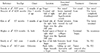
Only six cases of JXG of the fingers have been reported in the English literature; these cases are summarized in Table 1134678. Sonoda et al1 reviewed 57 patients with JXG, and reported the case of a 27-year-old woman with JXG involving a finger. Piraccini et al3 presented a patient with JXG on the proximal nail fold of the right thumbnail. Kim et al4 and Hughes et al6 each encountered a case of JXG mimicking a giant cell tumor of the tendon sheath (GCTTS), which had extended to the collateral ligaments. Esterly et al7 described a 2- year-old boy with an extensive facial eruption. Initially, two biopsies were taken from the right cheek, and pathology results were interpreted as being compatible with histiocytosis X. However, the clinical course, nature of the eruption, and laboratory findings all argued against a diagnosis of histiocytosis X. Upon further evaluation, a reddish papule was detected at the base of the left fifth finger, which was histologically diagnosed as JXG. Another report by Chang et al8 documented a 2.5-year-old Caucasian male with JXG in the nail bed beneath a fingernail, which was presented with progressive dystrophy and was elevated by the tumor in the nail bed
A histological study of JXG revealed an illdefined, unencapsulated, dense histiocytic infiltrate in the papillary and reticular dermis2. Neutrophils, eosinophils and lymphocytes were observed scattered within the lesion2. Mature lesions were reported to contain Touton giant cells, foreign body giant cells, and foamy cells6. Touton giant cells are characterized by a peripheral rim of vacuolated cytoplasm surrounding a ring of nuclei bordering a central zone of eosinophilic cytoplasm, a feature that is nearly pathognomonic for JXG6. Serum lipid profiles are usually normal and laboratory evaluations are not mandatory6. Radiologically, JXG does not involvement of the underlying bony structure6.
JXG on the finger is often misdiagnosed clinically as a giant cell tumor of the tendon sheath, dermatofibroma, or as infantile digital fibroma469. However, these diagnoses can be distinguished from JXG by the absence of Touton giant cells, which are the histological hallmark of JXG9. Notably, the clinical patterns of JXG and solitary reticulohistiocytoma are similar4. Solitary reticulohistiocytoma is a rare, benign disorder of the non-Langerhans cell histiocytic family, like JXG. Histologically, histiocytes form an abundant, smooth, eosinophilic "ground-glass" cytoplasm in solitary reticulohistiocytoma29. However, the histological findings of the lesion in our patient revealed few giant cells with ground glass cytoplasm, and therefore, we diagnosed JXG.
The cutaneous lesions of JXG regress spontaneously within 3 to 6 years6. However, hyperpigmentation, atrophy, or anetoderma may remain in up to 48% of cases after regression6. Nevertheless, despite the self-limited nature of JXG, surgical intervention is usually considered for cosmetic or diagnostic reasons4. For our patient, the lesion was totally removed with a 4 mm punch for diagnostic purposes.
In conclusion, the clinical diagnosis of JXG is easily made. However, JXG can develop in unusual sites with equally unusual shapes, distributions, or sizes6. JXG should be considered during the differential diagnosis of a soft tissue tumor of the finger in children4. If physical and radiology examinations do not suggest a diagnosis, a histopathological examination is mandatory. This case illustrates a rare JXG of the right fourth finger in a 4-year-old girl, which was diagnosed based on its characteristic histological findings.
 XML Download