

 PDF
PDF ePub
ePub Citation
 Print
Print


INTRODUCTION
Muscle herniation is not a new malady, but it has received little attention in the dermatology literature1. Anterior tibialis muscle herniation is the most common muscle herniation2, and this usually occurs in people whose occupations bring about a great strain on their legs, such as athletes and soldiers3. With the public's current increased interest in fitness and physical conditioning, dermatologists are most likely to encounter this disorder. Knowledge for making the correct diagnosis of this disorder is indispensable for its proper management and/or making the surgical referral1. We report here on the case of a young soldier who underwent dynamic ultrasonography to confirm the diagnosis of anterior tibialis muscle herniation.
CASE REPORT
A 21-year-old male Korean soldier was referred to our hospital for an evaluation of painful multiple nodules that were on his lower legs. The nodules were seen by the patient six months before the evaluation, and the patient denied any history of local trauma. He complained of the presence and accentuation of the nodules whenever he flexed his legs or when he engaged in any form of physical exercise. The physical examination revealed multiple skin-colored, round, soft, subcutaneous nodules at the anterior-lateral margin of the mid-tibiae of both lower legs. Angiolipomatosis was suspected clinically. Incisional biopsy was performed, but it revealed no specific findings. The subsequent magnetic resonance imaging (MRI) failed to document any significant change.
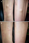
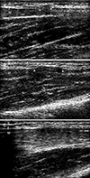
A "dynamic" physical examination was then performed. The nodules were more pronounced and firmer when the patient was standing with his hips,knees and ankle flexed (Fig. 1A). The lesions quickly disappeared at rest, i.e., with plantarflexion (Fig. 1B). Anterior tibialis muscle herniation was suspected, and the patient was evaluated by conducting an ultrasound examination with using a HDI-5000Ⓡ Scanner (ATL Ultrasound, Bothell, The Use of Dynamic Ultrasonography for the Confirmation of Lower Leg Muscle Herniation WA, USA) and a 7- to 12-MHz linear array transducer in the longitudinal and transverse planes. Both static and dynamic sonographic imaging examinations were conducted. The dynamic examination included imaging during rest, isometric muscle contraction and compression of the herniation for reduction. The longitudinal sonogram at rest showed that the fascia was thinned and elevated by a small muscle bulge (Fig. 2A). With active dorsiflexion of the feet, the anterior tibialis muscle was shown to slide under the thinned fascia, forming a greater degree of muscle protrusion (Fig. 2B). A reduction of the muscular herniation below the fascial defect was demonstrated with probe compression (Fig. 2C). The diagnosis of anterior tibialis muscle herniation was confirmed. The daily use of compression stockings and the avoidance of vigorous exercise were recommended. Follow-up was conducted after three months, and this revealed an increase in the size of the herniated muscles, which produced significant shin pain. The patient was then referred to the Orthopedic Surgery Department for surgical treatment.
DISCUSSION
Muscle herniation is defined as a protrusion of a portion of a muscle through a defect of the muscle fascia4. The problem is mainly cosmetic, but the disorder may cause spontaneous pain, cramps or local tenderness4. Muscle herniation is a relatively common complaint among athletes and it has been frequently reported in the orthopedic surgery literature56. Most dermatologists are unfamiliar with this condition, and they are likely to consider it as a peculiar kind of tumor17. For the case presented herein, the provisional diagnosis was angiolipomatosis, and an unnecessary skin biopsy was performed.
Although the diagnosis of muscle herniation can be suspected upon careful physical examination (with performing the "fencer's lunge" maneuver)1, ultrasonographic evaluation can be useful for making the definite diagnosis. Discontinuity at the site of the fascial defect with the associated muscle herniation can be demonstrated on the static sonographic images2. Examination during plantarflexion and dorsiflexion of the foot can demonstrate the varying size and shape of the herniation2, which is postulated to reflect an increased pressure within the anterior fascial compartment of the leg4. Transducer compression over the mass permits demonstrating the reduction of the herniated muscle4, but this procedure could potentially result in a false negative examination if one does not recognize the persistent underlying fascia defect2. Some authors2 have suggested that the addition of immediate post-exercise imaging could improve the conspicuity of the lesions and the confidence in making the proper diagnosis.
Other imaging techniques such as computerassisted tomography (CT) and MRI have been used to identify the fascial defect38. Muscles and the enclosing fascia have similar attenuation on CT and they are not easily separable. The CT diagnosis of a fascial defect is therefore presumptive, whereas with MRI, there is direct visualization of both the fascial rent and the muscle bulge8. Yet the depiction of the fascial defect with MRI is not always straightforward3. In the case presented herein, a fascial defect could not be identified with MRI, and this was possibly due to the close apposition of different structures in a reduced space. Moreover, it is not easy to perform a dynamic study with MRI as the patients are usually not able to cooperate properly3.
Ultrasound scanning has recently become an important diagnostic tool in dermatology because it is easy to use, completely safe and it provides important diagnostic information9. We believe that dermatologists should be familiar with anterior tibialis muscle herniation so that they can arrive at a proper diagnosis and make a surgical referral when they encounter positional subcutaneous nodules in the lower legs. We also suggest that dynamic ultrasonography is a non-invasive, highly accurate, readily available and cost-effective imaging technique for confirming muscle herniation.
 XML Download