

 PDF
PDF ePub
ePub Citation
 Print
Print


INTRODUCTION
Kaposi's sarcoma (KS) is a rare lympho-angioproliferative disease. Classical KS usually appears in elderly Eastern European Jewish or Mediterranean men, with male to female ratio of approximately 3:11. It is clinically characterized by single or multiple pea-sized bluish-red macules on the distal portions of the lower extremities. The lesion usually progresses very slowly and may coalesce to form large plaques, nodules or tumors. Histologically, KS evolves through stages of patches, plaques, and tumors. The characteristic histological features are spindle cell proliferation, an erythrocyte-filled, honeycomb-like pattern of vascular spaces and small vessels lined with prominent endothelial cells. The treatment of KS depends on the extent and the localization of lesions as well as on the clinical type of the disease, and includes ionizing radiation, cryotherapy, surgical excision, photodynamic therapy, laser irradiation, chemotherapy and interferon-α2.
We report herein a case of recurrent classical type of Kaposi's sarcoma successfully treated by interferon (INF)-α.
CASE REPORT
Patient: A 58 year-old male
Chief complaint: A tender, firm, erythematous nodule with surrounding small papules and macules on the right sole for 6 months
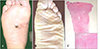
Physical examination: A 1.2 cm sized, firm,dome shaped, erythematous nodule and multiple, 7 ~8 mm-sized, satellite purplish macules and papules (Fig. 1A)
Histopathologic findings: The biopsy specimen showed numerous dilated, anastomosing slit-like vascular spaces through the entire dermis (Fig. 1C). The vascular structures were lined by a single layer of endothelium and there was diffuse extravasation of erythrocytes.
Associated findings: Laboratory examinations showed a negative anti HIV antibody, and the chest and abdominal CT and lymphangiography results were non-specific.
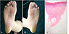
Treatment and progress: The patient was successfully treated with subcutaneous injections of recombinant human (rHu) INF-α three times a week for 6 months, leaving hyperpigmented macules on the right sole. After 3 years, the patient revisited our hospital due to an asymptomatic, solitary patch on the left sole for 5 months (Fig. 1B). The lesion was histopathologically confirmed to be KS. The patient was treated with fractional ionizing radiation with a total dose of 3,000 cGy, but there was no response. We then administrated six million units of rHu INF-α three times per week for 6 months. The lesion showed complete resolution and there has been no recurrence for 1 year after treatment (Fig. 2).
DISCUSSION
Kaposi's sarcoma (KS) is a lympho-angioproliferative disease, having four variants; epidemic or AIDS-associated KS, endemic or African KS, iatrogenic immunodepression-associated KS and Mediterranean or classical KS (CKS). Classical KS is characterized by single or multiple pea-sized bluish-red macules on the distal portions of the lower xtremities.
The treatment of KS depends on the extent and the localization of lesions as well as on the clinical type of the disease. Modalities include ionizing radiation, cryotherapy, surgical excision, photodynamic therapy, laser irradiation, chemotherapy and INF-α2. Chemotherapy has been used with fair to good results but often with high recurrence rates despite prolonged maintenance therapy. In cases of transplant-related KS, foscarnet4, anti-CD 205 , and sirolimus6 have been used successfully, yet sometimes with an increased risk of acute rejection. Radiotherapy is known to be effective in controlling KS in local disease but not with systemic involvement7. Photodynamic therapy is a promising modality for the treatment of diseases that involve uncontrolled growth of tissues, including several types of cancer and infections. It has been used in KS patients with a moderate amount of success, sometimes resulting in phototoxic reactions or poor cosmetic results8.
The interferons are a group of naturally occurring proteins that inhibit the growth of tumors in vivo and of many transformed cells in vitro. The antitumor mechanisms of action in vivo are complex and may involve direct inhibitory antiproliferative activity on tumor cells, antiviral effects, antiangiogenic action with early degeneration of the endothelial cells in the vessels of the tumor, and modulation of the cellular and humoral immune response. The most common side effects are flu-like symptoms, but weight loss, nausea, depression, anemia, and transient neutropenia may also occur. In classical KS, a low dose of INF-α treatment is effective. Some authors reported that relapse after INF treatment was delayed and very limited, and recurrences were often successfully treated with INF treatment2. Furthermore, it has been shown that rHu INF-α is particularly useful for AIDS patients with Kaposi' sarcoma who have relatively well-preserved immune function, resulting in a major remission rate, ranging from 3 to 67%9.
There are a myriad of modalities for the treatment of KS, but not one is 100% successful. Clinicians are often challenged in selecting the appropriate therapeutic plan, and such decisions must be made based on a thorough review of each individual patient. Based on the results of our case, we suggest that rHu INF-α treatment of patients with classical KS is a promising option.
 XML Download