

 PDF
PDF ePub
ePub Citation
 Print
Print


INTRODUCTION
Eruptive collagenoma is a rare acquired connective tissue hamartoma consisting predominantly of collagen without family history1. Eruptive collagenoma has been described as multiple small papules with areas of decreased or degenerated elastic fibers, usually on the trunk and arms1. We herein report a case of eruptive collagenoma which developed on the left calf in a 16-year-old boy.
CASE REPORT
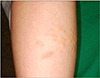
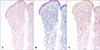
A 16-year-old boy presented with numerous asymptomatic papules on the left calf. The lesion first appeared at the age of 10 years and had been slowly increasing in number and size. He denied previous skin eruptions or trauma to the areas where the papules developed. He had no other significant problems in health and there was no family history of similar disorders. Physical examination revealed multiple slightly raised, yellowish grouped papules on the left calf (Fig. 1). The papules measured 3 to 5 mm in diameter. The initial clinical diagnosis was sebaceous hyperplasia, xanthoma or collagen disorders. The histological examination revealed unremarkable epidermis and slightly homogenized condensed collagen and decreased elastic fibers in the dermis (Fig. 2A). Dense fascicular bundles of collagen were highlighted by Masson's trichrome (Fig. 2B) and decreased and fragmented elastic fibers were revealed by Verhoeff-van Gieson stain (Fig. 2C). Based on these features, the skin lesion on the left calf was diagnosed as eruptive collagenoma. No specific treatment was given.
DISCUSSION
Connective tissue nevi of the skin are acquired hamartomatous lesions consisting predominantly of one of the components of the extracellular matrix, namely collagen, elastin, or glycosaminoglycans12. Among these, collagenomas are connective tissue nevi composed predominantly of collagen2. They have been classified as either inherited or acquired. Inherited collagenomas include familial cutaneous collagenoma and Shagreen patch of tuberous sclerosis2. Acquired collagenomas contain isolated collagenoma and eruptive collagenoma depending on the number of lesions but they cannot be differentiated clinically3.
Eruptive collagenoma appears in the first or second decades as raised, cutaneous nodules or scattered papules on the trunk and arms4. The lesions present as asymptomatic, skin-colored domeshaped papules as well as nodules of various size but usually less than 1 cm in diameter. And there is no established family history or associated systemic findings4.
Histopathologically the lesions are characterized by an excessive accumulation of dense, coarse collagen fibers in the dermis. Elastic fibers appear diminished in number, perhaps representing a dilution phenomenon due to excess collagen accumulation14.
Eruptive collagenoma should be differentiated from other diseases with focal absence of elastic fibers such as nevus anelasticus and papular elastorrhexis. Nevus anelasticus has been defined as acquired perifollicular papules with a paucity or lack of elastic tissue5. Papular elastorrhexis is a variant of nevus anelasticus and it occurs in the twenties as multiple asymptomatic small, white papules scattered over the trunk and extremities with no predilection for the perifollicular areas6. Nevus anelasticus and papular elastorrhexis show histologically focal area of decreased and fragmented elastic fibers6 and most cases are sporadic but some familiar occurrence has been described3. Some authors have mentioned these three entities represent a single disease or disease spectrum because of similar clinical and histopathologic features789. They also have common features in terms of peak age of onset, distribution of lesion involving the trunk and upper extremities, and a lack of history of trauma, inflammation, family history, or extracutaneous manifestations7.
The pathogenesis of eruptive collagenoma is unknown. Uitto et al10 showed that collagenoma almost exclusively consists of type I collagen and the underlying defect seemed to be a reduced production of collagenase in that location, and therefore a decreased local degradation of collagen. And some reports that the growth of collagenoma was influenced during pregnancy or puberty implythat hormone may be involved in the pathogenesis of this disorder111.
No specific treatment is given in most cases11. Two cases of eruptive collagenomas were reported to be treated with intralesional steroids with transient flattening of the lesions79. Transient but exaggerated dermal atrophy after intralesional steroid injections may have resulted from the absence of elastic fibers in this disorder9.
Clinically, our patient's lesion could have been confused with sebaceus hyperplasia, xanthoma and collagen disorder. However, typical elastic fibers supported the diagnosis of collagenoma. In addition to its clinical features, the facts that it was acquired without a family history and associated disorders, were in favor of the diagnosis of eruptive collagenoma. Our case has a different characteristic from the previously reported cases. Nine cases4911121314151617, in the English literature and two cases1819 in Korean literature have been reported to date. Most patients developed eruptive collagenoma mainly on the trunk, abdomen and upper extremities, whereas one patient developed lesions localized on the left back16. To our knowledge, this is the first report of the eruptive collagenoma localized characteristically on the left calf. But the cause of distribution was not known.
In conclusion, we describe a case of eruptive collagenoma that occurred on the left calf, an area which is seldom affected in isolation.
 XML Download