

 PDF
PDF ePub
ePub Citation
 Print
Print


INTRODUCTION
Port-wine stain (PWS) represents a type of congenital malformation involving mature dermal capillaries resulting in irreversible dilatation of capillaries. PWS is not only congenital but also acquired. All types are pathologically indistinguishable and represent progressive ectasia of vessels located in the superficial dermal plexus. In contrast to well-known congenital port-wine stain (CPWS), acquired port-wine stain (APWS) is rare and its onset is generally after one year of age. The exact patho-mechanism of APWS is unknown, but trauma 123 hormonal change1, medication4, and solar damage56 may contribute to its development. Pulsed dye laser (PDL) therapy is regarded as the treatment of choice in PWS. Some scientific researchers reported that APWS patients responded more favorably to PDL therapy, and required fewer treatments than those with CPWS12. They proposed telangiectatic nature, relatively sparse number and superficial location of ectatic vessels as a cause of better response in APWS2; however, at the same time, poor results in several patients were also reported by some authors12. Generally, pathological parameters like vessel diameter, depth, and luminal erythrocytes contents were provided as evidence that could explain the difference in therapeutic outcome789. Therefore it was thought that there might be some pathological differences between APWS and CPWS; however, no research has been conducted on this discrepancy to date. Thus, we undertook a project to reveal such histopathologic differences between APWS and CPWS, using a computer assisted image analysis.
MATERIALS AND METHODS
Materials
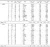
The skin biopsies of 31 patients before treatment were included in this study. The patients consisted of 14 APWS and 17 CPWS. The mean age was 26.7 years in APWS and 25.7 years in CPWS. In APWS, the ages of the onset were from 2 to 40. Confirmation of the acquired nature of the lesion was obtained in each case by reviewing photographs of the patient taken prior to onset of the lesion. If not available, then family members confirmed the time of onset. The details of these patients are shown in Table 1.
Methods
1) Histopathologic assessment
Punch biopsies taken using local anesthesia were fixed with a formalin-mucriculide-acetic acid (FMA) solution embedded in paraffin. Immunohistochemical stain using factor VIII-related antigen was obtained from the biopsy specimens to better visualize the vessels. Detailed histopathologic analyses of each specimen were made with the assistance of a computer assisted image analyzer (Image & Microscope Technology™, USA). Using this program, variable parameters of selected objects from the scanned image of the histologic slide were measured automatically. Vessel area, percentage of vascular area (the percentage of dermis occupied by vessels) and vessel depth were measured down to a depth of 1 mm from the dermo-epidermal junction in all slides. For the purposes of analysis, we regarded measured area from the scanned vessel image as actual vessel area. The percentage of vascular area refers to the percentage of summation of vessels over dermal area. The statistical analysis was performed with an SPSS 10.0 statistical program. The Mann-Whitney test was used to compare differences between APWS and CPWS and a p-value of less than 0.05 was considered to be significant.
RESULTS
The Table 1 shows the summary of the mean data of vessel area, percentage of vascular area and vessel depth of each patient.
Vessel area
The mean vessel area in APWS was 1014.7 ± 782.5µm2 and CPWS, 1341.5 ± 689.9µm2. The mean data was higher in CPWS, but there was no significant statistical difference between APWS and CPWS.
DISCUSSION
In capillary malformations, there are two types: port-wine stain (PWS) and telangiectasia. PWS is one of the most common types of capillary malformation and occurs as pink to red macules or patches, usually on a unilateral side. The color of the lesion tends to gradually deepen with time. The lesion grows proportionately and becomes raised and nodular as a result. Although the initial nature is similar to PWS, fading macular stains, referred to as stork bite or salmon patch, are located most commonly on the nape of the neck, the eyelids, and the glabella and may disappear spontaneously between 1 and 3 years of age. In these cases, there is no need for treatment.
In all cases of PWS, regardless of their onset, pulsed dye laser (PDL) therapy is the treatment of choice. Not all patients however will respond to laser therapy and many studies have been done to investigate the variables influencing the response of PWS to the PDL. The possible variables include clinical features such as lesion color, location and the age of patient and pathologic parameters such as vessel diameter, vascular area, vessel depth, vessel wall thickness, and the amount of erythrocytes in vessels. To demonstrate correlation between clinical features and therapeutic response, many investigations were done with no unanimity1011121314. Histopathologic examinations of PWS have also been done to establish the relationship between pathologic parameters and therapeutic responses789151617. Hohenleutner et al15 confirmed histochemically in post-PDL treated PWS biopsies, superficial PWS vessels of a diameter up to 150µm were completely coagulated. With increasing vessel diameter, strong superficial hemoglobin absorption led only to partial vessel wall coagulation. Also, deeper vessels were not coagulated because of shadowing from superficial vessels. In addition, the overall coagulated depth was limited to a maximum of only 0.65 mm. Fiskerstrand et al8 examined pretreatment biopsies in 30 patients with PWS. They found that the vessels of the good responders were located significantly more superficially than the vessel of the moderate and poor responders, and the poor responders had significantly smaller vessels than those of the moderate and good responders. The authors concluded that the therapeutic result was dependent on both the vessel diameter and its depth. Eubanks and McBurney16 used videomicroscopy and found that PWS in areas that typically respond well to laser treatment (V3 dermatome, neck, and trunk) were more likely to have a superficial pattern and PWS in areas that have a poorer response to therapy (V2 dermatome, distal extremities) were more likely to have a deeper pattern. Hence, it was suggested that both the depth and the diameter of the ectatic blood vessels in PWS have influence on the response to PDL.
Most cases of port-wine stain (PWS) are congenital, but the acquired form of PWS has only recently been described. In 1939, Traub18 reported the first case of acquired port-wine stain (APWS). The average age of onset of APWS is usually after 1 year. Many cases of acquired PWS have since been reported123518 but there has been little discussion about treatment12. There have been only two reports investigating the efficacy of laser therapy in APWS12. Dinehart et al1 found that APWS had generally a faster response to the PDL treatment than CPWS, however, they could not explain the exact mechanism because some patients responded rather poorly to laser treatment. Lanigan2 supposed that APWS's telangiectatic nature, relatively sparse number and superficial site of ectatic vessels probably explained why the response was better than CPWS to laser therapy. Therefore, he concluded that patients with APWS could be expected to respond well to PDL therapy and fewer treatments were required than those patients with CPWS. But in these articles, only small numbers of patients were studied and the therapeutic results were not same in all patients with APWS. As for our experience of long pulsed dye laser in APWS therapy, the result was rather similar to that of Dinehart et al1. The response to PDL in patients with APWS was controversial, so we undertook this study to find out whether there are some differences of histopathologic features of the two entities or not.
To demonstrate histopathologic differences between the two groups by using image analyzer, we compared APWS with CPWS by three parameters such as vessel area, percentage of vascular area, and vessel depth. This is not objective data, but we thought it not a problem when viewing comparison of the two groups. In our study, we measured vessels to a depth of 1 mm from the dermoepidermal junction. The reason for selecting this upper depth is based on mathematical modeling which predicts that only ectatic blood vessels at a depth of less than 800–900µm contribute to the visual appearance of the color of PWS919. Another problem in measuring depth was that the epidermal base was not a straight line. Therefore we regarded that epidermal base was averaged into a straight line by roughly bisecting the line of papillary dermal tips and line of tip of rete ridges.
There were no statistically significant differences between APWS and CPWS. Limitations of this study such as small numbers of patients, only one biopsy specimen in each person, bias in method and so forth may affect the result.
In conclusion there were no significant histopathologic differences of variables between APWS and CPWS. We could neither find any histopathologic differences that had an influence on therapeutic results, nor suggest any factors that explain the possibility of better response in APWS to the laser. In order to establish, therefore, whether there is a difference in the therapeutic response of PDL between the two entities, further investigation of other factors not involved in this study such as vessel wall thickness, luminal erythrocytes contents and so on, will be needed.
 XML Download