

 PDF
PDF ePub
ePub Citation
 Print
Print


Abstract
Background and Objectives:
To review our experience with lacrimal sac tumors in an effort to identify features, to evalu-ate the results of various methods of management, and to compare our data with previous studies.
Methods:
We reviewed the medical records of all patients with lacrimal sac tumors who were managed in our institution between January 1990 and December 2015. The preoperative clinical data, imaging, operation notes, and follow-up records were reviewed for each patient.
Results:
The study group consisted of four men and six women with a mean age of 47.6 years. Most patients experienced long-standing epiphora, for a mean period of 20 months. Two of the tumors were benign, and eight of them were malignant. The benign tumors were treated with dacryocystectomy. All but one malignant tumor were treated with medial or total maxillectomy. Adjuvant radiotherapy was administered to four patients with malignant tumors. In the eight patients with malignant tumors, the mean follow-up period was 65 months.
Conclusions:
Important characteristics of lacrimal sac tumors include dacryocystitis, epiphora, and in some cases, a palpable medial canthal area mass. Wide en bloc resection via medial or total maxillectomy and/or postoperative radiotherapy are proper treatments for malignant lesions of the lacrimal sac.
Go to : 
REFERENCES
1). Schenck NL, Ogura JH, Pratt LL. Cancer of the lacrimal sac. Pre-sentation of five cases and review of the literature. Ann Otol Rhinol Laryngol. 1973; 82:153–61.
2). Hornblass A, Jakobiec FA, Bosniak S, Flanagan J. The diagnosis and management of epithelial tumors of the lacrimal sac. Ophthalmolo-gy. 1980; 87:476–90.

3). Ni C, D'Amico DJ, Fan CQ, Kuo PK. Tumors of the lacrimal sac: a clinicopathological analysis of 82 cases. Int Ophthalmol Clin. 1982; 22:121–40.
4). Spira R, Mondshine R. Demonstration of nasolacrimal duct carcinoma by computed tomography. Ophthal Plast Reconstr Surg. 1986; 2:159–61.
5). Stefanyszyn MA, Hidayat AA, Pe'er JJ, Flanagan JC. Lacrimal sac tumors. Ophthal Plast Reconstr Surg. 1994; 10:169–84.
6). Saccogna PW, Strauss M, Bardenstein DS. Lymphoma of the nasolacrimal drainage system. Otolaryngol Head Neck Surg. 1994; 111:647–51.
7). Pe'er J. Lacrimal Sac Tumors. Clinical Ophthalmic Oncology. Springer;2014. p. 115–21.
8). Parmar DN, Rose GE. Management of lacrimal sac tumours. Eye (Lond). 2003; 17:599–606.
9). Montalban A, Liétin B, Louvrier C, Russier M, Kemeny J-L, Mom T, et al. Malignant lacrimal sac tumors. European annals of otorhino-laryngology, head and neck diseases. 2010; 127:165–72.
10). Ryan SJ, Font RL. Primary epithelial neoplasms of the lacrimal sac. Am J Ophthalmol. 1973; 76:73–88.
11). Skinner HD, Garden AS, Rosenthal DI, Ang KK, Morrison WH, Esmaeli B, et al. Outcomes of malignant tumors of the lacrimal ap-paratus: the University of Texas MD Anderson Cancer Center experience. Cancer. 2011; 117:2801–10.
12). Rootman J, White VA. Changes in the 7th edition of the AJCC TNM classification and recommendations for pathologic analysis of lacrimal gland tumors. Arch Pathol Lab Med. 2009; 133:1268–71.
Go to :
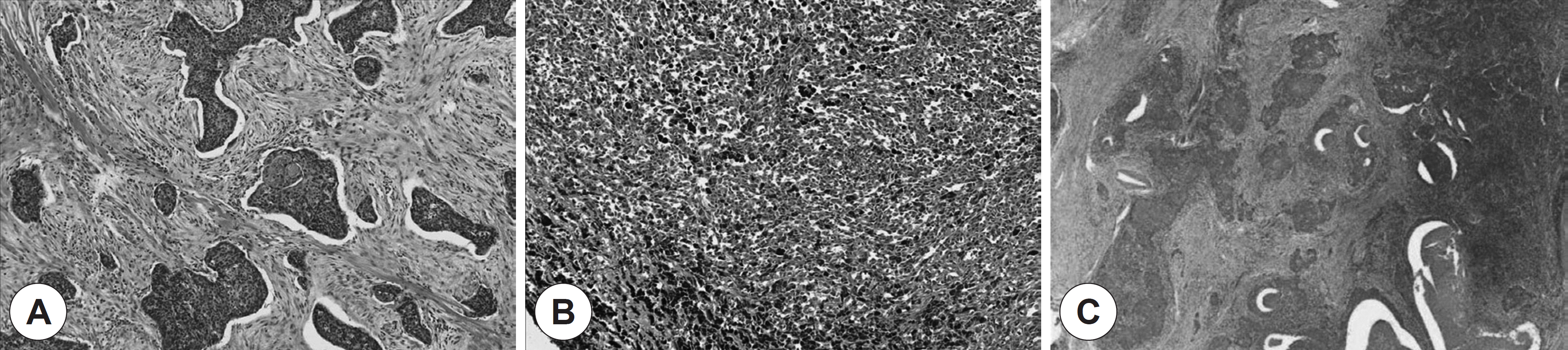 | Fig. 1.Histopathologic findings of lacrimal sac tumors. ( A) Squamous cell carcinoma ( Case 10). The tumor shows a squamous nest containing keratin pearls and hyperchromatic cells with nuclear atypia (×100, H&E stain). ( B) Malignant melanoma ( Case 8). The tumor shows diffuse sheets of melanoma cells, with melanin pigments that show hyperchromatic nuclei and prominent nucleoli (×100, H&E stain). ( C) Undifferentiated carcinoma ( Case 9). The tumor shows a nested pattern with comedonecrosis without squamous or glandular differentiation (×40, H&E stain). |
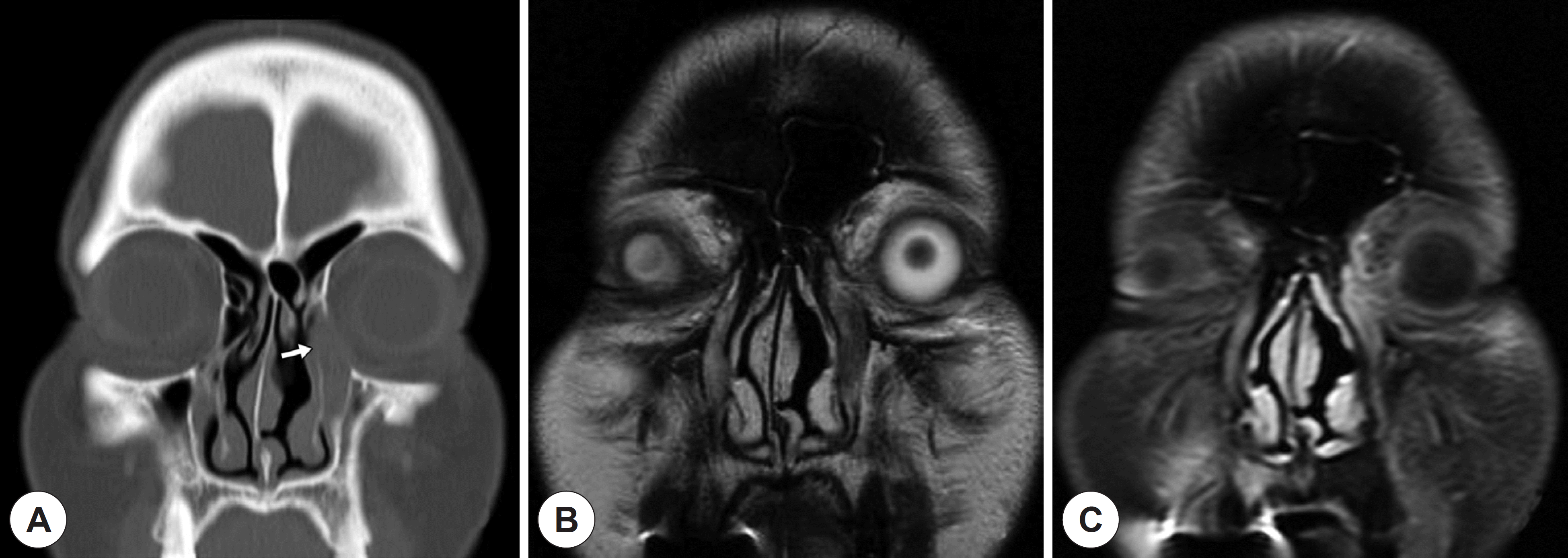 | Fig. 2.Radiologic findings of the patient with undifferentiated carcinoma ( Case 9). ( A) Computed tomogram shows expansion of the lacrimal sac fossa with bony erosion ( white arrow). ( B) Magnetic resonance imaging shows a low signal intensity lesion on T2 weighted image, extending to upper nasolacrimal duct. ( C) The mass shows heterogeneously enhancement on contrast en-hanced T1 weighted image. These findings provide more accurate soft-tissue differentiation. |
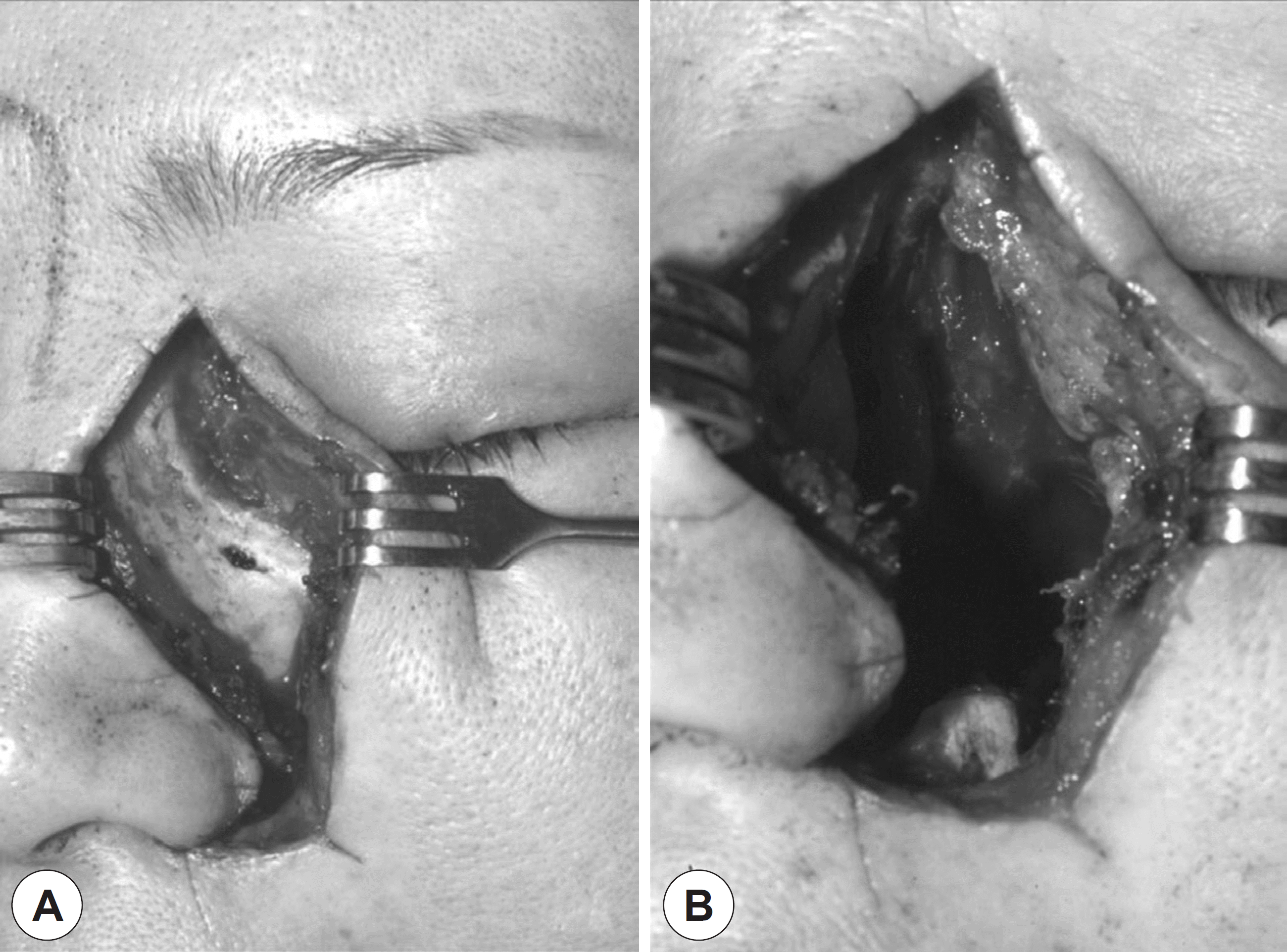 | Fig. 3.Intra-operative findings ( Case 9). ( A) Exposure of the lesion was done with a lat-eral rhinotomy incision. ( B) The tumor and surrounding structures were excise d with a medial maxillectomy. |
Table 1.
Clinical characteristics of lacrimal sac tumors
 XML Download