

 PDF
PDF ePub
ePub Citation
 Print
Print


INTRODUCTION
Honey has been used for thousands of years as a medicinal agent and dietary supplement. Contents of honey vary but generally consist of sugars, vitamins, minerals and bee proteins, along with phytochemicals. The use of honey as a therapeutic agent is one of the earliest reported methods of wound-care which accelerates and improves wound healing.1) There is a growing body of evidence indicating that honey exhibits beneficial immunomodulatory activities for the treatment of inflammation and inhibition of many bacterial pathogens.2) Honey also has antioxidant, anti-tumor, anti-inflammatory, anti-mutagenic and anti-viral properties, with the observed physiological effects dependent on the nutritional composition, osmolality, pH, and hydrogen peroxide activity of the honey consumed.3)4) There is a demonstrated variation in antibacterial activity between honeys obtained from different floral origins and geographical sources, which confirm that different honey types will have different antibacterial properties. Antibacterial effect of honey is determined by hydrogen peroxide, polyphenoic acid and flavonoids components.5) Oral administration of honey has been shown to decrease inflammatory bowel disease, indomethacin-induced gastritis, and radiation induced oral mucositis.6)7)8)
Black locust honey is one of the most popular unifloral types of honey and commonly produced during spring season in Korea. Korean black locust honey suppressed bacterial growth and their antibacterial activity was associated with the content of hydrogen peroxide.9) New Zealand honeys, such as manuka and kanuka, have been studied for a long time and they have anti-inflammatory and anti-bacterial activities. Manuka honey is active against a broad spectrum of gram-positive and gram-negative bacteria, with biocidal activity at 33% v/v2. Both manuka and kanuka honey exhibited potent, dose dependent reduction of inflammation. The antibacterial property of New Zealand honey has been attributed to the presence of methylglyoxal and diacyl compounds which may provide anti-inflammatory effect.3)4)5) However, the immunologic characteristics of honey were not commonly studied. There is a variation in immunologic activity between honeys obtained from different floral origins and geographical sources, which confirm different honey types will have different properties. The aim of this study was to compare the anti-inflammatory effects and immunologic characteristics of manuka, kanuka and black locust honey.
METHODS
Preparation of honey solution
Manuka and kanuka honey were obtained from the Honey NZ International Ltd (Hamilton, New Zealand) and Black Locust honey was obtained from a beekeeper in Yongchon (South Korea) area. The honey samples were gamma irradiated using a cobalt-60 irradiator at 25 kGy then diluted by weighing out 1.37 g of honey and diluting with 19 ml of RPMI 1640 to achieve a V/V concentration of 5%. To remove pollen and bee tissues, honey was filtrated with Minisart Sartorius filter (Millipore Corp., Seattle WA, USA). Honey was kept at 4℃ in dark containers to prevent enzyme denaturation and degradation.
Culture of peripheral blood mononuclear cells (PBMCs) with honey
Heparinized venous blood was collected from ten healthy volunteers (6 men and 4 women; 35.3±9.6 years) after obtaining informed consent which was approved by the Institutional Review Board at the Daegu Catholic University Hospital. PBMCs were isolated by density gradient centrifugation on Histopaque (Sigma, St. Louis, MO, USA). Cells from the interphase were harvested, washed and resuspended at 2×10⁶ cells/ml in RPMI-1640 medium (Gibco, Rockville, MD, USA) supplemented with 10% calf-serum, 2 mmol/L glutamine and 50 µM 2-mercaptoethanol. The cell suspension was distributed in each well in duplicate on a 96-well culture plate and cultured.
PBMCs were incubated at 37℃, 5% CO2 in 96 well plates in complete RPMI-1640 supplemented with 10 ug/ml of lipopolysaccharide (LPS) in the absence or presence of various concentration of honey (0, 0.05, 0.1, 0.5, and 1%) for 72 hours at 37℃, 5% CO2. Cell free culture supernatants were harvested, aliquoted and frozen at -70℃ for further experiments. Cell cytotoxicities were measured using a CellTiter-96® aqueous cell proliferation assay kit (Promega, Madison WI, USA). For CellTiter-96® aqueous cell proliferation assay, tetrazolium compound and Owen's reagent were added to each well and incubated for 4 hours at 37℃ in humidified 5% CO2 chamber. Color intensities were assessed with a microplate reader at 490 nm wavelength. And to determine whether the LPS enhance the toxicity of honey on PBMC, cytotoxicities were measured after 48 hours that PBMCs cultured with LPS and three kinds of honeys.
Interleukin-5 (IL-5), IL-10, interferon-γ (INF-γ), and tumor necrosis factor-α (TNF-α) levels in supernatant were measured to determine the anti-inflammatory effect of honey in duplicate using commercially available enzyme-linked immunosorbent assay kit (Endogen, Wobrun, MA, USA) according to the manufacturer's instruction. The sensitivities of this kit were 2 pg/mL for these cytokines.
Statistical analysis
Results are expressed as means±SD of five separate experiments using different donors. Honey cytotoxicity was analyzed using the one-factor repeated measure analysis. One-way analysis of variance and Dunnett's test for multiple comparison was performed to determine the overall effect of honey pretreatment on PBMC (SPSS ver. 14.0; SPSS Inc., Chicago, IL, USA). p values of less than 0.05 were considered to be statistically significant.
RESULTS
Effect of honeys on the proliferation of PBMC
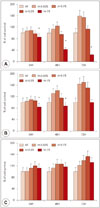
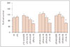
When PBMC was treated with three different honeys, PBMC showed increase in cell proliferation with time dependent manner. At high concentration, 1% of manuka and kanuka honey significantly suppressed cell proliferation, which seems to be a cytotoxic effect of these two honeys. However, Black Locust honey did not show the cytotoxic effect. The cytotoxic effect of 1% manuka honey showed time dependent manner (Fig. 1). When the PBMC was treated with three kinds of honeys and LPS, 1% of Manuka, kanuka, and black locus honey significantly suppressed cell proliferation (Fig. 2). Since honeys showed cytotoxic effects, further experiments were performed under 0.5% of honeys.
Effect of honeys on the production of chemical mediators
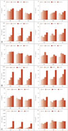
When PBMCs were cultured with various concentrations of three different kinds of honeys, chemical mediator production tended to increase at dose dependent manner. Among four different kinds of chemical mediators, TNF-α and IL-10 were significantly increased by manuka and kanuka honey compared with non-treated group. TNF-α production was significantly increased as a dose dependent manner. TNF-α production was highest after 24 hours culture with 0.5% of manuka honey (2225.6±378.5 pg/mL) and after 48 hours culture with 0.5% of kanuka honey (2551.6±472.6 pg/mL). IL-10 production was only significantly increased at 0.5% of manuka and kanuka honey. 0.5% of black locust honey, only enhanced TNF-α production from PBMCs (Fig. 3).
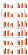
PBMCs were pretreated with honeys for 1 hour and then stimulated with LPS. LPS induced INF-γ production (example; 81.2±23.5 pg/ml after 48 hours) was significantly inhibited with 0.5% of Manuka (47.2±13.6 pg/mL) and kanuka (53.4±17.9 pg/mL) honey, and LPS induced IL-10 production (example; 525.1±69.7 pg/mL after 48 hours) was significantly inhibited with 0.5% of three kinds of honeys (manuka, 142.0±32.8 pg/mL; kanuka 2582.2±117.3 pg/ml; black locust, 213.6±83.2 pg/mL). However, honeys did not influence the LPS induced IL-5 and TNF-α production (Fig. 4).
DISCUSSION
Honey has various biological and pharmacological activities with anti-inflammatory, antioxidative, antibacterial, antihypertensive, and hypoglycemic effects. Honey exhibits immunomodulatory activities, it not only inhibits but also stimulates immune responses.3)4)10) Honey has various source of contamination with microorganisms, which include pollen, the digestive tract of honey bee, dust, nectar and human contamination by air, food handlers, equipment and cross-contamination.11) To prevent contamination and improve the quality of honey, gamma irradiation as a phytosanitary treatment had performed and 25 kGy of gamma-irradiation was sufficient to achieve sterility for honey.12) In this study, three honey samples, derived predominantly from manuka, kanuka, and black locust were found to exhibit differences in their ability to induce or suppress the production of cytokines from PBMC.
PBMCs contain lymphocytes, monocytes, and granulocytes and can produce various chemical mediators. Among these chemical mediators, we measured INF-γ, which represents Th1 response; IL-5, which represents Th2 response; IL-10, which represents anti-inflammatory response; TNF-α; which represents acute phase inflammation. Honeys itself induced the production of TNF-α and IL-10 from PBMCs. Both manuka and kanuka honey stimulated similar amounts of TNF-α and IL-10, but black locust honey only enhanced the production of TNF-α. These two cytokine production was increased in concentration dependent manner, without time dependency. Manuka and kanuka may have more immunostimulatory properties than the black locust honey. Although honeys stimulated the production of TNF-α in PBMC when the PBMCs were pretreated with honeys then activated with LPS, TNF-α production was not enhanced by honeys. This may not be a direct effect of honey on LPS induced TNF-α production, but other anti-inflammatory cytokines, which produced by honeys may influence TNF-α production through various feedback mechanisms. All the honeys contained endotoxin, but the concentration of endotoxin in the honeys were not sufficient to explain immunostimulatory activity. When we measured the concentration of endotoxin in honeys, black locus honey contained the highest (1.84±0.38 EU/mL) than manuka (1.64±0.65 EU/mL) and kanuka (1.66±0.72 EU/mL) honey. Hydrogen peroxide has been proven to stimulate inflammation and these components in honeys may be associated with the immunostimulatory property.13) The immunostimulatory activity of kanuka honey explained by the presence of the arabinogalactan protein.4) Honey components may differentially impact the immune modulatory function. Honeys also induce TNF-α, IL-1β, and IL-6 release via TLR4 from monocytes, and these pro and anti-inflammatory cytokines modulate the activity of many immune cells.14)
LPS is a cell wall component of gram negative bacteria which interact with toll-like receptor 4 and has a potent immunostimulatory property. LPS induced IL-10 production from PBMC was significantly suppressed by 0.5% of three kinds of honeys. LPS induced INF-γ production was significantly suppressed by 0.5% of manuka and kanuka honeys. IL-10 production was most strongly suppressed by 0.5% of kanuka honey (67.5% to 73.1%) than other honeys (manuka honey; 21.4% to 52.0%, black locust honey; 3% to 59.4%). INF-γ production was more strongly suppressed by 0.5% of kanuka honey (24.4% to 50.0%) than manuka honey (31.8% to 41.8%). There is a demonstrated variation in immunologic characteristics between honeys obtained from different floral origins and geographical sources, which confirm that different honey types have different immunological properties.3)4)15) IL-10 is an anti-inflammatory cytokine which inhibits the infiltration of neutrophils and macrophages toward the site of inflammation as well as inhibiting the expression of several chemokines and pro-inflammatory cytokines. INF-γ is a pro-inflammatory cytokine with immunostimulatory and immunomodulatory effect. Honeys maintain immune surveillance system of PBMCs by suppressing LPS induced inflammatory cytokine production, such as IL-10 and INF-γ.
Honey include both enzymatic and non-enzymatic substances, in addition to more than 150 polyphenolic compounds including flavonoids, flavonols, phenolic acids, catechins, cinnamic acid derivatives, and small amounts of other constituents such as minerals, proteins, vitamins, organic acid and other phytochemicals. Phenolic compounds exhibits anti-inflammatory effects with antioxidant properties which could be used for the treatment of inflammatory diseases.16) The components responsible for the inhibition of chemical mediator production from PBMC observed in this study remain to be determined. We used three kinds of unifloral honeys and honeys of different botanical origin must not be equated when comparing their components. Botanical factors have the greatest influence on the trace element content of honey and this difference may influence the anti-inflammatory effect of honey.
Honeys had influenced the production of chemical mediators from PBMC. Honey itself had immunostimulatory characters such as enhancing the production of TNF-α and IL-10 and also inhibiting the LPS induced IL-10 and INF-γ production with 0.5% of honeys. These results may suggest that honey has immunosuppressive effect in the local or systemic pathologic inflammation. On the other hand, without inflammation, honey may enhance local or system immune responses. Among three honeys, manuka and kanuka honey more strongly influenced the production of chemical mediators from PBMC. For the clinical use of honeys, we need to determine the optimal concentration of honey to use and to understand the immunomodulatory effects or immunologic characteristics of different kinds of honeys.
 XML Download