Abstract
The “nasal swell body” (NSB) or septal turbinate is a distinct structure of the anterior nasal septum that is observed on endoscopic and radiographic examination. It is primarily a glandular rather than a venous formation that is comprised of septal cartilage, bone, and thick mucosal lining. It is commonly found in patients with symptoms of chronic sinusitis and allergic rhinitis, and is linked to septal deviation. Space occupying lesions of the septum such as tumors, mucoceles, and pneumatization of the septum can lead to anatomical and functional disorders such as nasal obstruction and sinusitis, while more serious clinical conditions can develop when these lesions are combined with the NSB. Recently, there has been emphasis on the functional aspects of the NSB. It is especially being emphasized for clinicians to pay attention to the NSB and its connection with the stuffy nose. We report an interesting case of the NSB combined with pneumatization of the perpendicular plate of the ethmoid bone causing severe nasal obstruction and repetitive sinusitis along with a literature review.
Go to : 
References
1). Yenigun A, Ozturan O, Buyukpinarbasili N. Pneumatized septal turbinate. Auris Nasus Larynx. 2014; 41:310–2.

2). Wotman M, Kacker A. Should otolaryngologists pay more attention to nasal swell bodies? Laryngoscope. 2015; 125(8):1759–60.

3). Wexler D, Braverman I, Amar M. Histology of the nasal septal swell body(septal turbinate). Otolaryngol Head Neck Surg. 2006; 134(4):596–600.
4). Chao TK. Uncommon anatomic variations in patients with chronic paranasal sinusitis. Otolaryngol Head Neck Surg. 2005; 132:221–5.

5). Arslan M, Muderris T, Muderris S. Radiological study of the intu-mescentia septi nasi anterior. J Laryngol Otol. 2004; 118:199–201.

6). Costa DJ, Sanford T, Janney C, Cooper M, Sindwani R. Radiographic and anatomic characterization of the nasal septal swell body. Arch Otolaryngol Head Neck Surg. 2010; 136(11):1107–10.

7). Elwany S, Salam SA, Soliman A, Medanni A, Talaat E. The septal body revisited. J Laryngol Otol. 2009; 123(3):303–8.

8). Unlu HH, Altuntas A, Aslan A, Eskiizmir G, Yucel A. Inferior concha bullosa. J Otolaryngol. 2002; 31:62–4.

9). Lei L, Wang R, Han D. Pneumatization of perpendicular plate of the ethmoid bone and nasal septal mucocele. Acta Otolaryngol. 2004; 124:221–2.

10). Setlur J, Goyal P. Relationship between septal body size and septal deviation. Laryngoscope. 2010; 120(Suppl 4):S246.

11). Arslan M, Muderris T, Muderris S. Radiologic stusy of the intumescentia septi nasi anterior. J Laryngol Otol. 2004; 118(3):199–201.
Go to : 
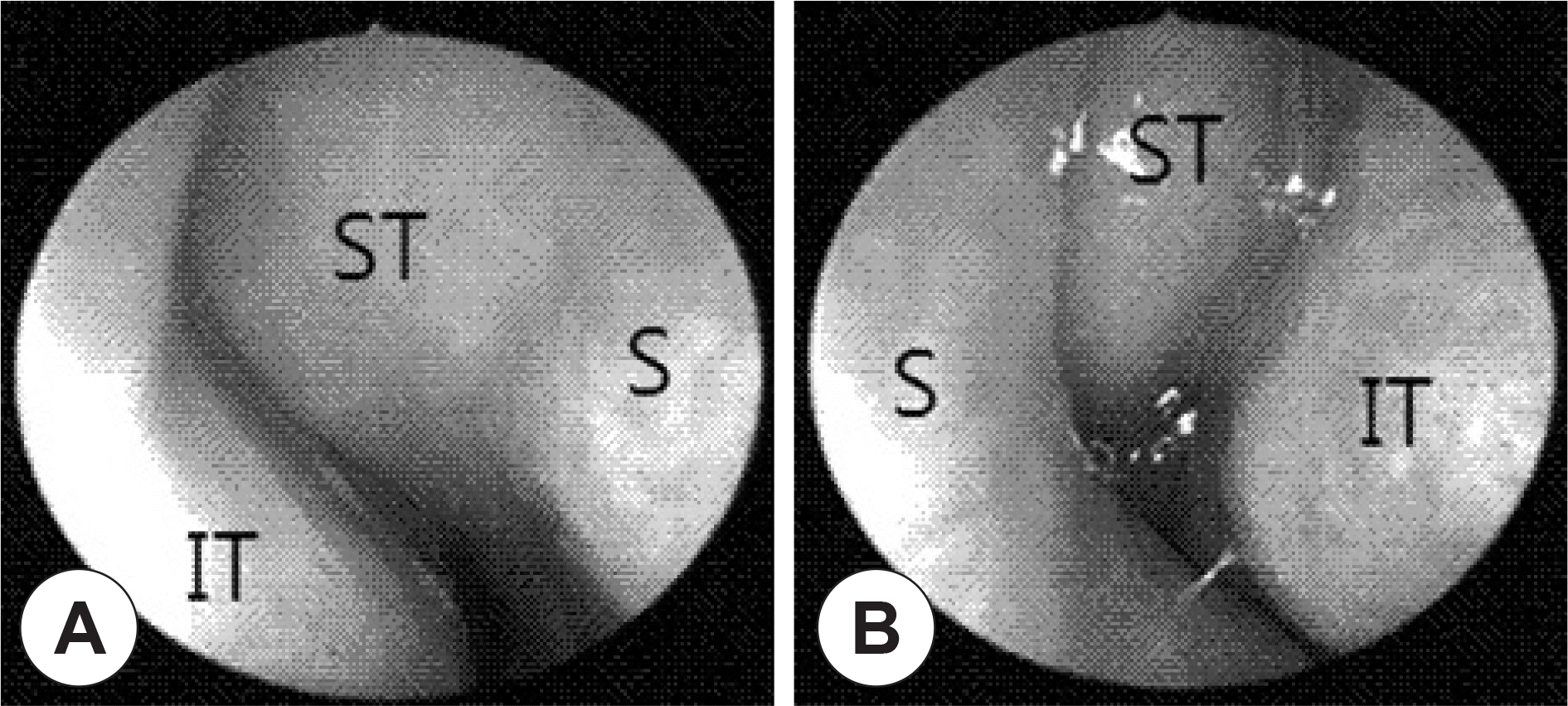
 | Fig. 1.Pre-operative nasal endoscopic findings. It showed bulging of both antero-superior portions of the septum and middle turbinates on both sides and previous surgical site could not be observed. Right side of the nasal cavity (A), left side of the nasal cavity (B). S: nasal septum, IT: inferior turbinate, ST: septal turbinate. |
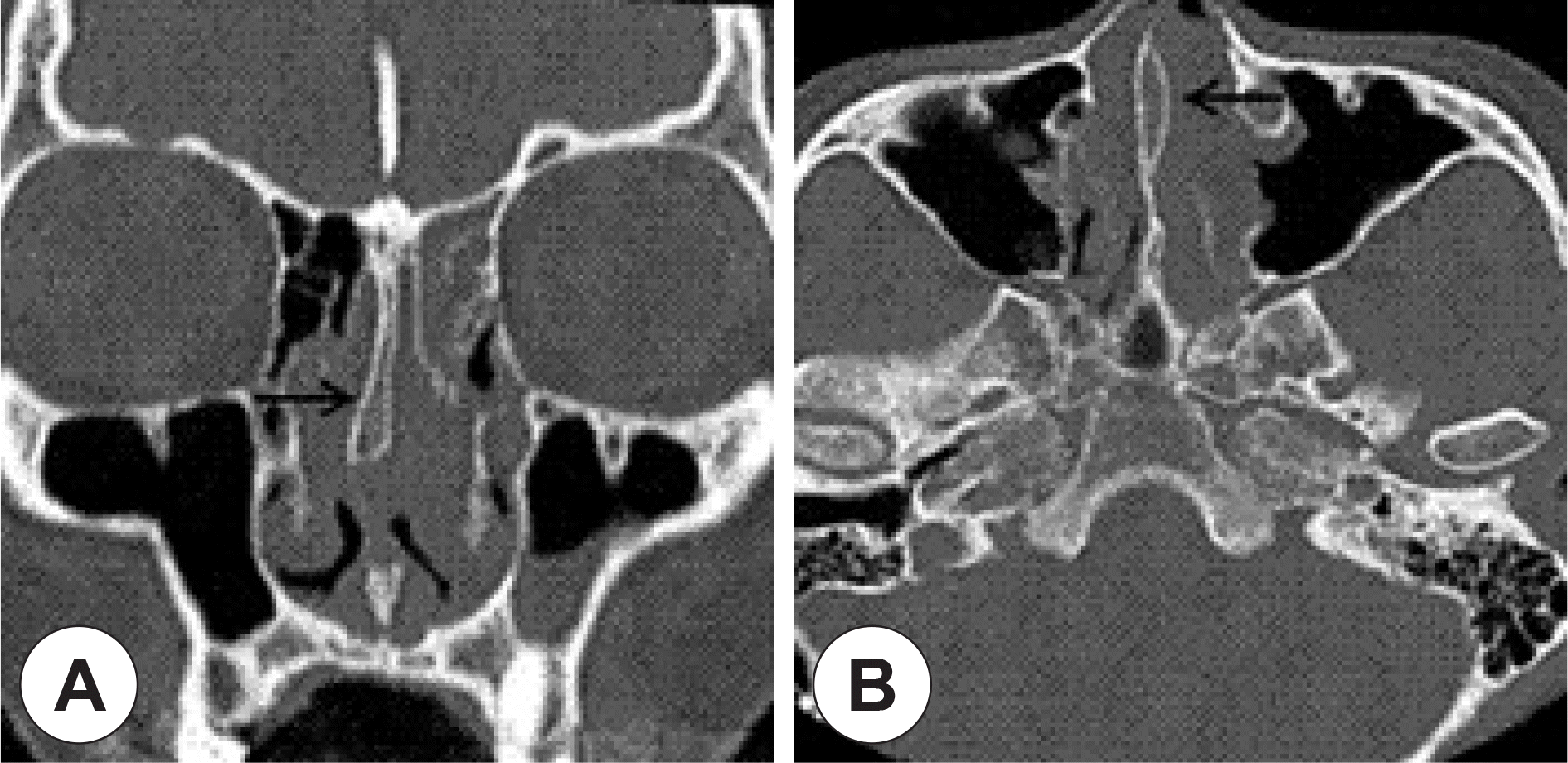
 | Fig. 2.CT images of the patient. Coronal view (A), Axial view (B). These CT images showed pneumatization of perpendicular plate of the ethmoid bone and mucosal thickening in both the antero-superior portions of the septum (Black arrow). Total opacity was observed in the right middle turbinate and the left frontal, ethmoid, and maxillary sinuses. |
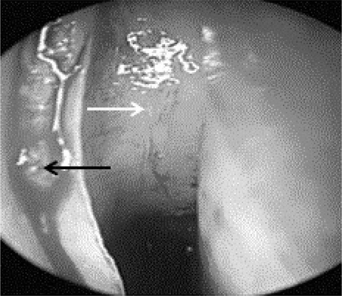
 | Fig. 3.Intraoperative finding. This figure shows a huge round mass on the bony septum. Black arrow: Bony cartilage junction of septum. White arrow: Huge round mass. |
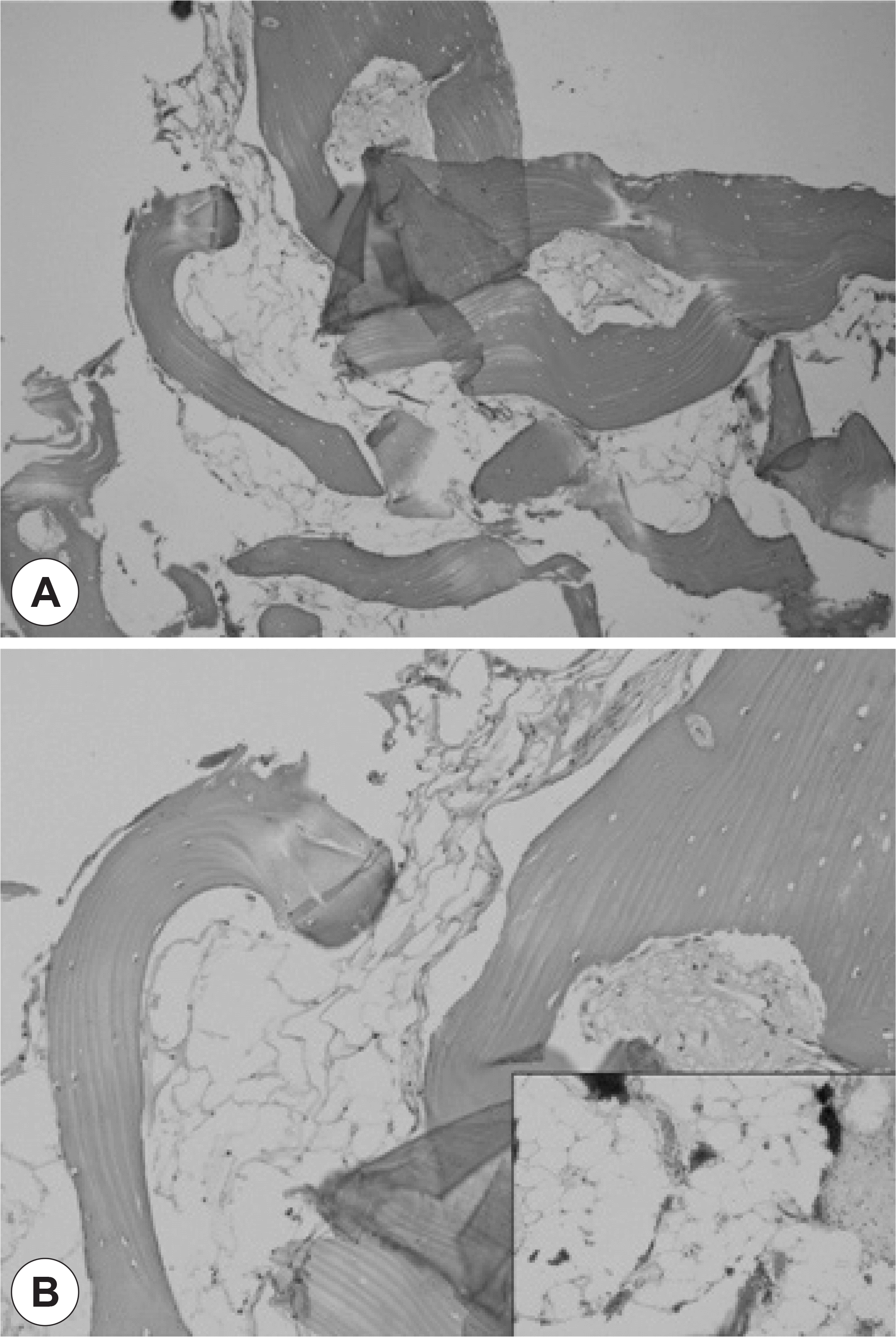
 | Fig. 4.A: An osseous nasal septum showing focal fibrous changes and fatty reduction (upper middle part) in the marrow part (H-E stain, ×40). B: A higher power view of perpendicular plate of the ethmoid bone shows variably-sized empty and collapsed air spaces in the marrow cavity. The spaces replace normal white marrow components. Inset shows comparable normal white marrow interrupted by thin fibrovascular tissue (H-E stain, ×200). |


 PDF
PDF ePub
ePub Citation
 Print
Print


 XML Download