

 PDF
PDF ePub
ePub Citation
 Print
Print


Abstract
Orbital wall reconstruction after maxillectomy for maxillary malignancies is very challenging for surgeons. Preservation of the orbit is a current trend in this procedure in order to increase the quality of life of patients. If the patients have not achieved a proper and adequate rigid frame of the orbital floor after maxillectomy, they can suffer from enophthalmos, diplopia, and/or decreased vision. The authors report one case of immediate orbital floor reconstruction using a bioreabsorbable panel and titanium mesh after successful suprastructure maxillectomy. The functional and plastic outcomes of the orbit were excellent 4 years after surgery, without complications or recurrence.
References
1). Carrau RL, Segas J, Nuss DW, Snyderman CH, Janecka IP, Myers EN, et al. Squamous cell carcinoma of the sinonasal tract invading the orbit. Laryngoscope. 1999; 109:230–5.

2). Ketcham AS, Chretien PB, Van Buren JM, Hoye RC, Beazley RM, Herdt JR. The ethmoid sinuses: A reevaluation of surgical resection. Am J Surg. 1973; 126(4):469–76.
3). McCary WS, Levine PA, Cantrell RW. Preservation of the eye in the treatment of sinonasal malignant neoplasms with orbital involvement: A confirmation of the original treatise. Arch Otolaryngol Head Neck Surg. 1996; 122(6):657–9.
4). Perry C, Levine PA, Williamson BR, Cantrell RW. Preservation of the eye in paranasal sinus cancer surgery. Arch Otolaryngol Head Neck Surg. 1988; 114(6):632–4.
5). Larson DL, Christ JE, Jesse RH. Preservation of the orbital contents in cancer of the maxillary sinus. Arch Otolaryngol. 1982; 108(6):370–2.
6). Litting L, Dan L, Quiyun G. Quality of life in advanced maxillary sinus cancer after radical versus conservative maxillectomy. J Craniofac Surg. 2013; 24(4):1368–72.
7). Imola MJ, Schramm VL Jr. Orbitalpreservation in surgical management of sinonasal malignancy. Laryngoscope. 2002; 112(8):1357–65.
8). Stern Sj, Goepfert H, Clayman G, Byers R, Wolf P. Orbital preservation in maxillectomy. Arch otolaryngol Head Neck Surg. 1993; 109:111–5.
9). Hanasono MM, Silva AK, Yu P, Skoracki RJ. A comprehensive algorithm for oncologic maxillary reconstruction. Plast Reconstr Surg. 2013; 131(1):47–60.
10). Jaquiéry C, Aeppli C, Cornelius P, Palmowsky A, Kunz C, Hammer B. Reconstruction of orbital wall defects: Critical review of 72 patients. Int J Oral Maxillofac Surg. 2007; 36(3):193–9.
11). Holmes RE, Cohen SR, Cornwall GB, Thomas KA, Klenihenz KK, Beckett MZ. Macropore resorbable devices in craniofacial surgery. Clin Plast Surg. 2004; 31:393–406.
12). Earley MJ. Primary maxillary reconstruction after cancer excision. Br J Plast Surg. 1989; 42(6):628–37.
13). Ji YB, Song MN, Sung YS, Lee YS, Kim KR, Tae K. The significance of orbital preservation in surgery of the sinonasal malignancies. Korean J Otorhinolaryngol-Head Neck Surg. 2009; 52(4):);. 349–53.
Fig. 1.
Facial CT scans shows mass which destructs anterior wall, medial wall and orbital floor in right maxillary sinus. A: Axial scan. B: Coronal scan.
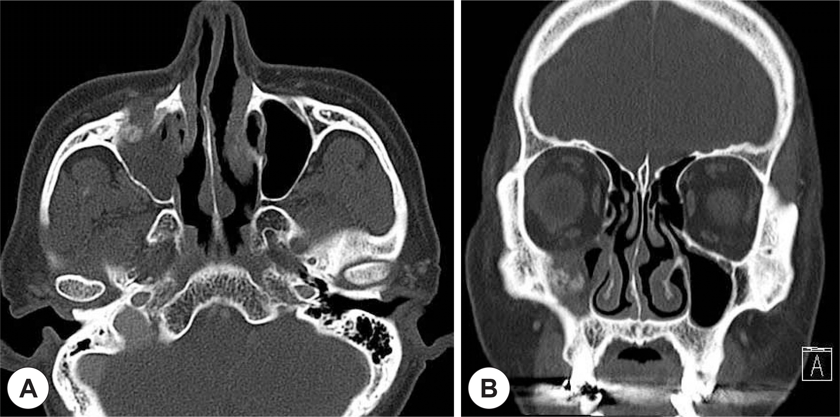
Fig. 2.
Facial MRI scans shows mass which invade facial soft tissue. None-theless, subcutaneous fat boundaries remain intact. A: Axial scan. B: Coronal scan.
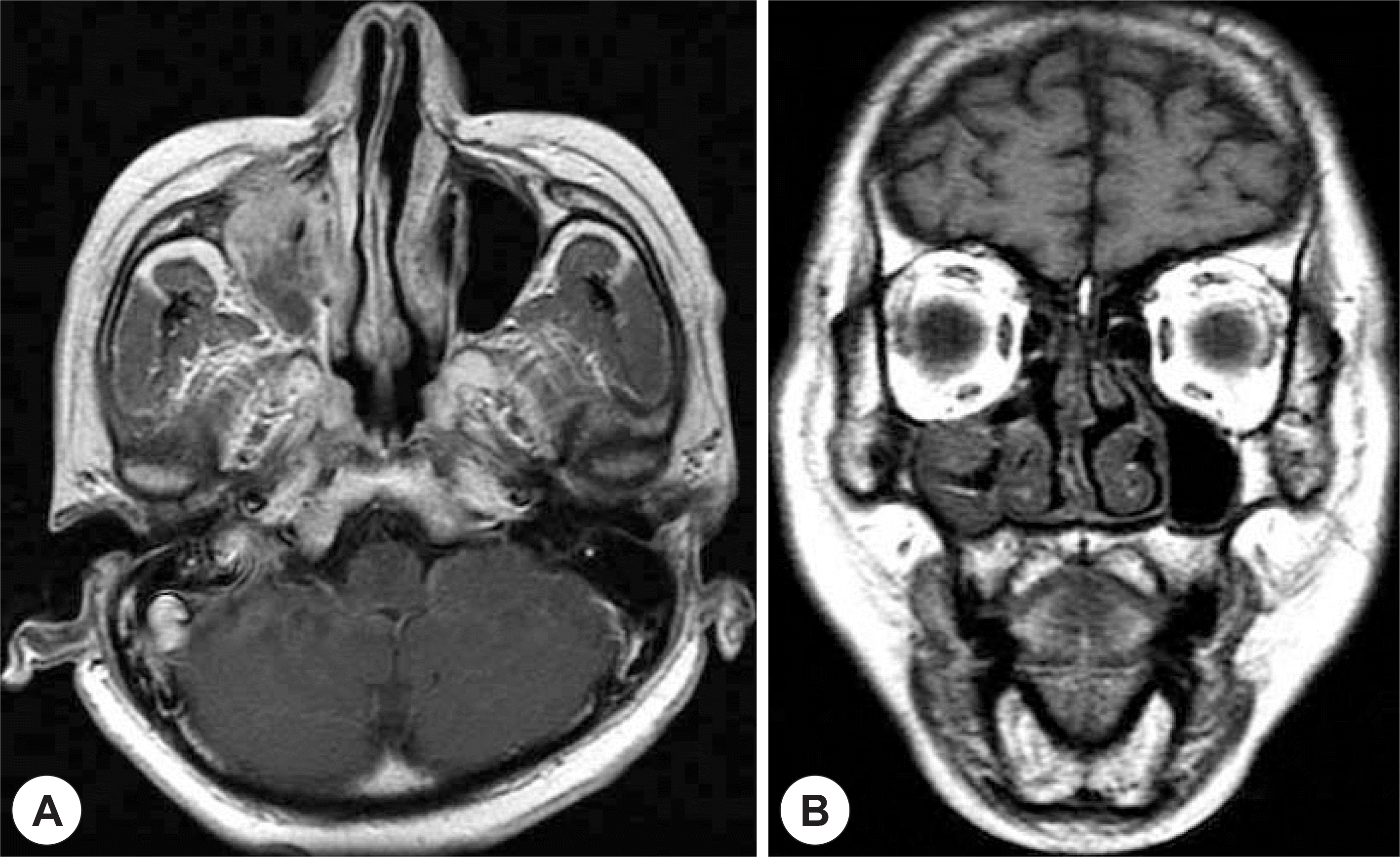
Fig. 3.
Bioresorbable panel (arrow head) was placed around the periorbital tissue in order to protect the soft tissue during reconstruction and to facilitate accurate placement of reconstructive material. Titanium mesh (arrow) used to provide stability in reconstruction inferior of orbit and orbital floor, respectively. A: Before reconstruction. B: After reconstruction.
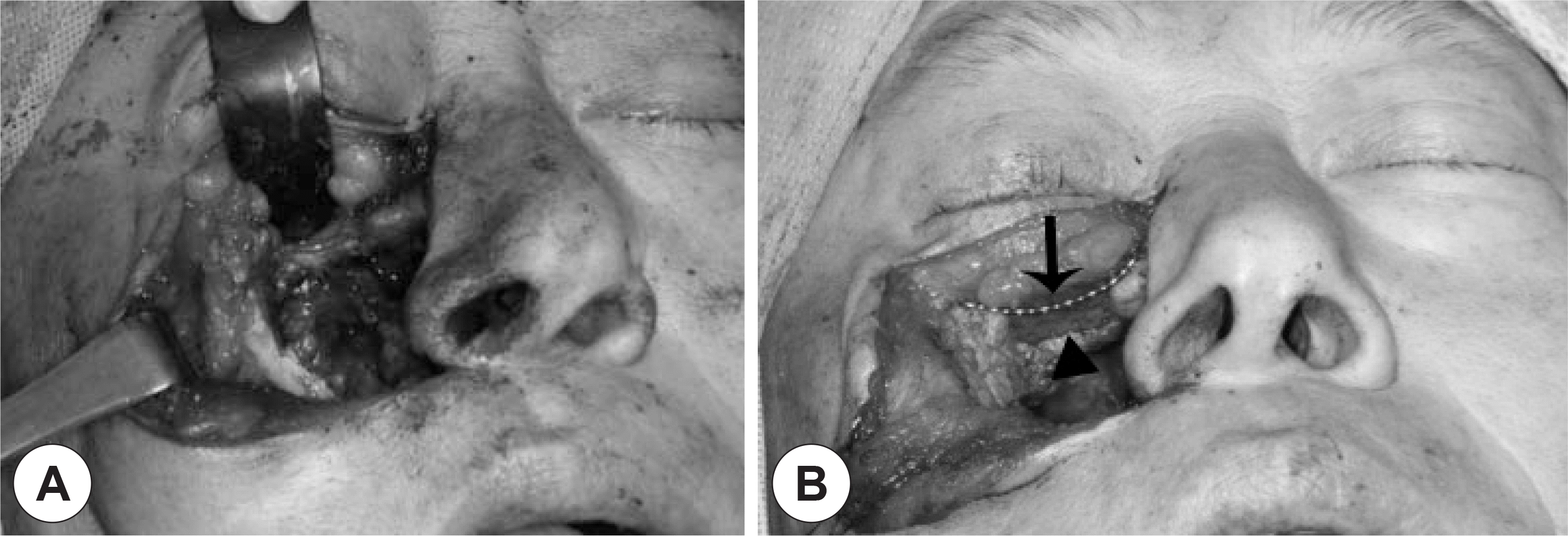
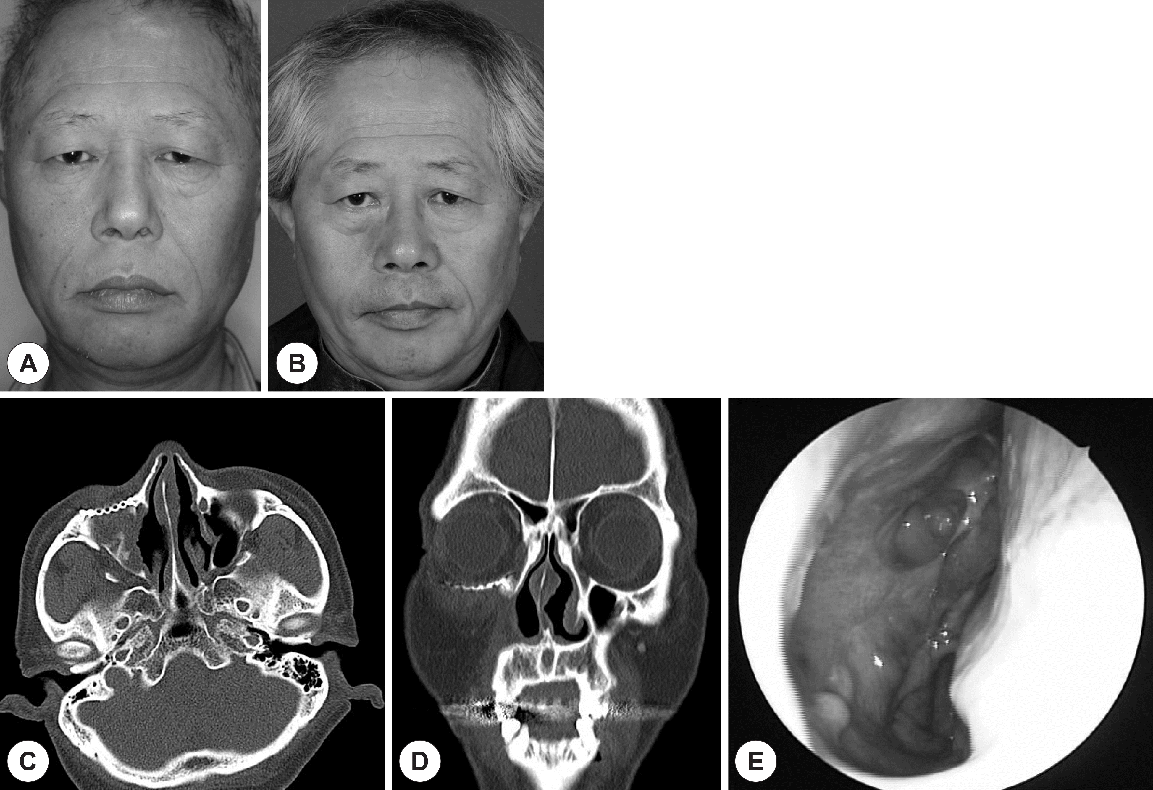
Fig. 4.
Postoperative facial photographs, facial CT scan and endoscopic finding, 6 month after surgery, show well reconstructed inferior orbital rim and orbital floor without enophthalmos. A: Preoperative facial photograph. B: Postoperative facial photograph. C: axial scan. D: Coronal scan. E: Endoscopic finding.
 XML Download