Abstract
Mucormycosis is a rare invasive fungal infection of the nose and paranasal sinus, which often has an acute fulminant course and distinctive clinical findings. It usually occurs in diabetics or immunocompromised patients and shows rapid progression with a high mortality rate. Slow, silent progression is a highly unusual presentation of this disease. Herein, we report a case of mucormycosis with a chronic course of invasion into the hard palate and the maxillary sinus.
References
1). Jeon EJ, Park Y, Park YS, Jeong I. Two cases of Rhino-orbito-Ce-rebral Mucormycosis. Korean J Otolaryngol. 2001; 44:666–70.
2). Harril WC, Stewart MG, Lee AG, Cernoch P. Chronic rhinocerebral mucormycosis. The Laryngoscope. 1996; 106:1292–7.
3). Chakrabarti A, Denning DW, Ferguson BJ, Ponikau J, Buxina W, Kita H, et al. Fungal rhinosinusitis: A categorization and definitional schema addressing current controversies. The Laryngoscope. 2009; 119:1809–18.
4). Kim HJ, Choung YH, Cho MJ, Baik SS. Clinical Analysis of Eight Cases of Mucormycosis. Korean J Otolaryngol. 2004; 15:134–8.

5). Strasser MD, Kennedy RJ, Adam RD. Rhinocerebral mucormycosis: Therapy with amphotericin B lipid complex. Arch Intern Med. 1996; 156:337–9.

6). Scharf JL, Soliman AM. Chronic rhizopus invasive fungal rhinosinusitis in an immunocompetent host. The Laryngoscope. 2004; 114:1533–5.

7). Waitzman AA, Birt BD. Fungal sinusitis. J Otolaryngol. 1994; 23:244–9.
8). Blitzer A, Lawson W. Fungal infections of the nose and paranasal sinuses: Part I. Otolaryngol Clin North Am. 1993; 26:1007–35.
9). Mohindra S, Mohindra S, Gupta R, Bakshi J, Gupta SK. Rhinocerebral mucormycosis: The disease spectrum in 27 patients. Mycoses. 2007; 50:290–6.

10). Waizel-Haiat S, Cohn-Zurita F, Vargas-Aguayo AM, Ramirez-Acev-es R, Vivar-Acevedo E. [Chronic invasive rhinocerebral mucormy-cosis]. Cir Cir. 2003; 71:145–9.
11). Ferguson BJ. Mucormycosis of the nose and paranasal sinuses. Otolaryngol Clin North Am. 2000; 33:349–65.

12). McLean FM, Ginsberg LE, Stanton CA. Perineural spread of rhinocerebral mucormycosis. Am J Neuroradiol. 1996; 17:114–6.
13). Busaba NY, Colden DG, Faquin WC, Salman SD. Chronic invasive fungal sinusitis: A report of two atypical cases. Ear Nose Throat J. 2002; 81:462–6.

15). Yun DH, Chung YS, LEE BJ. Comparison fo inflammatory Cells infiltrating the Maxillary Sinus Mucosa between Chronic Sinusitis and Noninvasive Fungal Sinusitis. Korean J Otolaryngol. 2001; 44:47–51.
Fig. 1.
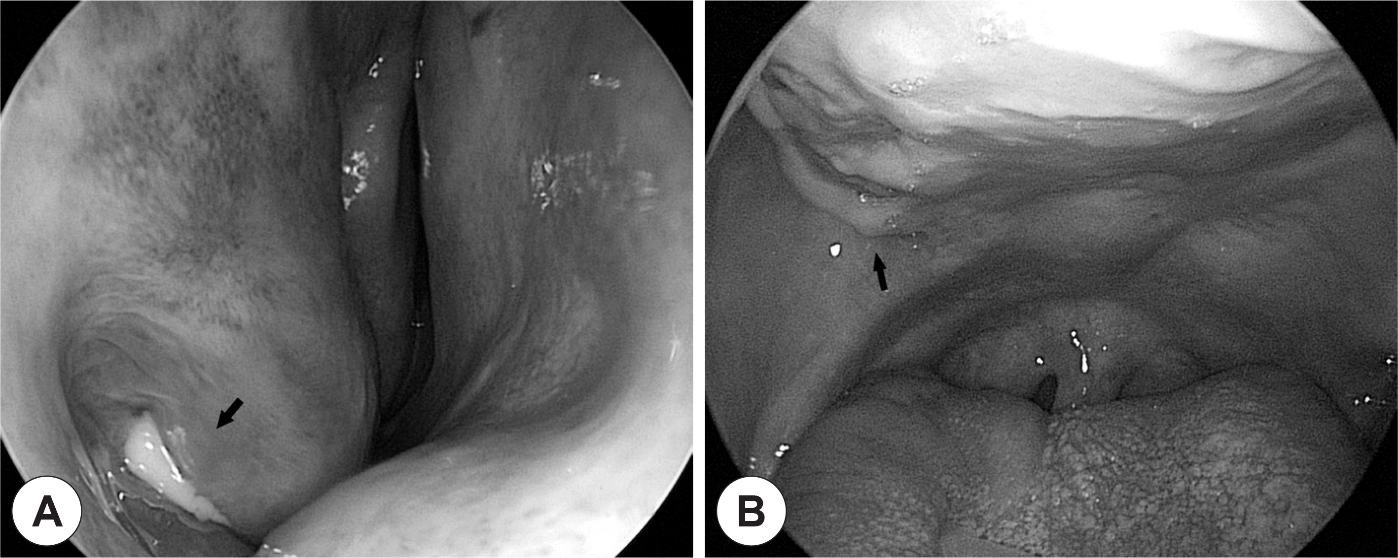
Preoperative local findings. Endoscopic findings of right nasal cavity showed (A) edematous inferior turbinate with pus, and (B) right hard palatal bulging and growing granulomatous tissue from extraction area of molars.

Fig. 2.
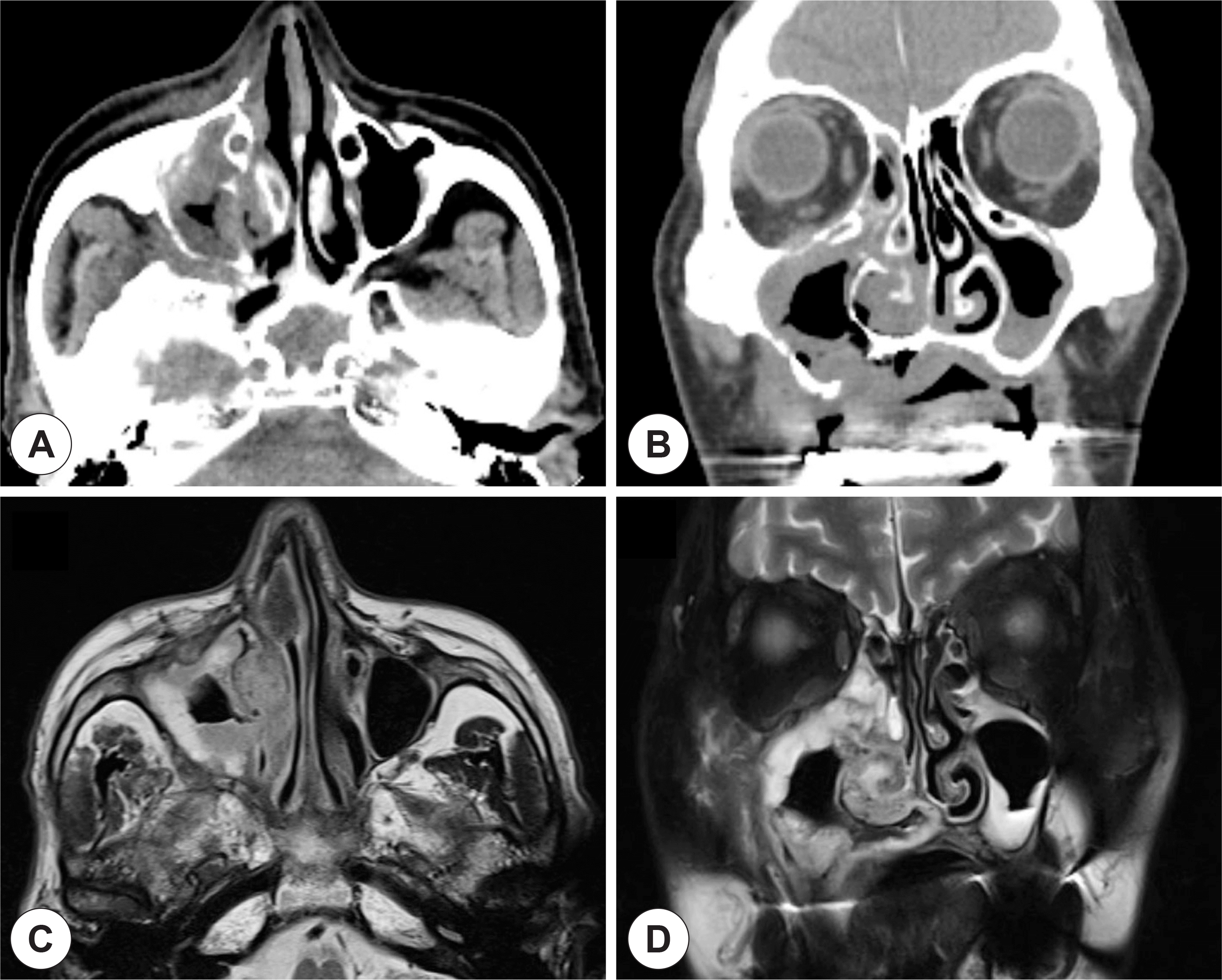
Preoperative imaging studies. Preoperative computed tomography revealed soft tissue density and bony destruction of the right maxillary sinus and mucosal abnormalities of the right inferior turbinate and the hard palate (A and B). Magnetic resonance images showed mass-like lesion along with internal necrosis at the right maxillary sinus, nasal cavity and hard palate (C and D).

Fig. 3.
Pathologic findings of the specimens (hematoxylin & eosin stain). Extensive tissue necrosis with granulomatous inflammation (center) was displayed under the overlying respiratory epithelium, which was accompanied with bone destruction (left lower) (H&E, ×12.5) (A). At high magnification, the entrapped necrotic bony spicules were observed in the edge of the granuloma (H&E, ×100) (B). Fungal organisms, showing broad ribbon-like, non-septate hyphae with branching at right angles, morphologically compatible with mucormycosis were found with angioinvasion (H&E, ×200, ×400, respectively) (C and D).


 PDF
PDF ePub
ePub Citation
 Print
Print


 XML Download