

 PDF
PDF ePub
ePub Citation
 Print
Print


Abstract
Undifferentiated pleomorphic sarcoma (UPS) is a rare soft tissue sarcoma of the sinonasal area. Here, we present two primary cases of UPS and three post-irradiation sinonasal UPS cases. Imaging findings were misinterpreted by radiologists as representing other malignant tumors or recurrence of the primary tumor. Our cases indicate that post-irradiation UPS can originate within any part of the radiation field. Treatment outcomes of primary sinonasal UPS seem to be favorable if the tumor is treated ag-gressively, but the outcomes of post-irradiation sinonasal UPS may be poor if appropriate surgical margins cannot be obtained.
Go to : 
References
1). Weiss SW, Enzinger FM. Malignant fibrous histiocytoma: an analysis of 200 cases. Cancer. 1978; 41:2250–66.
2). Barnes L, Kanbour A. Malignant fibrous histiocytoma of the head and neck. A report of 12 cases. Arch Otolaryngol Head Neck Surg. 1988; 114:1149–56.

3). Park SW, Kim HJ, Lee JH, Ko YH. Malignant fibrous histiocytoma of the head and neck: CT and MR imaging findings. AJNR Am J Neuroradiol. 2009; 30:71–6.
4). Wang CP, Chang YL, Ting LL, Yang TL, Ko JY, Lou PJ. Malignant fibrous histiocytoma of the sinonasal tract. Head & Neck. 2009; 31:85–93.
5). Sheppard DG, Libshitz HI. Postradiation sarcomas: a review of the clinical and imaging features in 63 cases. Clinical Radiology. 2001; 56:22–9.
6). Arlen M, Higinbotham NL, Huvos AG, Marcove RC, Miller T, Shah IC. Radiation-induced sarcoma of bone. Cancer. 1971; 28:1087–99.
7). Goldblum JR, Folpe AL, Weiss SW. Undifferentiated Pleomorphic Sarcoma. Goldblum JR, editor. editor.Enzinger and Weiss's Soft Tissue Tumors. 6th ed.Philadelphia: Elsevier;2013. p. 421–40.
8). Guillou L, Folpe AL. Fibroblastic and Fibrohistiocytic Tumors. Folpe AL, editor. editor.Bone and Soft Tissue Pathology. Philadelphia: Saunders & Elsevier;2010. p. 43–96.
9). Agaimy A, Gaumann A, Schroeder J, Dietmaier W, Hartmann A, Hofstaedter F, et al. Primary and metastatic high-grade pleomorphic sarcoma/malignant fibrous histiocytoma of the gastrointestinal tract: an approach to the differential diagnosis in a series of five cases with emphasis on myofibroblastic differentiation. Virchows Arch. 2007; 451:949–57.
10). Shahoon H, Esmaeili M, Nematollahi M. Eight-year Follow-up of Malignant Fibrous Histiocytoma (Undifferentiated High-grade Pleomorphic Sarcoma) of the Maxilla: Case Report and Review of the Literature. J Dent Res Dent Clin Dent Prospects. 2009; 3:32–5.
11). Vuity D, Bogdan S, Csurgay K, Sapi Z. Malignant fibrous histiocy-toma/undifferentiated high-grade pleomorphic sarcoma of the maxillary sinus: report of a case and review of the literature. Pathol Oncol Res. 2013; 19:605–9.
12). Blitzer A, Lawson W, Zak FG, Biller HF, Soon ML. Clinical-pathological determinants in prognosis of fibrous histiocytomas of head and neck. Laryngoscope. 1981; 91:2053–70.
13). Weber RS, Benjamin RS, Peters LJ, Ro JY, Achon O, Goepfert H. Soft tissue sarcomas of the head and neck in adolescents and adults. Am J Surg. 1986; 152:386–92.
14). Rodrigo JP1. Fernández JA, Suárez C, Gómez J, Llorente JL, Herrero A. Malignant fibrous histiocytoma of the nasal cavity and paranasal sinuses. Am J Rhinol. 2000; 14:427–31.
15). Park SW, Kim HJ, Lee JH, Ko YH. Malignant fibrous histiocytoma of the head and neck: CT and MR imaging findings. AJNR Am J Neuroradiol. 2009; 30:71–6.
16). Bentz BG, Singh B, Woodruff J, Brennan M, Shah JP, Kraus D. Head and neck soft tissue sarcomas: a multivariate analysis of outcomes. Ann Surg Oncol. 2004; 11:619–28.
17). Jang TY, Kim CH, Kim YM, Chu YC. Two cases of malignant fibrous histiocytoma in the nasal cavity and paranasal sinus. J Rhinol. 1998; 5:72–5.
18). Seo BS, Choi SJ, Jang YJ, Chung YS, Lee BJ. Recurrence patterns of the maxillary sinus cancer after total maxillectomy. J Rhinol. 2008; 15:39–43.
19). Hsu HC, Huang EY, Wang CJ. Treatment results and prognostic factors in patients with malignant fibrous histiocytoma. Acta Oncol. 2004; 43:530–5.
20). Le Doussal V, Coindre JM, Leroux A, Hacene K, Terrier P, Bui NB, et al. Prognostic factors for patients with localized primary malignant fibrous histiocytoma: a multicenter study of 216 patients with multivariate analysis. Cancer. 1996; 77:1823–30.
Go to :
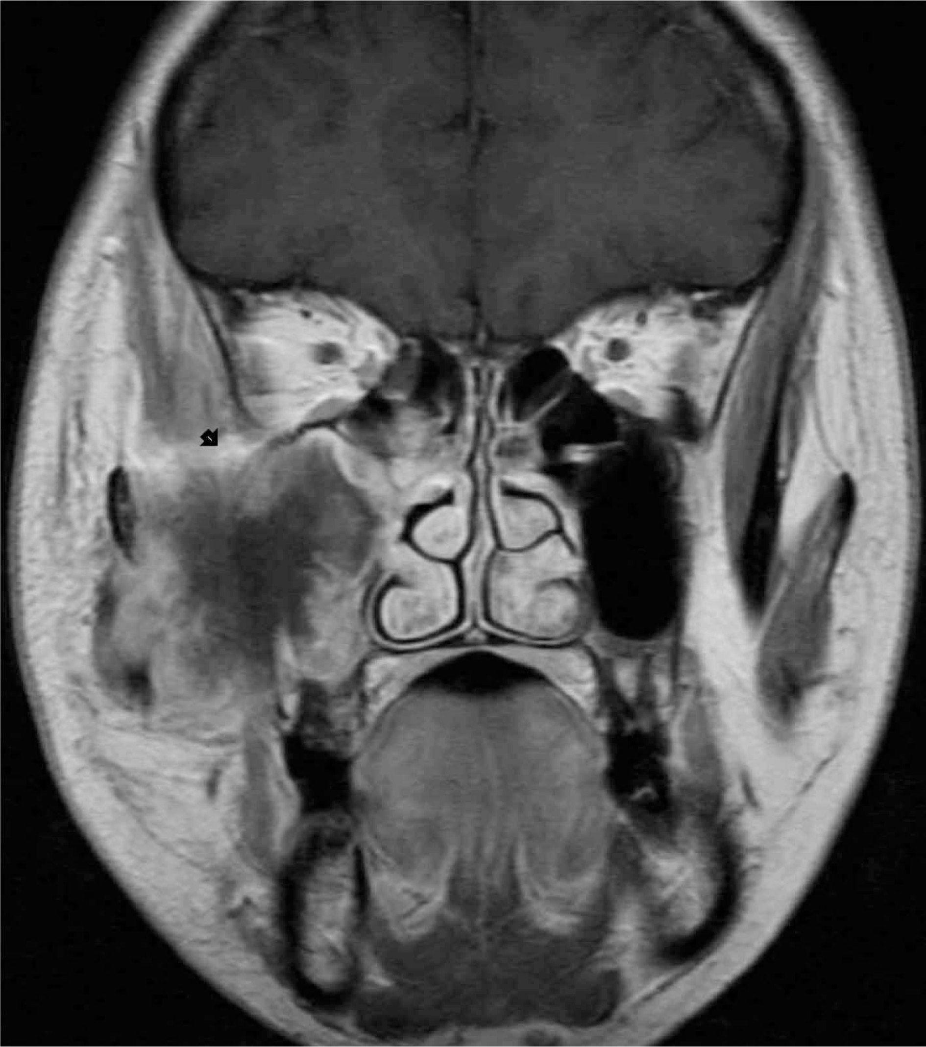 | Fig. 1.A bulky soft tissue mass in the right maxillary sinus with destruction of the maxillary walls and extension to the buccal space in gadolinium-enhanced T1-weighted MR image of primary UPS. |
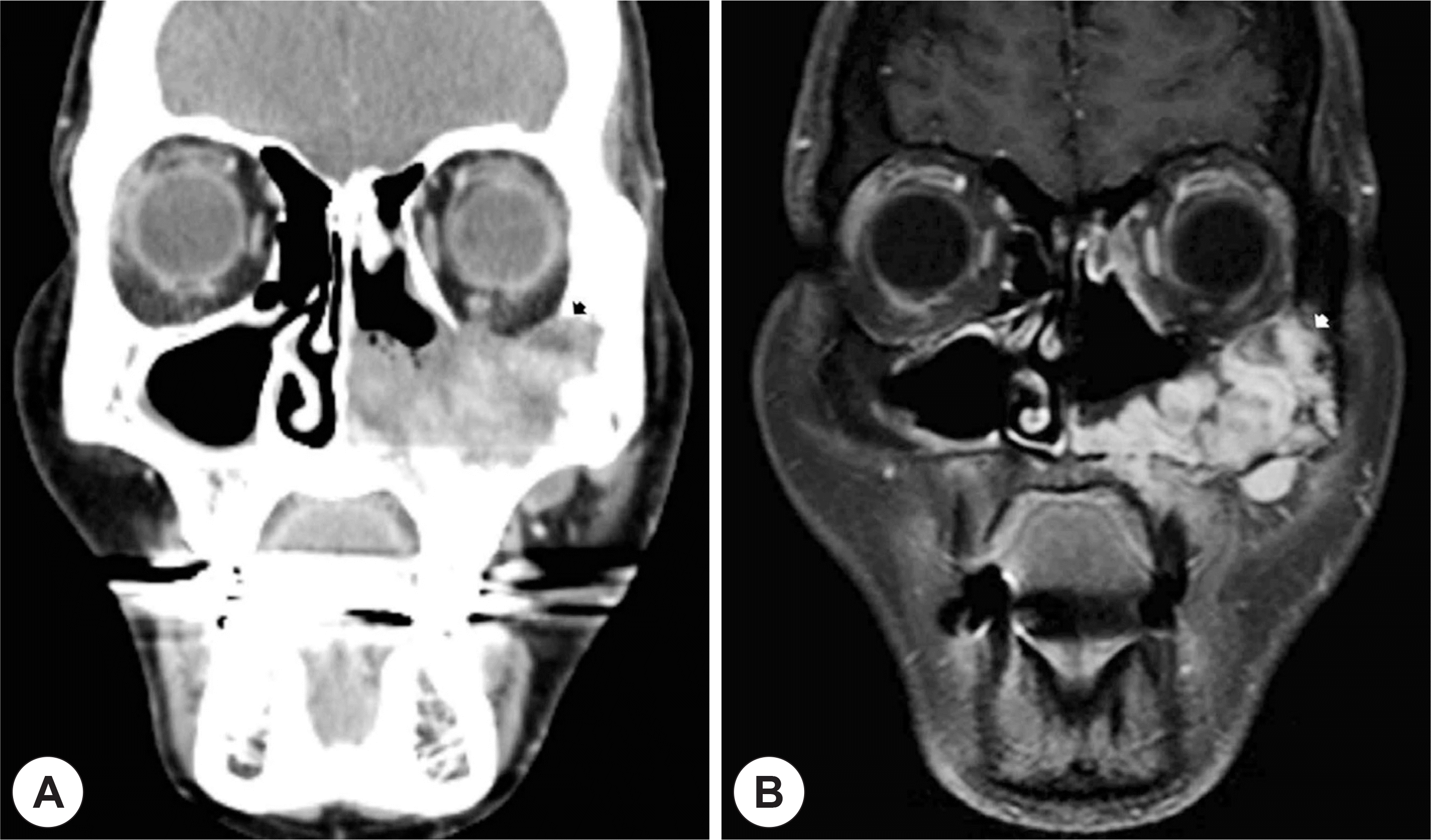 | Fig. 2.Heterogeneously enhancing large mass with bony destruction of the maxillary walls and nasal cavity in CT scan (A) and MR image (B) of post-irradiated UPS. |
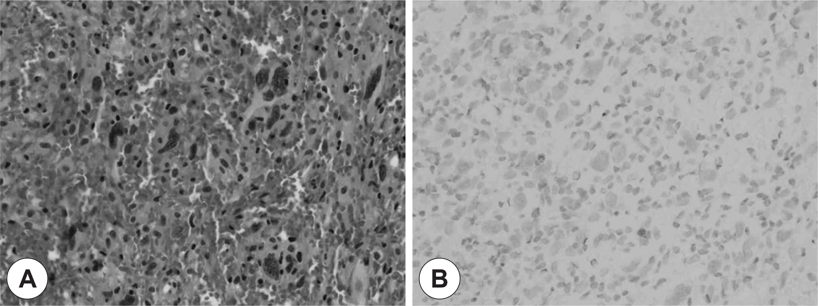 | Fig. 3.Histopathology of radiation-induced undifferentiated pleomorphic sarcoma. The tumor consists of mixed spindle cells and roundedcells arranged in a stori-form pattern. A: Hematoxylin-eo-sin (×400). B: Negative staining for cytokeratin by immunohistochemistry (×400). |
Table 1.
Clinical features of the current series of undifferentiated pleomorphic sarcoma of the sinonasal tract
 XML Download