Abstract
Small cell carcinoma commonly originates in the lung, with only about 4% of cases arising at extrapulmonary sites. Further-more, small cell carcinoma of the sinonasal tract is extremely rare. In Korea, only 2 cases of primary sinonasal small cell carcinoma have been reported in the nasal cavity and the nasal septum, respectively. Recently, we have experienced a rare case of small cell carcinoma arising from the right maxillary sinus coexisting with a fungal ball lesion. Herein, we report this case with a review of the literature.
References
1). Kang JM, Lee HY, Lee KS, Ko SY. Small cell Neuroendocrine Carcinoma of the Nasal cavity. Korean J Otolaryngol. 2003; 46:164–7.
2). Kim YS, Bae CH, Song SY, Kim YD. A case of a small cell neuroendocrine carcinoma of the nasal septum. Korean J Otorhinolaryn-gol-Head Neck Surg. 2009; 52(6):529–32.

3). Park CH, Roh JL, Park YH, Rha KS. A Case of Primary Small Cell Carcinoma of the Larynx. Korean J Otolaryngol. 2005; 48:124–6.
4). Tang IP, Singh S, Krishnan G, Looi LM. Small cell neuroendocrine carcinoma of the nasal cavity and paranasal sinuses: a rare case. J Laryngol Otol. 2012; 126(12):1284–6.

5). Tsukahara K, Nakamura K, Motohashi R, Sato H. Two Cases of Small Cell Cancer of the Maxillary Sunus Treated with Cisplatin plus Irinotecan and Radiotherapy. Case Rep Otolaryngol. 2013; 2013:893638.
6). Zhu Q, Zhu W, Wu J, Zhang H. The CT and MRI observations of small cell neuroendocrine carcinoma in paranasal sinuses. World J Surg Oncol. 2015; 13:54.

7). Babin E, Rouleau V, Vedrine PO, Toussaint B, de Raucourt D, Ma-lard O, et al. Small cell neuroendocrine carcinoma of the nasal cavity and paranasal sinuses. J Laryngol Otol. 2006; 120(4):289–97.

8). Travis WD, Brambilla E, Nicholson AG, Yatabe Y, Austin JHM, Beasley MB, et al. The 2015 WHO classification of lung tumors. J Thorac Oncol. 2015; 10:1243–60.
9). Terada T. Primary small cell carcinoma of the maxillary sinus: a case report with immunohistochemical and molecular genetic study involving KIT and PDGFRA. Int J Clin Exp Pathol. 2012; 5(3):264–9.
10). Han G, Wang Z, Guo X, Wang M, Wu H, Liu D. Extrapulmonary small cell neuroendocrine carcinoma of the paranasal sinuses: a case report and review of the literature. J Oral Maxillofac Surg. 2012; 70(10):2347–51.

11). Kim KO, Lee HY, Chun SH, Shin SJ, Lim MK, Lee KY, et al. Clinical Overview of Extrapulmonary Small cell carcinoma. J Korean Med Sci. 2006; 21:833–7.

12). Segawa Y, Nakashima T, Shiratsuchi H, Tanaka R, Mitsugi K, et al. Small Cell Carcinoma of the Tonsil Treated with Irinotecan and Cisplatin: A Case Report and Literature Review. Case Rep Oncol. 2011; 4:587–91.

13). Jin S, Wang T, Chen X, Xu B, Sun J, Guo R, et al. Phase II Study of Weekly Irinotecan plus Cisplatin in Patients with Previously Untreated Extensive-Stage Extrapulmonary Small Cell Carcinoma. Onkol-ogie. 2011; 34:378–81.

Fig. 1.
Endoscopic findings of the Rt. nasal cavity. Well demarcated mass with soft surface ranged from the middle meaus to inferior meatus. IT: Inferior turbinate, S: nasal septum.
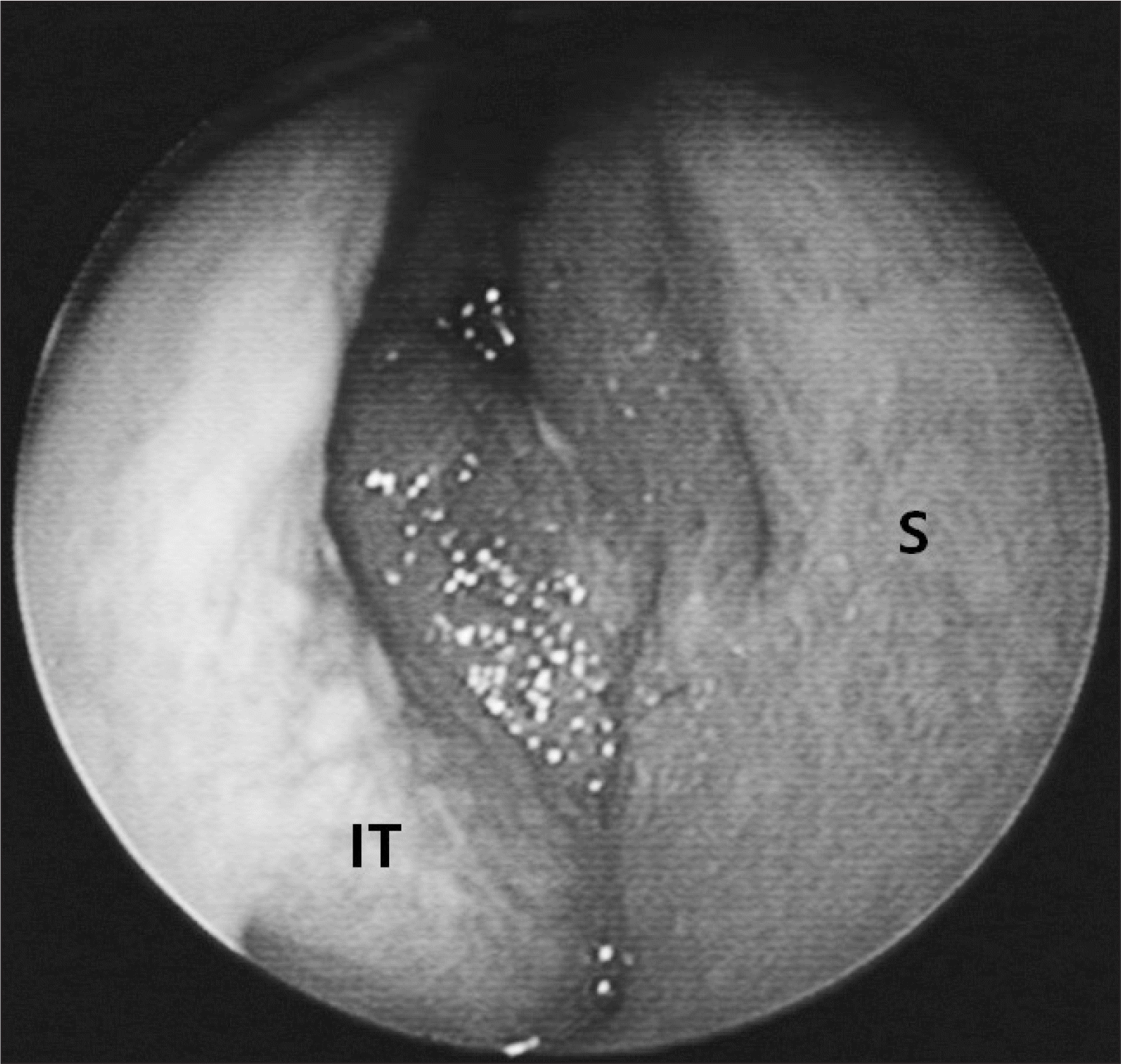
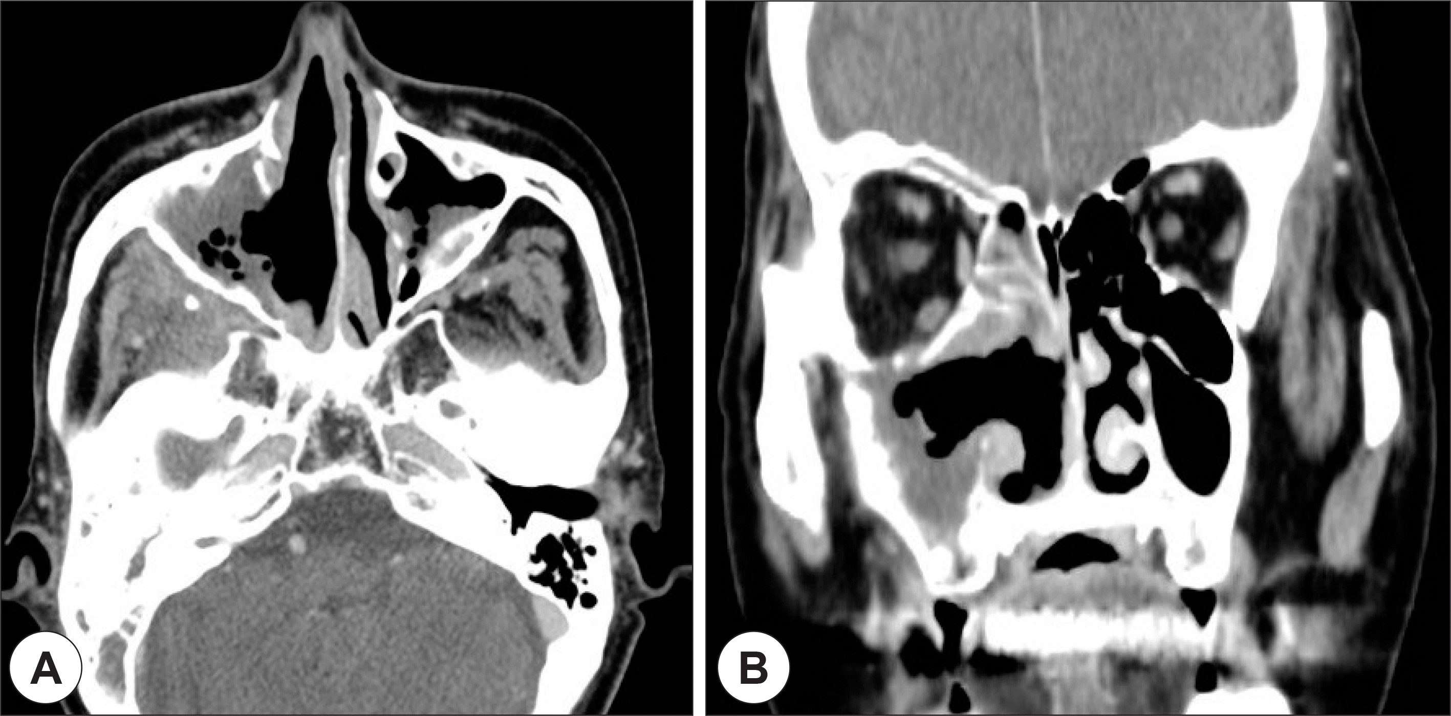
Fig. 2.
Axial (A) and coronal (B) PNS CT scan. Soft tissue density lesion at Rt. maxillary, ethmoid, sphenoid sinus and nasal cavity with bony destruction of anterior and posterior maxillary sinus wall (astrix) and orbit (arrow).
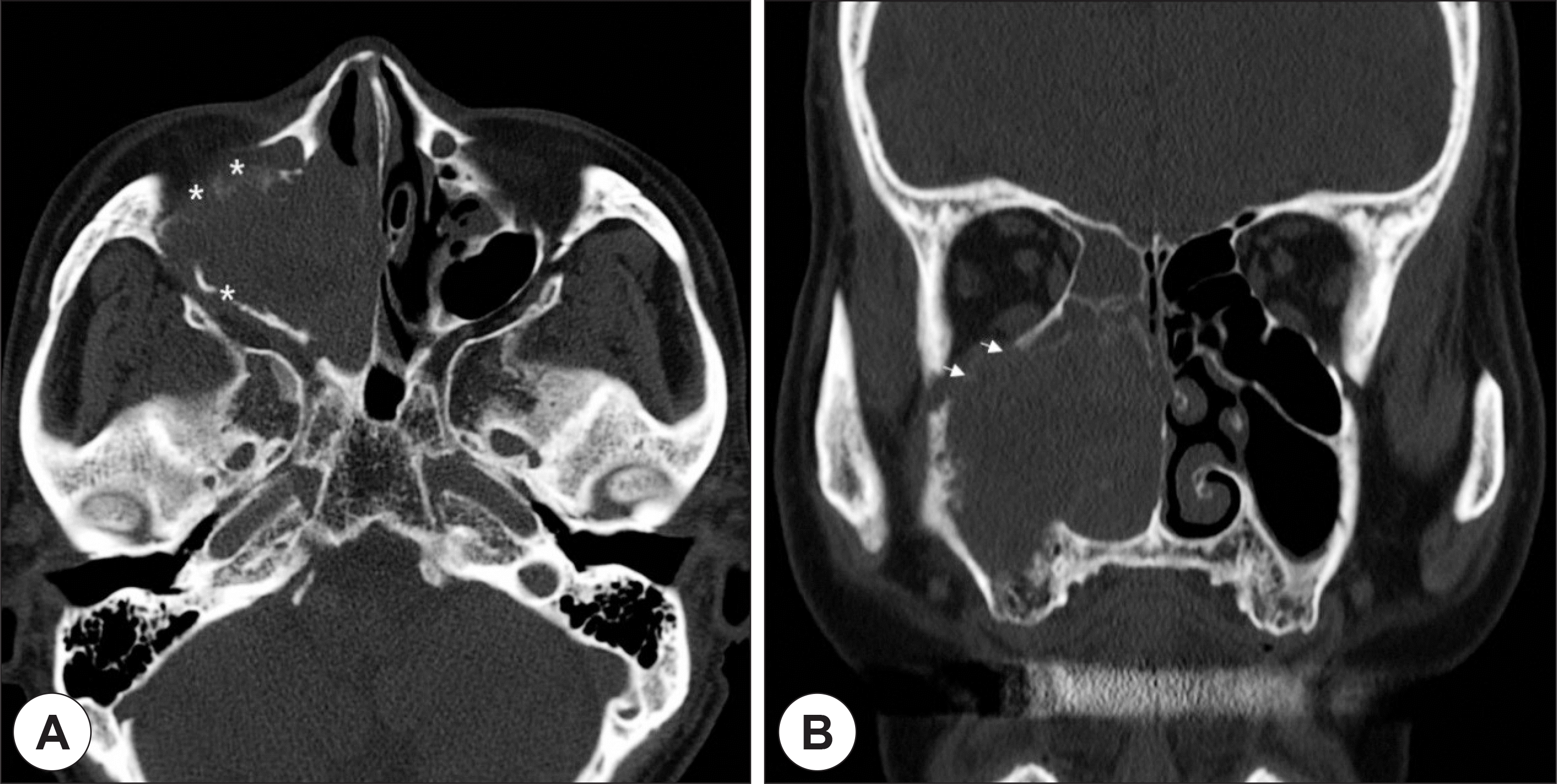
Fig. 3.
T1 enhance (A) and T2 weighted (B) MR image. Heterogeneous signal intensity mass lesion at Rt. maxillary sinus extent to nasal cavity and invade the orbit (astrix) and buccal fat pad (arrow).
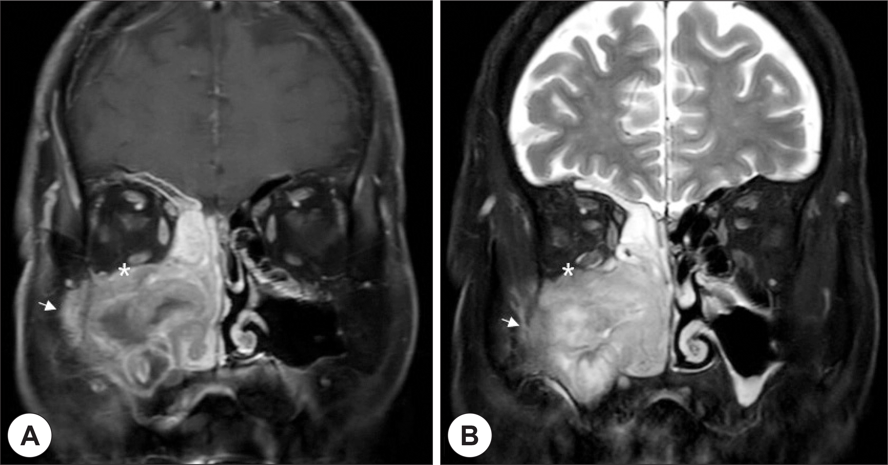
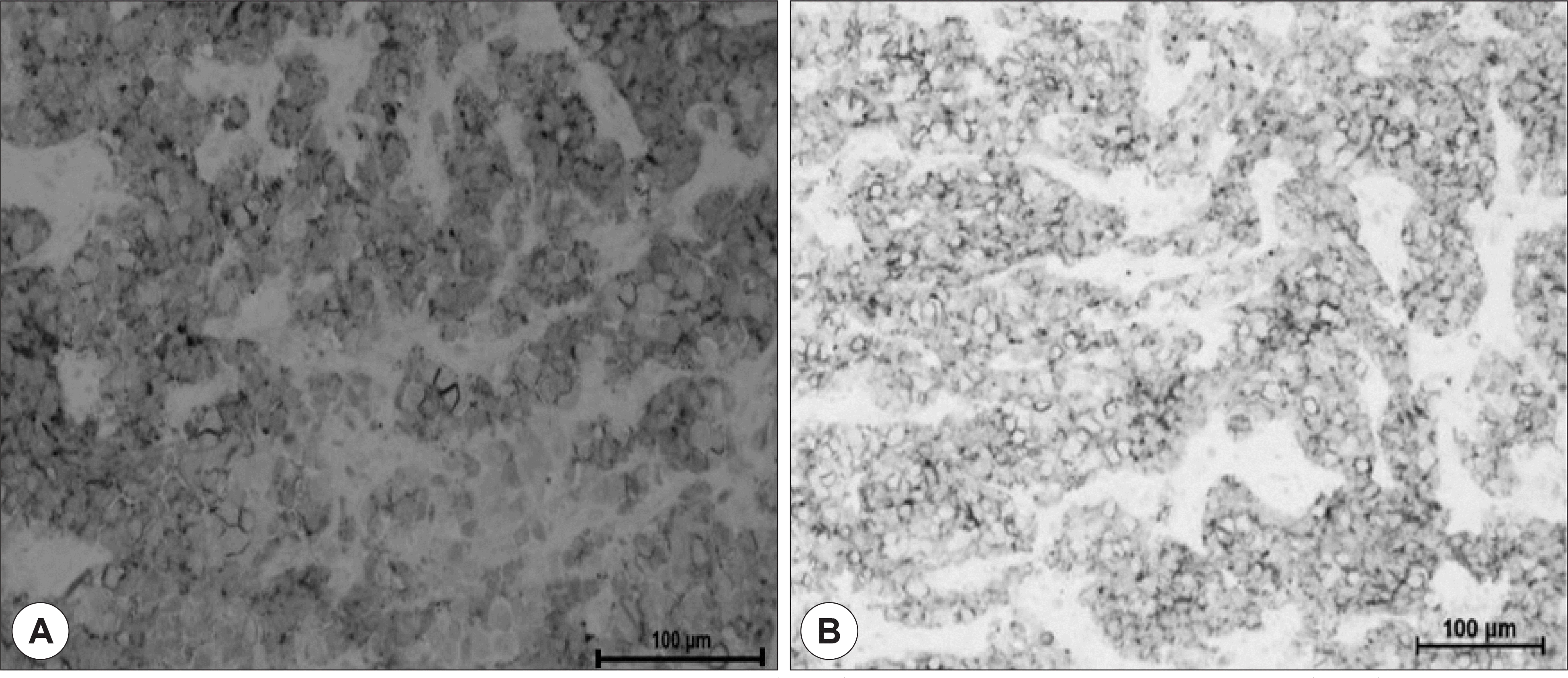
Fig. 4.
Histopathologic findings. A: Round and spindle shaped cell with small amount of cytoplasm and abundent nucleus showing abundent cell division (H-E stain, ×400). B: Staining for aspergillus fungus ball (H-E stain, ×400).


 PDF
PDF ePub
ePub Citation
 Print
Print


 XML Download