

 PDF
PDF ePub
ePub Citation
 Print
Print


Abstract
In recent years, endoscopic sinus marsupialization has become the treatment of choice for the treatment of paranasal sinus mucoceles due to its noninvasiveness and successful outcome. However, mucoceles located at the lateral portion of the frontal sinus and protruding into the orbit with erosion of the frontal sinus floor arestill difficult to address with standard endoscopic sinus surgery techniques. Here, we report a case of a mucocele located atthe lateral side of the frontal sinus and successfully marsupialized with a transblepharoplasty approach combined with an endoscopic approach.
References
1). Lund VJ, Milroy CM. Fronto-ethmoidal mucocoeles: a histopathological analysis. J Laryngol Otol. 1991; 105:921–3.

2). Constantinidis J, Steinhart H, Schwerdtfeger K, Zenk J, Iro H. Therapy of invasive mucoceles of the frontal sinus. Rhinology. 2001; 39:33–8.
3). Kennedy DW, Josephson JS, Zinreich SJ, Mattox DE, Goldsmith MM. Endoscopic sinus surgery for mucoceles: a viable alternative. Laryngoscope. 1989; 99:885–95.
4). Bockmuhl U, Kratzsch B, Benda K, Draf W. Surgery for paranasal sinus mucocoeles: efficacy of endonasal micro-endoscopic management and longterm results of 185 patients. Rhinology. 2006; 44:62–7.
5). Stumpe MR, Sindwani R, Chandra RK. Endoscopic management of sinus disease with frontal lobe displacement. Am J Rhinol. 2007; 21:324–9.
6). Shikowitz MJ, Goldstein MN, Stegnjajic A. Sphenoid sinus mucocele masquerading as a skull base malignancy. Laryngoscope. 1986; 96:1405–10.
7). Har-El G. Endoscopic management of 108 sinus mucoceles. Laryngoscope. 2001; 111:2131–4.
8). Weber R, Draf W, Kratzsch B, Hosemann W, Schaefer SD. Modern concepts of frontal sinus surgery. Laryngoscope. 2001; 111:137–46.
9). Ulualp SO, Carlson TK, Toohill RJ. Osteoplastic flap versus modified endoscopic Lothrop procedure in patients with frontal sinus disease. Am J Rhinol. 2000; 14:21–6.
10). Knipe TA, Gandhi PD, Fleming JC, Chandra RK. Transblepharoplasty approach to sequestered disease of the lateral frontal sinus with ophthalmologic manifestations. Am J Rhinol. 2007; 21:100–4.
Fig. 1.
Extraocular movements of the patient. Preoperative examination shows restriction with superior gaze (A). Postoperative examination shows recovered superior gaze (B).
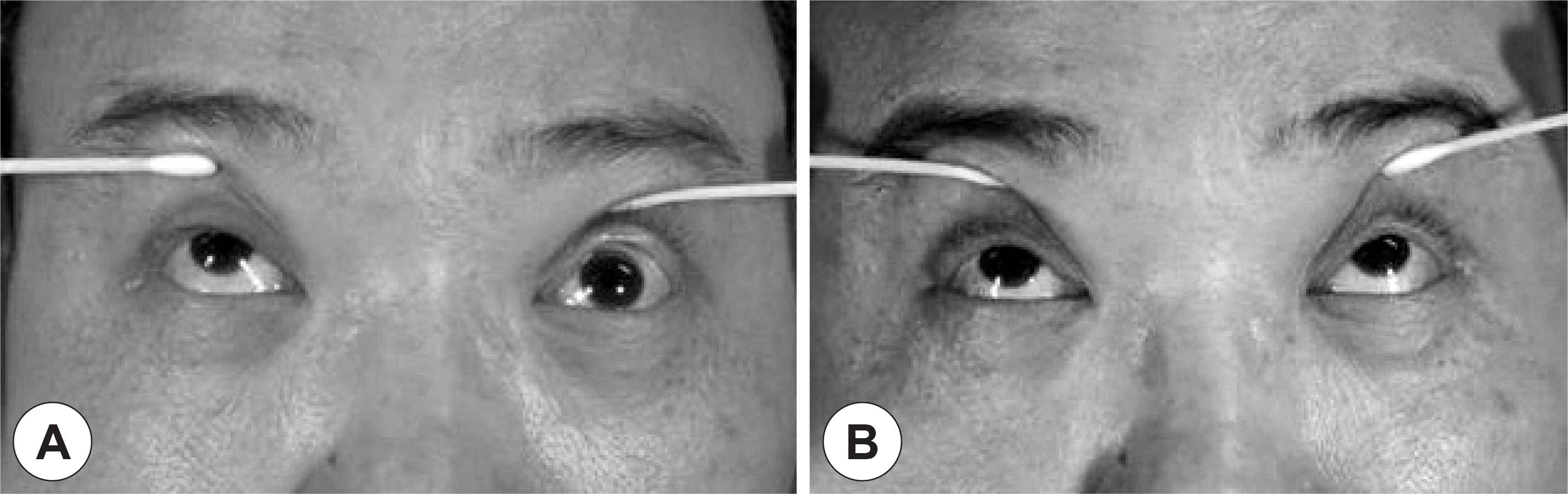
Fig. 2.
Preoperative coronal CT scan of paranasal sinuses shows expansile heterogeneous soft tissue density at lateral portion of left frontal sinus with bony erosion of superior orbital rim (A). Preoerative T1-weighted Gadolinium enhanced coronal image reveals a heterogeneously expansile enhancing mass within in the left frontal sinus extending to the orbit (B).
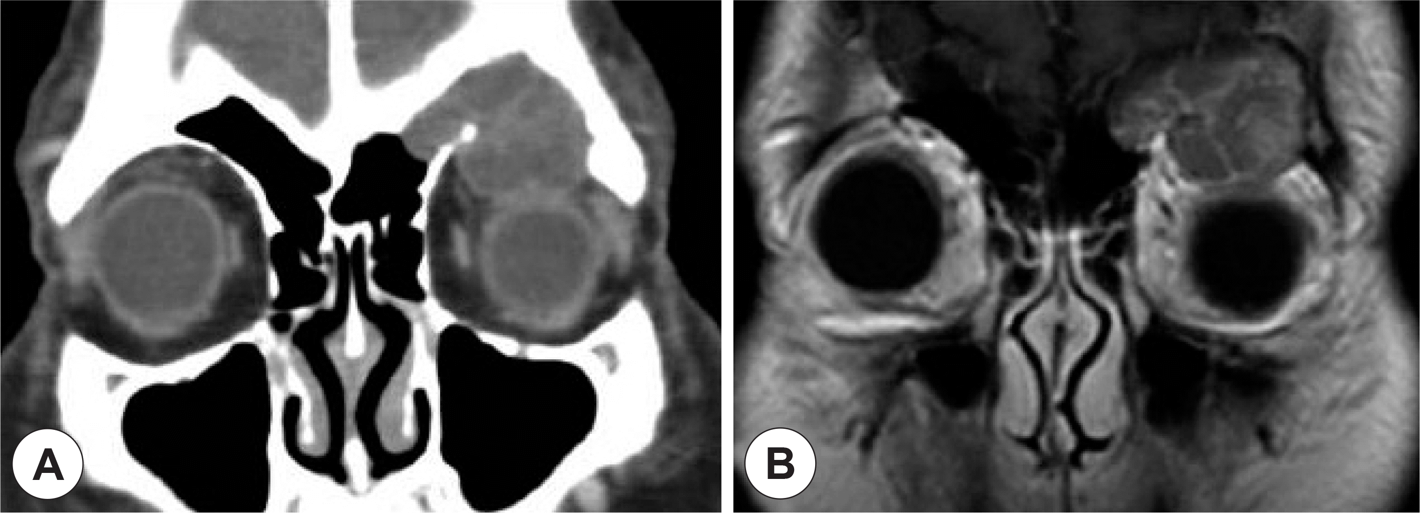
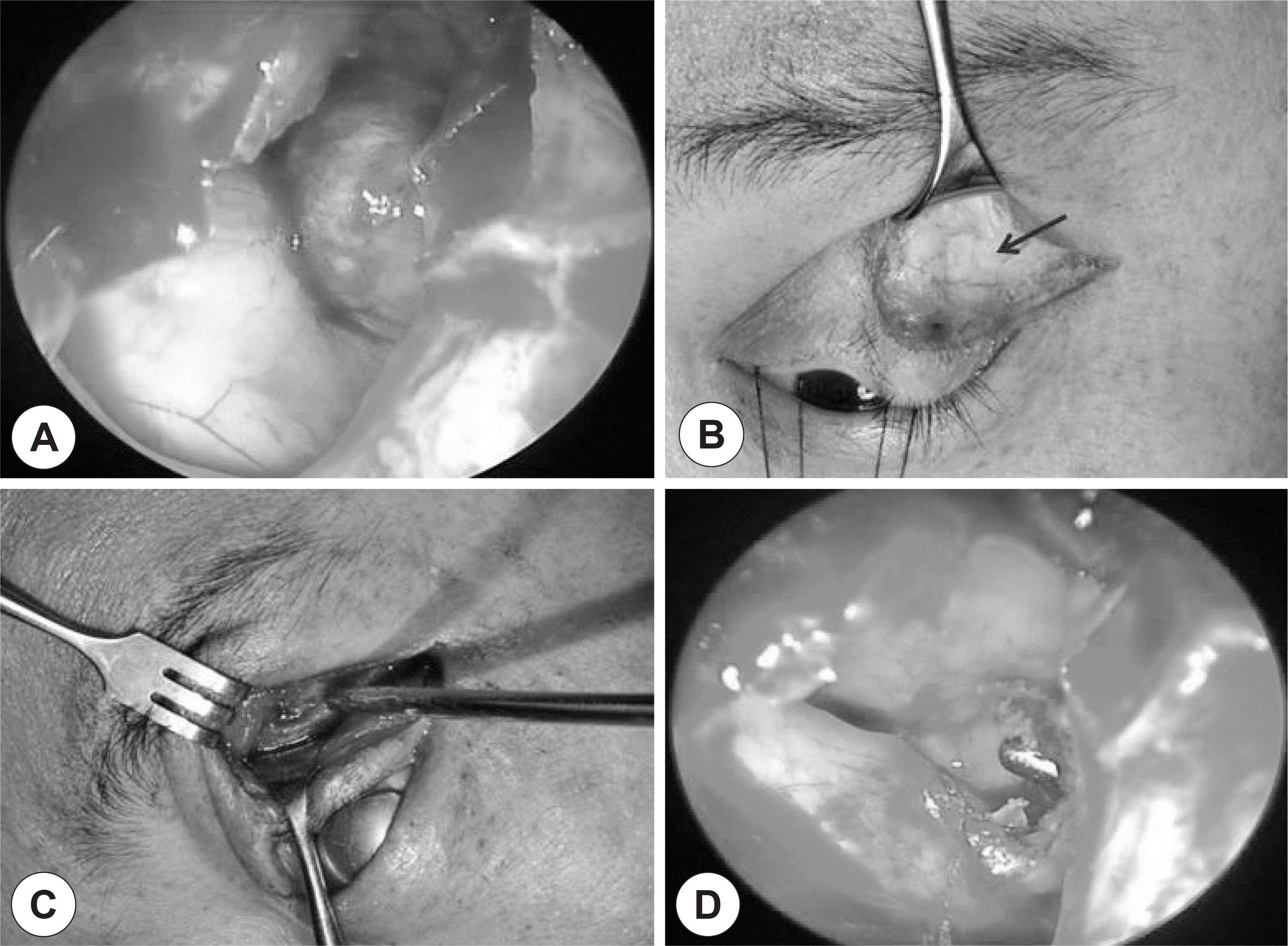
Fig. 3.
Intraoperative findings. After anterior ethmoidectomy, frontal recess was widened under 70° endoscope and brown colored cystic mass was observed at lateral portion of left frontal sinus (A). Left upper lid incision was placed in a skin fold above the tarsal plate and myocutaneous flap was raised superficial to the orbital septum (arrow). The orbital periosteum was incised at the superior orbital rim and retracted inferiorly along with the orbital contents (C). The preexisting dehiscence of superior orbital rim was exposed and cystic wall of mucocele was removed (D).
 XML Download