

 PDF
PDF ePub
ePub Citation
 Print
Print


Abstract
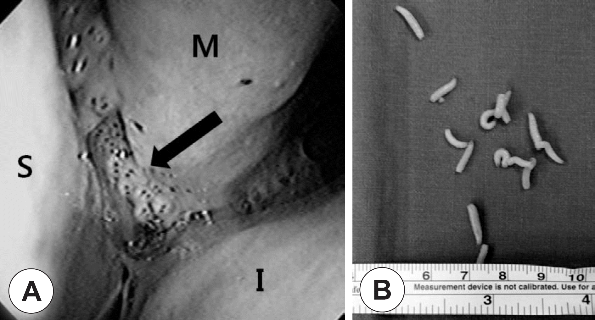
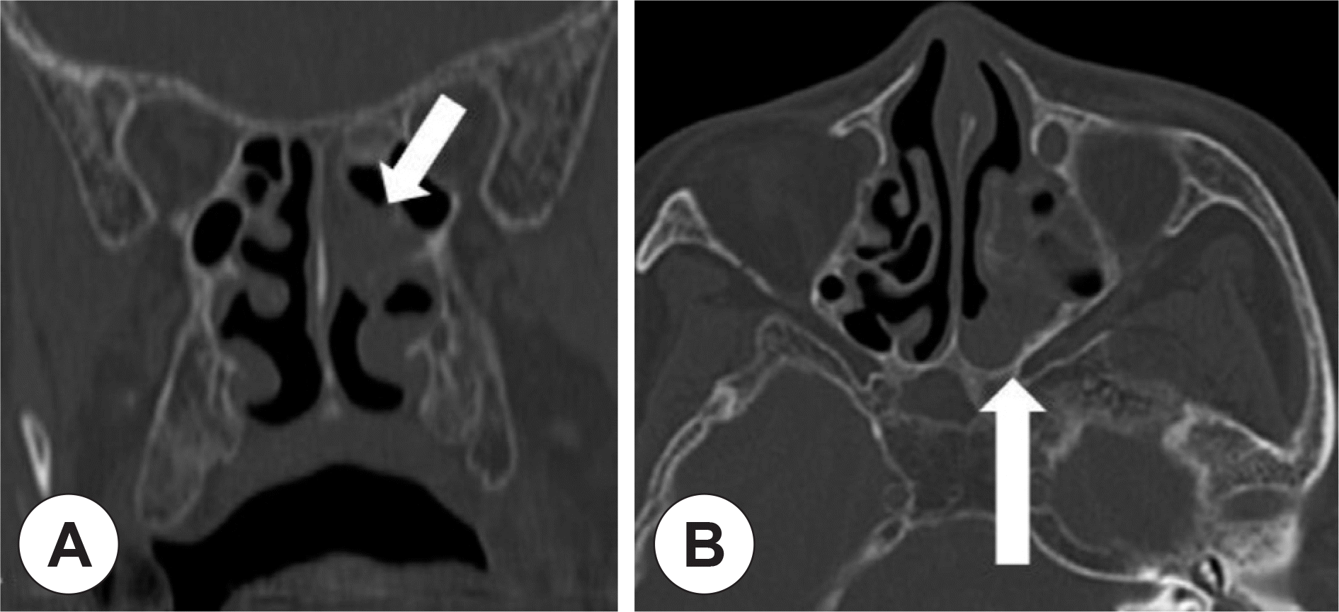
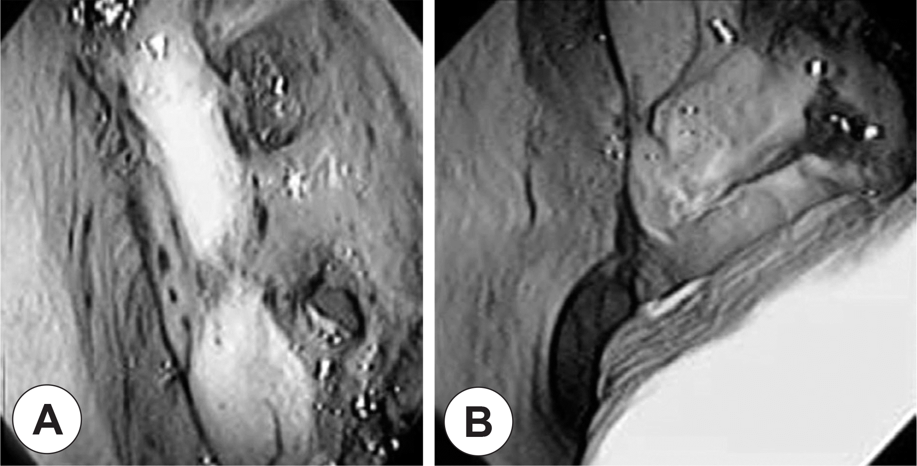
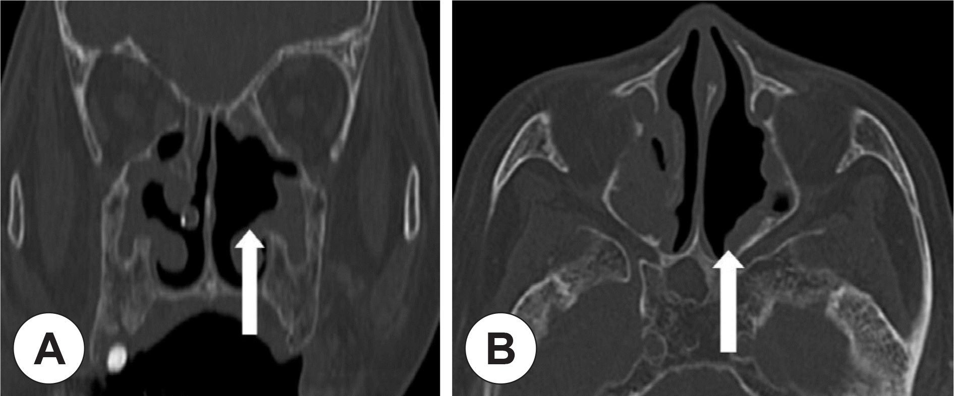
Infestation of tissue by fly larvae is termed myiasis, and it is unusual in humans. Nasal myiasis is common in low socioeconomic status individuals due to poor nasal hygiene. It commonly affects the skin and rarely the nasal and paranasal sinuses. Recently an 82-year-old female was admitted to the emergency department because of discharge of live maggots from the nasal cavity. She had been diagnosed with brain infarction and Alzheimer's disease several years previous. We successfully removed all the maggots from the patient's nasal cavity and sinuses via endoscopic surgery under local anesthesia. Subsequently, the patient's nasal problem resolved completely.
References
1). Pampiglione S, Bettoli V, Cestari G, Staffa M. Furuncular myiasis due to Cordylobia anthropophaga, endemic in the same locality for over 130 years Ann Trop Med Parasitol. 1993; 87(2):219–20.
2). Droma EB, Wilamowski A, Schnur H, Yarom N, Scheuer E, Schwartz E, et al. Oral myiasis: a case report and literature review. Oral Surg Oral Med Oral Pathol Oral Radiol Endod. 2007; 103(1):92–6.

3). Caumes E, Carrière J, Guermonprez G, Bricaire F, Danis M, Gentili-ni M, et al. Dermatoses associated with travel to tropical countries: a prospective study of the diagnosis and management of 269 patients presenting to a tropical disease unit. Clin Infect Dis. 1995; 20(3):542–8.
4). Lee YT, Chen TL, Lin YC, Fung CP, Cho WL. Nosocomial nasal myiasis in an intubated patient. J Chin Med Assoc. 2011; 74(8):369–71.
5). Kim JS, Seo PW, Kim JW, Go JH, Jang SC, Lee HJ, et al. A nasal myiasis in a 76-year-old female in Korea. Korean J Parasitol. 2009; 47(4):405–7.
6). Nazni WA, Jeffery J, Lee HL, Lailatul AM, Chew WK, Heo CC, et al. Nosocomial nasal myiasis in an intensive care unit. Malays J Pathol. 2011; 33(1):53–6.
7). McGraw TA, Turiansky GW. Cutaneous myiasis. J Am Acad Dermatol. 2008; 58(6):907–26.
8). Magnarelli LA, Andreadis TG. Human cases of furuncular, traumatic, and nasal myiasis in Connecticut. Am J Trop Med Hyg. 1981; 30(4):894–6.
9). Kuruvilla G, Albert RR, Job A, Ranjith VT, Selvakumar P. Pneumocephalus: a rare complication of nasal myiasis. Am J Otolaryngol. 2006; 27(2):133–5.
10). Adisa CA1. Mbanaso A. Furuncular myiasis of the breast caused by the larvae of the Tumbu fly (Cordylobia anthropophaga). BMC Surg. 2004; 4:5.
11). Jang M, Ryu SM, Kwon SC, Ha JO, Kim YH, Kim DH, et al. A case of oral myiasis caused by Lucilia sericata (Diptera: Calliphoridae) in Korea. Korean J Parasitol. 2013; 51(1):119–23.
12). Duque CS, Mosquera CA, Casiano RR, Abreu CE. Radiologic findings in sinonasal myiasis. Otolaryngol Head Neck Surg. 2006; 135(4):638–9.
13). Babamahmoudi F, Rafinejhad J, Enayati A. Nasal myiasis due to Lucilia sericata (Meigen, 1826) from Iran: a case report. Trop Biomed. 2012; 29(1):175–9.
14). Bosmia AN, Zimmermann TM, Griessenauer CJ, Shane Tubbs R, Rosenthal EL. Nasal Myiasis in Hinduism and Contemporary Otorhinolaryngology. J Relig Health. 2014; 3.
15). McIntosh MD, Merritt RW, Kolar RE, Kimbirauskas RK. Effectiveness of wound cleansing treatments on maggot (Diptera, Calliphori-dae) mortality. Forensic Sci Int. 2011; 210(1–3):12–5.
16). Soni NK. Endoscopy in nasal myiasis. Trop Doct. 2000; 30(4):225–7.
17). Verettas DA, Chatzipapas CN, Drosos GI, Xarchas KC, Staikos C, Chloropoulou P, et al. Maggot infestation (myiasis) of external fixation pin sites in diabetic patients. Trans R Soc Trop Med Hyg. 2008; 102(9):950–2.
 XML Download