

 PDF
PDF ePub
ePub Citation
 Print
Print


Abstract
Allergic fungal rhinosinusitis (AFRS) is a noninvasive fungal infection of the paranasal sinuses that are usually seen in young immunocompetent patients with atopy and/or asthma. Fungus balls can grow in moist cavities of the paranasal sinuses of a host with normal immunologic status. Cases of AFRS with concurrent fungus balls is very rare. We present a case of a patient who had AFRS on one side of the paranasal sinus and allergic fungal sinusitis on the other side. A 51-year-old female with atopy presented with a few-year history of nasal obstruction and rhinorrhea, as well as a history of high-dose systemic steroid therapy. The patient had nasal polyps and showed an elevated level of total IgE and positive MAST to fungal antigens. Endoscopic sinus surgery was performed. Allergic mucin from the right maxillary sinus contained sheets of eosinophils and Charcot-Leyden crystals. Also, a clay-like dark brown material from the left maxillary sinus was revealed to be a fungus ball.
References
1). McClay JE, Marple B, Kapadia L, Biavati MJ, Nussenbaum B, Newcomer M, et al. Clinical presentation of allergic fungal sinusitis in children. Laryngoscope. 2002; 112:565–9.

2). Marple BF. Allergic fungal rhinosinusitis: current theories and management strategies. Laryngoscope. 2001; 111:1006–19.
3). Nicolai P, Lombardi D, Tomenzoli D, Villaret AB, Piccioni M, Mensi M, et al. Fungus ball of the paranasal sinuses: experience in 160 patients treated with endoscopic surgery. Laryngoscope. 2009; 119:2275–9.
4). Dufour X, Kauffmann-Lacroix C, Ferrie JC, Goujon JM, Rodier MH, Karkas A, et al. Paranasal sinus fungus ball and surgery: a review of 175 cases. Rhinology. 2005; 43:34–9.
5). Silva MP, Baroody FM. Allergic fungal rhinosinusitis. Ann Allergy Asthma Immunol. 2013; 110(4):217–22.
6). Manning SC, Holman M. Further evidence for allergic pathophysiology in allergic fungal sinusitis. Laryngoscope. 1998; 108:1485–96.
7). Bent JP 3rd, Kuhn FA. Diagnosis of allergic fungal sinusitis. Otolaryngol Head Neck Surg. 1994; 111:580–8.
8). Collins M, Nair S, Smith W, Kette F, Gillis D, Wormald PJ. Role of local immunoglobulin E production in the pathophysiology of non-invasive fungal sinusitis. Laryngoscope. 2004; 114(7):1242–6.
Fig. 1.
Preoperative axial (A) and coronal (B) CT scan showing total opacifi-cation of both maxillary, ethmoid sinuses with hyperattenuation in central portion of right maxillary sinus and calcification in central area of left maxillary sinus.
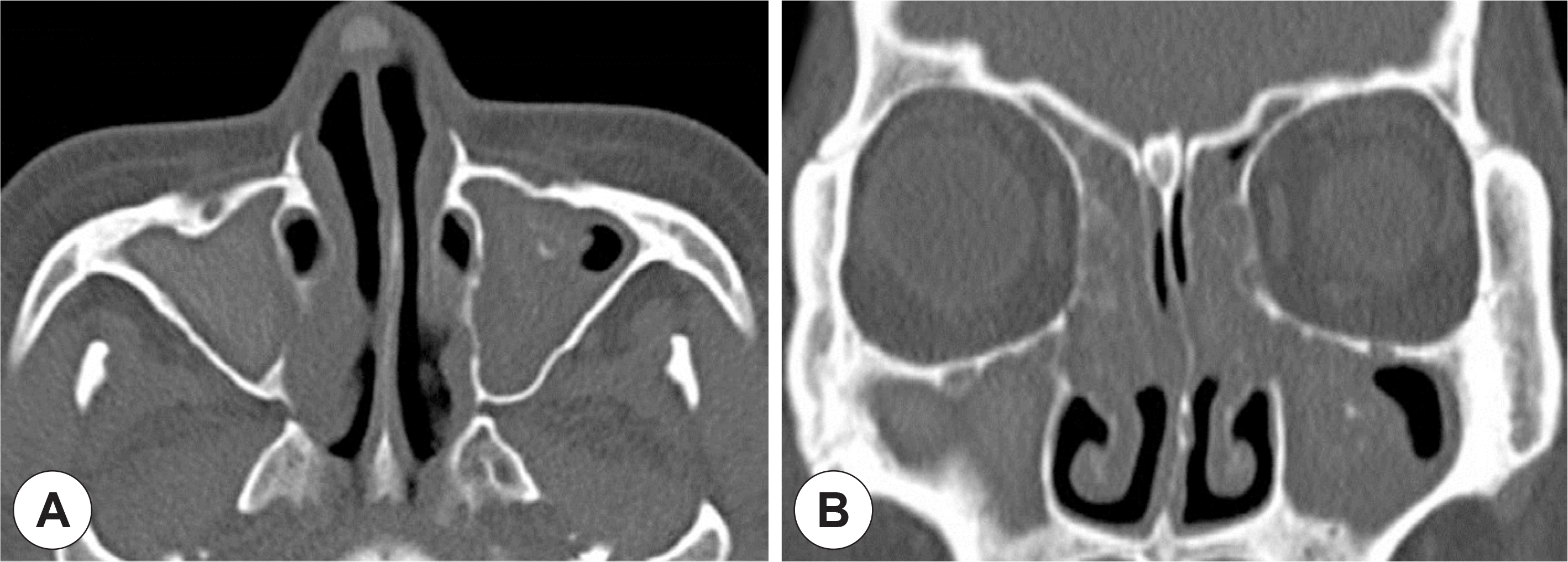
Fig. 2.
Thick, inspissated greenish mucoid secretion was removed from right maxillary sinus (A). Dark brown-colored clay-like material was brought out from left maxillary sinus (B).
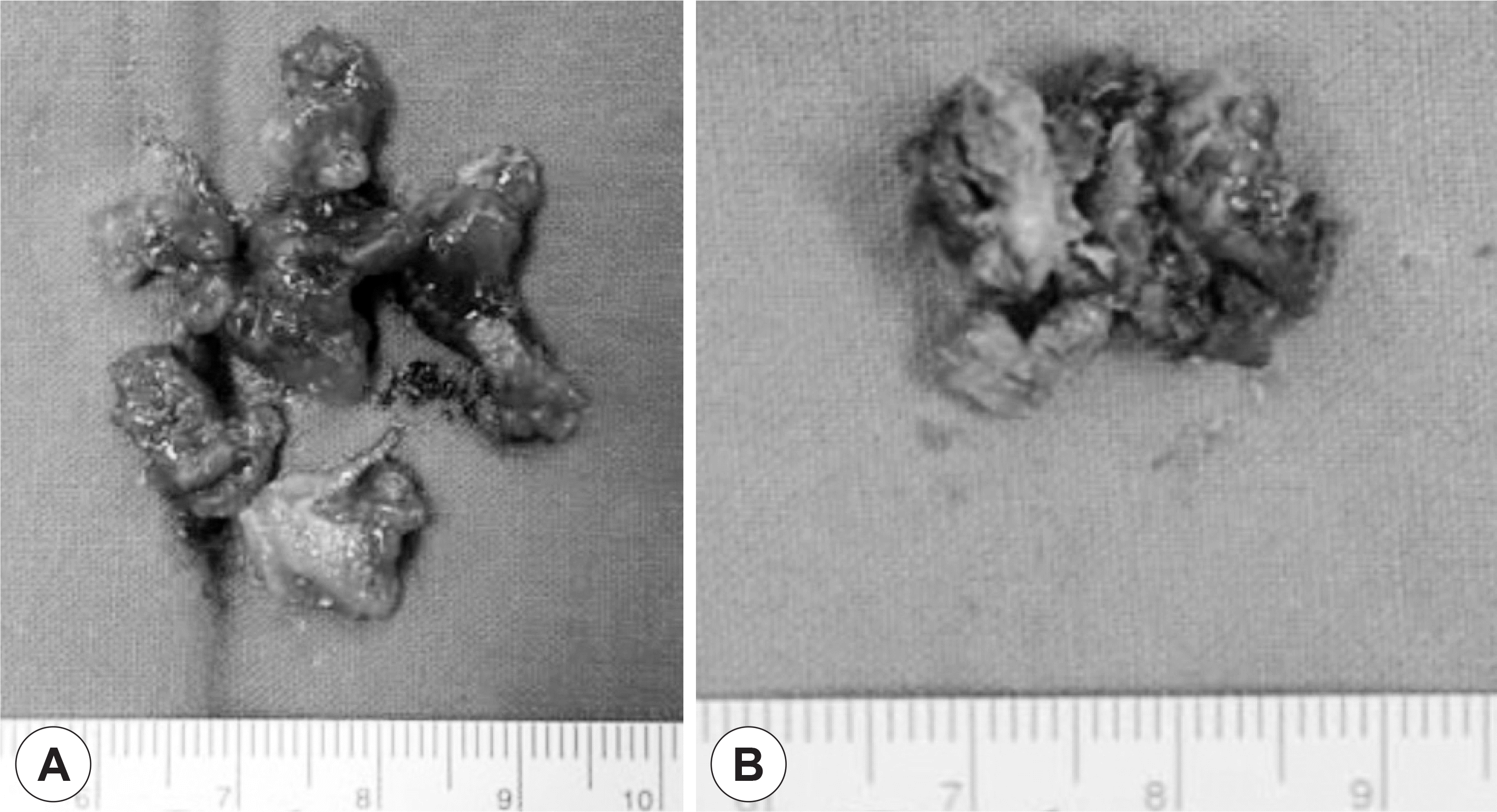
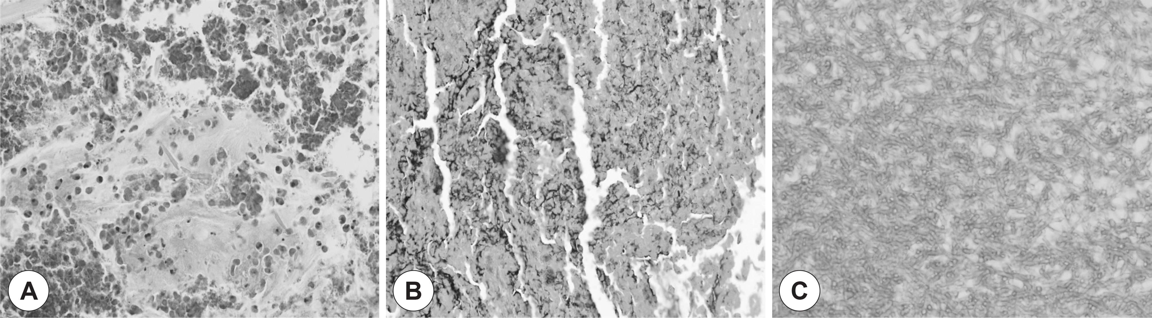
Fig. 3.
H&E stain of right maxillary sinus lesion showing inflammatory cells composed predominantly eosinophils and Charcot-Leden crystals (A). GMS stain of right maxillary sinus lesion was positive for fungal hyphae (B). H&E stain of left maxillary sinus lesion showing aggregation of fungal hyphae, consistent with Aspergillosis (C).
 XML Download