

 PDF
PDF ePub
ePub Citation
 Print
Print


Abstract
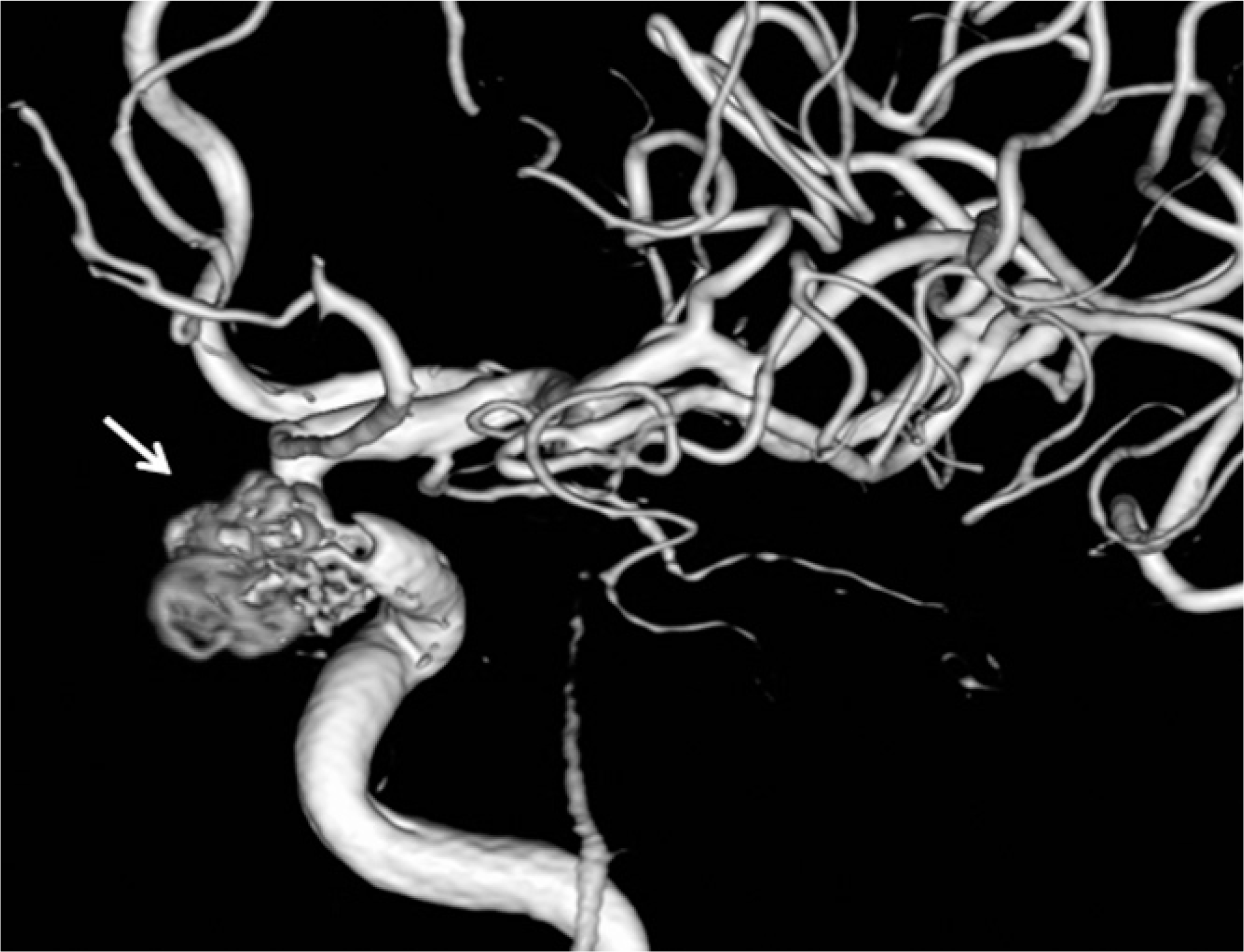
Rupture of the internal carotid artery (ICA) during endoscopic sinus surgery is a rare complication. However, it can potentially result in death within minutes. In the event of a traumatic injury to the ICA during sphenoid sinus exploration, it is very difficult to control the bleeding. We present a case of carotid-cavernous fistula after an accidentally-developed ICA bleed during endoscopic sphenoidotomy. The patient was successfully treated with endovascular embolization techniques that included detachable microcoils.
Go to : 
References
1). May M, Levine HL, Mester SJ, Schaitkin B. Complication of endoscopic sinus surgery: Analysis of 2108 patients-incidence and prevention. Laryngoscope. 1994; 104(9):1080–3.
2). Schaefer SD, Manning S, Close LG. Endoscopic paranasal sinus surgery: Indications and considerations. Laryngoscope. 1989; 99(1):1–5.
3). Laws ER Jr. Vascular complications of transsphenoidal surgery. Pituitary. 1999; 2(2):163–70.
4). Fujii K, Chambers SM, Rhoton AL Jr. Neurovascular relationships of the sphenoid sinus. A microsurgical study. J Neurosurg. 1979; 50(1):31–9.
5). Lee SK, Park YS, Cho JH, Park YJ, Kang JM, Jeon EJ, et al. Anatomic variation of sphenoid sinus and related neurovascular structures: A study of CT analysis. Korean J Otolaryngol. 2004; 47:978–82.
6). Sirikci A, Bayazit YA, Bayram M, Mumbuc S, Gungor K, Kan-likama M. Variations of sphenoid and related structures. Eur Radiol. 2000; 10(5):844–8.
7). Kinnman J. Surgical aspects of the anatomy of the sphenoidal sinuses and the sella turcica. J Anat. 1977; 124:541–53.
8). Elwany S, Yacout YM, Talaat M, El-Nahass M, Gunied A, Talaat M. Surgical anatomy of the sphenoid sinus. J Laryngol Otol. 1983; 97(3):227–41.

9). Kim HU, Kim SS, Kim IS, Chung IH, Yoon JH. Morphology of midline septum and accessory septum of the sphenoid sinus. Korean J Otolaryngol. 2001; 44(2):153–6.
10). Raymod J, Hardy J, Czepko R, Roy D. Arterial injuries in transsphenoidal surgery for pituitary adenoma; the role of angiography and endovascular treatment. Am J Neuroradiol. 1997; 18(4):655–65.
11). Lippert BM, Ringel K, Stoeter P, Hey O, Mann WJ. Stentgraft-implantation for treatment of internal carotid artery injury during endonasal sinus surgery. Am J Rhinol. 2007; 21(4):520–4.
12). 12) Valentine R, Wormald PJ. Carotid artery injury after endonasal surgery. Otolaryngol Clin North Am. 2011; 44(5):1059–79.
13). Goodwin JR, Johnson MH. Carotid injury secondary to blunt head trauma: case report. J Trauma. 1994; 37(1):119–22.
14). Barrow DL, Spector RH, Braun IF, Landman JA, Tindall SC, Tindall GT. Classification and treatment of spontaneous carotid-cav-ernous sinus fisulas. J Neurosurg. 1985; 62(2):248–56.
15). Iida K, Uozumi T, Arita K, Nakahara T, Ohba S, Satoh H. Steal phenomenon in a traumatic carotid-carvernous fistula. J Trauma. 1995; 39(5):1015–7.
16). Higashida RT, Halbach VV, Tsai FY, Norman D, Pribram HF, Mehringer CM, et al. Interventional neurovascular treatment of traumatic carotid and vertebral artery lesions: results in 234 cases. AJR Am J Roentgenol. 1989; 153(3):577–82.
17). Kim JK, Seo JJ, Kim YH, Kang HK, Lee JH. Traumatic bilateral cartotid-carvernous fistulas treated with detachable balloon. A case report. Acta Radiol. 1996; 37(1):46–8.
18). Pierot L, Moret J, Boulin A, Castaings L. Endovascular treatment of posttraumatic complex carotid-cavernous fistulas, using the arterial approach. J Neuroradiol. 1992; 19(2):79–87.
19). Halbach VV, Higashida RT, Barnwell SL, Dowd CF, Hieshima GB. Transarterial platinum coil embolization of cartotid-cavern-ous fistulas. Am J Neuroradiol. 1991; 12(3):429–33.
20). Yang PJ, Halbach VV, Higashida RT, Hieshima GB. Platinum wire: a new transvascular embolic agent. Am J Neuroradiol. 1988; 9(3):547–50.
Go to :
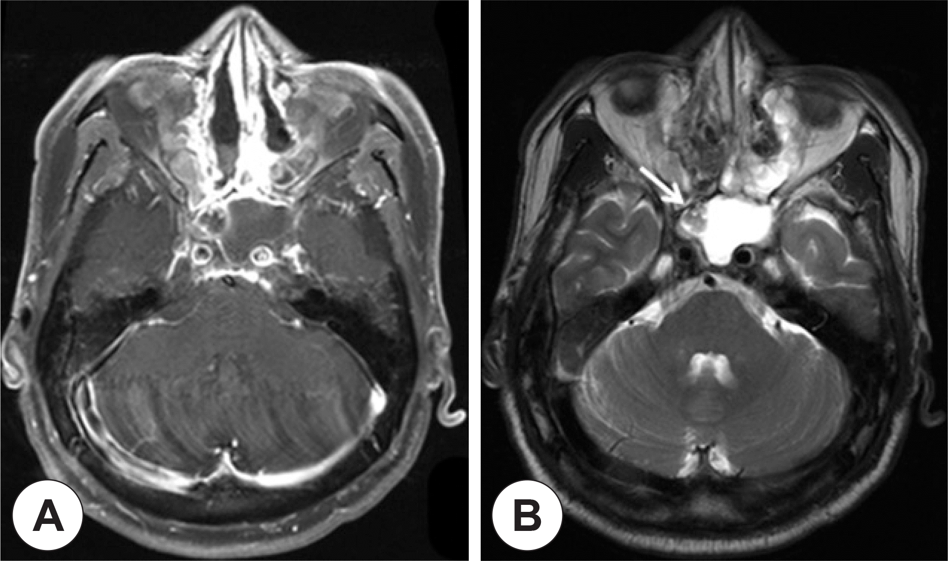
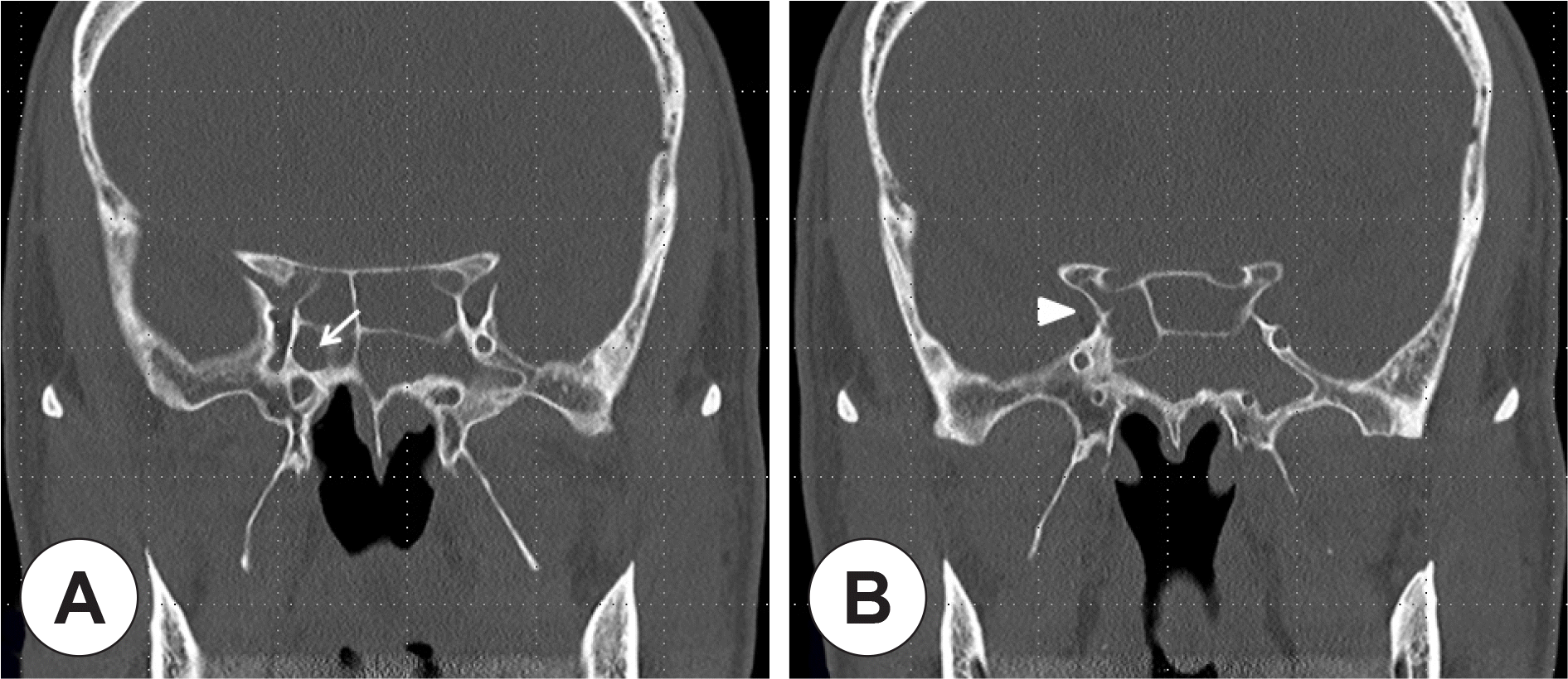 | Fig. 1.(A) Coronal CT scan of the paranasal sinues shows the right sphenoid sinus is less developed (arrow) than the left sphenoid sinus. (B) Coronal CT scan is more posterior section than. The right internal carotid artery (arrow head) is deviated to the medial side due to the less developed right spenoid sinus. |
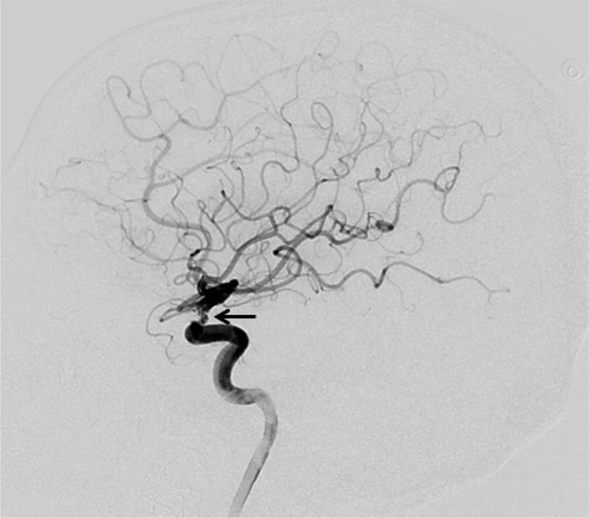 | Fig. 2.The right internal carotid angiography (lateral view) shows irregular narrowing of the supraclinoid segment of the right ICA (arrow) due to probably vasospasm after ICA injury. However, active bleeding is not observed. ICA: internal carotid artery. |
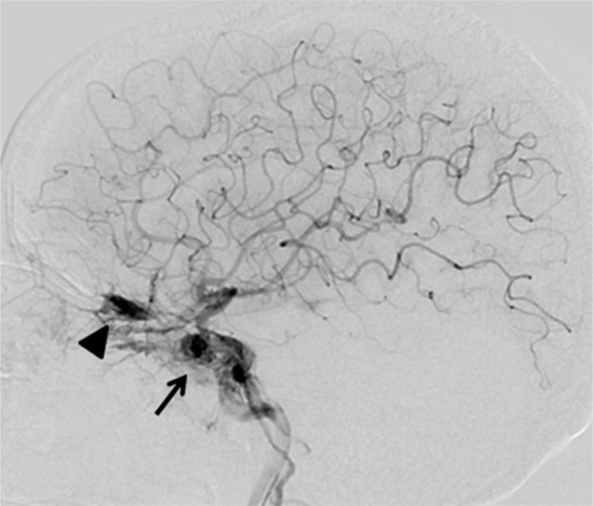 | Fig. 3.Transfemoral carotid angiography (lateral view) shows the early filling of cavernous sinus (arrow) and the venous drainage into the superior ophthalmic vein (arrow head) due to ca-rotid-cavernous fistula. |
 XML Download