

 PDF
PDF ePub
ePub Citation
 Print
Print


Abstract
Mucoceles are relatively common cystic lesions of the paranasal sinuses. However, mucocele of the nasal septum is extremely rare. We report a case of a mucocele present in this unusual location. Mucocele of the nasal septum should be considered in the differential diagnosis of a mass of the nasal septum and/or median canthal region. Nasal septal mucocele can be effectively treated with endoscopic marsupialization or complete excision.
References
1). Lee YB, Lee KC, Lee JW, Jin SM. A case of a sphenoid sinus mucocele protruding into both nasal cavities: transnasal endoscopic marsupialization. J Rhinol. 1999; 6(1):75–8.
2). Gall R, Witterick I. Mucocele of the nasal septum. J Otolaryngol. 2002; 31(4):246–7.

3). Lei L, Wang R, Han D. Pneumatization of perpendicular plate of the ethmoid bone and nasal septal mucocele. Acta Otolaryngol. 2004; 124(2):221–2.
4). Taskin U, Korkut YA, Aydin S, Oktay FM. Atypical presentation of primary giant nasal septal mucopyocele. J Craniofac Surg. 2012; 23(1):5–7.
5). Friedmann DR, Roman B, Lebowitz RA, Bloom JD. Radiology quiz case 2. Diagnosis: intraseptal mucocele. JAMA Otolaryngol Head Neck Surg. 2013; 139(6):647–8.
6). Lee KC, Lee NH. Comparison of clinical characteristics between primary and secondary paranasal mucoceles. Yonsei Med J. 2010; 51(5):735–9.
7). Stankiewicz JA, Newell DJ, Park AH. Complications of inflammatory diseases of the sinuses. Otolaryngol Clin North Am. 1993; 26(4):639–55.
8). Lund VJ, Milroy CM. Fronto-ethmoidal mucoceles: a histopathological analysis. J Laryngol Otol. 1991; 105(11):921–3.
9). Soon SR, Lim CM, Singh H, Sethi DS. Sphenoid sinus mucocele: 10 cases and literature review. J Laryngol Otol. 2010; 124(1):44–7.
Fig. 2.
Enhanced CT of the paranasal sinuses (A) revealed a 1.5 cm sized high attenuated cystic mass (arrow) in the left nasal septum. On MRI, a mass (arrow) on the left nasal septum had high signal on T1-weighted image (B) and low signal on T2-weighted image (C), suggesting a mucocele.
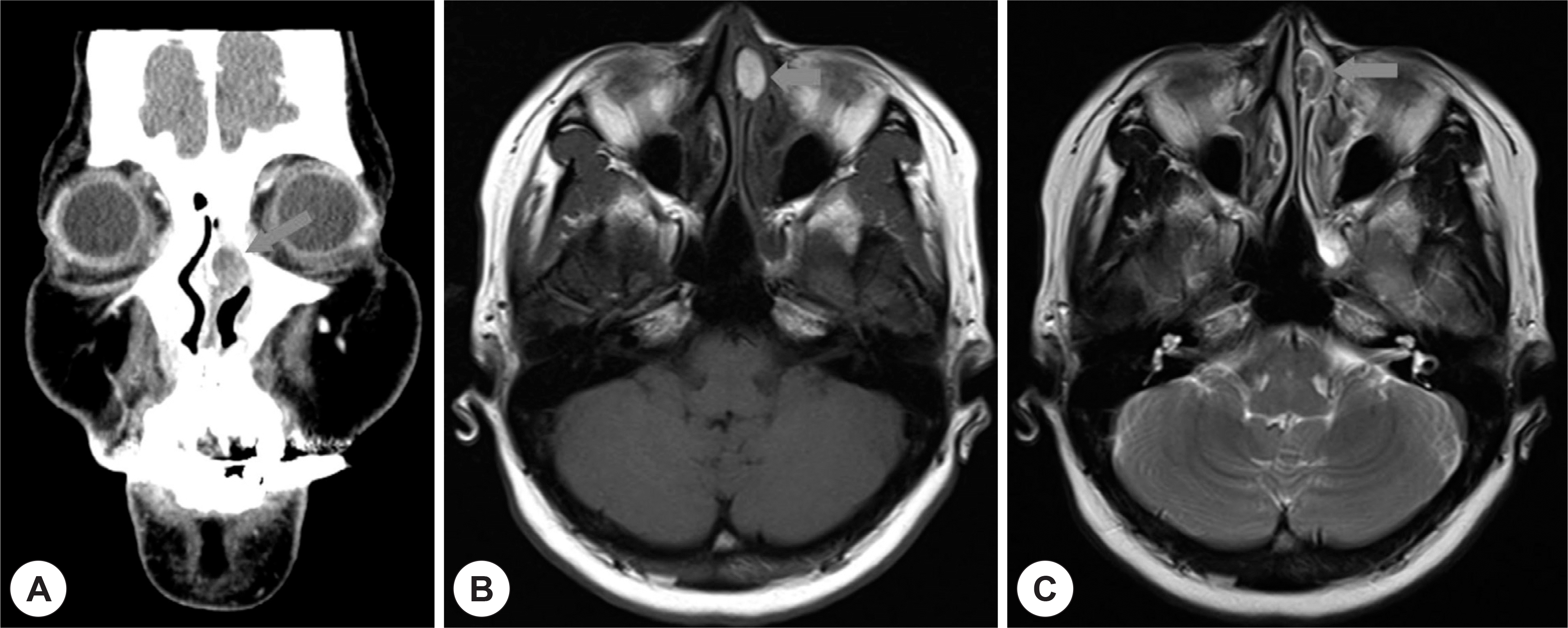
Fig. 3.
(A) Intraoperative appearance of left nasal cavity following endoscopic marsupialization (B) At 3months’ follow up, endoscopic examination shows a well marsupialized cavity on the left nasal septum.
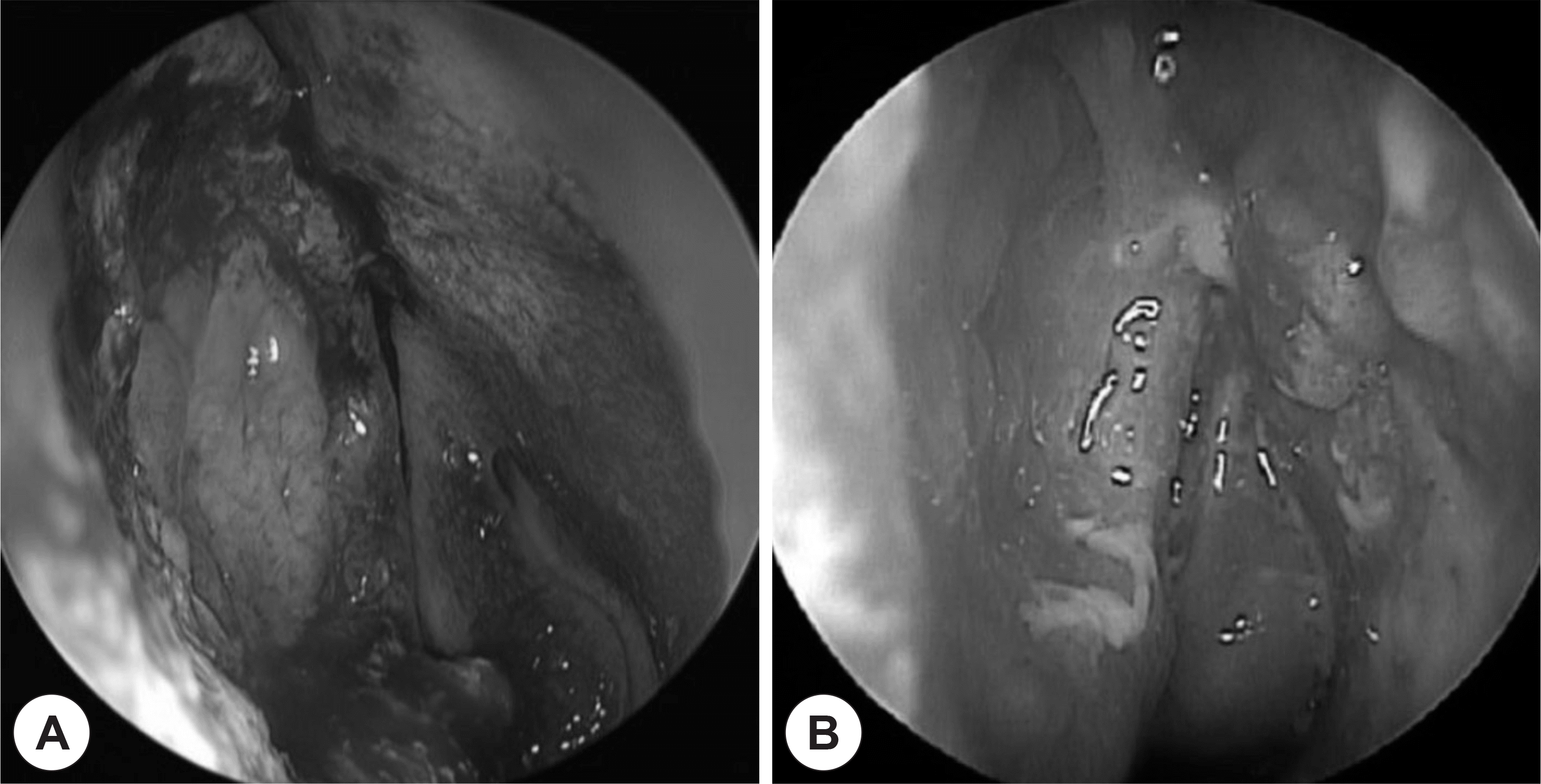
Fig. 4.
Histopatholgoic examination shows that a cyst is lined by low cuboidal epithelium and little inflammatory cells (Hematoxylin & Eosin staining, ×200).
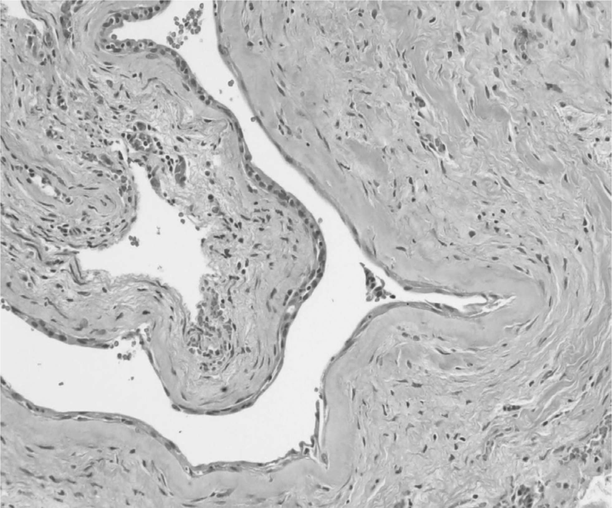
Table 1.
Summary of previously reported cases with nasal septal mucocele
 XML Download