Abstract
Nasal extranodal natural killer/T cell (NK/T cell) lymphoma is more common in East Asia than in the United States, comprising up to 7–10% of all non-Hodgkin's lymphoma. Early nasal symptoms are nonspecific and similar to chronic rhinosinusitis, such as nasal obstruction and nasal bleeding. With disease progression, inflammation and necrosis of the mucosa increase, hindering pathologic diagnosis. We experienced a case of nasal extranodal NK/T cell lymphoma in a 58-year-old woman who presented with recurrent periorbital swelling.
Go to : 
References
1). Choi CY, Jo YK, Lee BH, Lee YW, Lee KD, YU TH. Analysis of treatment in the patient with non-Hodgkin's lymphoma of the head and Neck. Korean J Otolaryngol-Head Neck Surg. 1997; 40:1820–5.
2). Au WY, Ma SY, Chim CS, Choy C, Loong F, Lie AK, et al. Clinicopathologic features and treatment outcome of mature T-cell and natural killer-cell lymphomas diagnosed according to the World Health Organization classification scheme: a single center experience of 10 years. Ann Oncol. 2005; 16:206–14.

3). Han KW, Choi SJ, Pae KH, Chung YS, Jang YJ, Lee BJ. Comparison of clinical characteristics of B cell lymphoma and NK/T cell lymphoma of the nose and paranasal sinuses. J Rhinol. 2005; 12:101–4.
4). Pine RR, Clark JD, Sokol JA. CD56 Negative extranodal NK/T-cell lymphoma of the orbit mimicking orbital cellulitis. Orbit. 2013; 32:45–8.

5). Kuwanara H, Tsuji M, Yoshi Y, Kakuno Y, Akioka T, Kotani T, et el. Nasal-type NK/T cell lymphoma of the orbit with distant metastasis. Human Path. 2003; 34:290–2.
6). McLean IW, Burneir MN, Zimmerman LF. Atlas of tumor pathology: Tumor of the eye and ocular adnexa. Washington DC, Armed Forces Institute of Pathology. 1994. 263–87.
7). King AD, Lei KI, Ahuja AT, Lam WW, Metreweli C. MR imaging of nasal T-cell/natural killer cell lymphoma. Am J Roentgenol. 2000; 174:209–11.

8). Jang SY, Jung JS, Kang JW, Yoon JH. Two Cases of Diffuse Large B-cell Lymphoma of Sinonasal Tract. J Rhinol. 2009; 16:169–72.
9). Sheahan P, Donnelly M, O'Reilly S, Murphy M. Pathology in focus: T/NK cell non-Hodgkin's lymphoma of the sinonasal tract. J Laryngol Otol. 2001; 115:1032–5.
10). Emile J-F, Boulland M-L, Haioun C, Kanavaros P, Petrella T, Del-fau-Larue MH, et al. CD5-CD56+ T-cell receptor silent peripheral T-cell lymphomas are natural killer cell lymphomas. Blood. 1996; 87:1466–73.
Go to : 
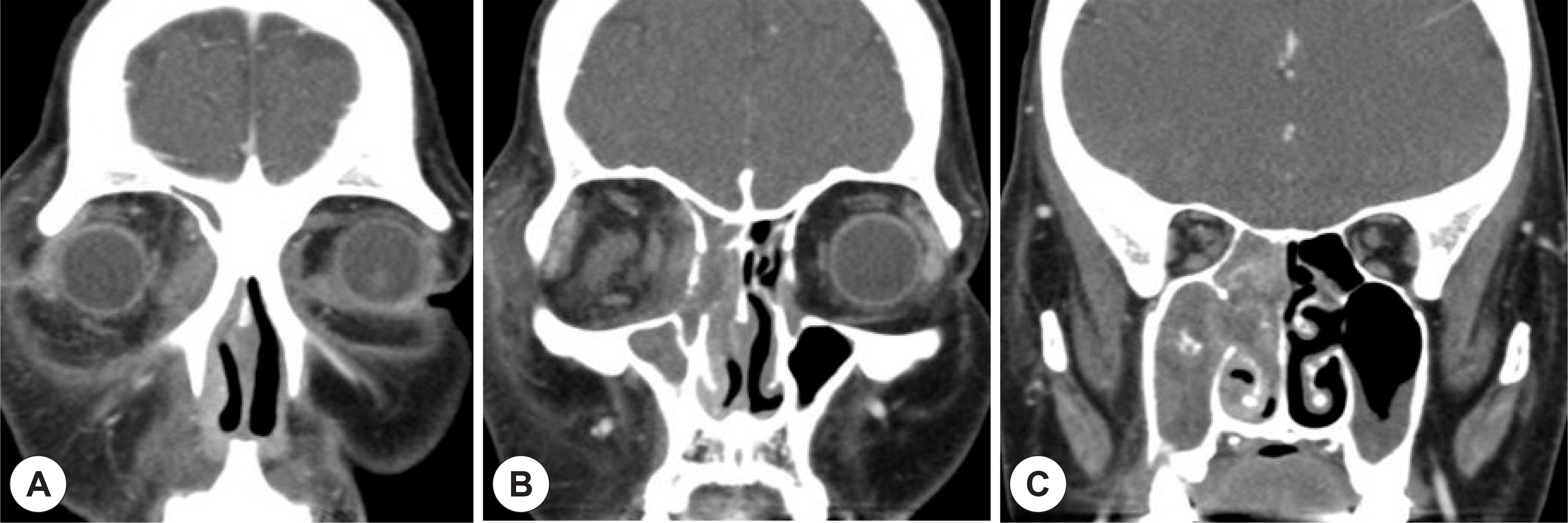
 | Fig. 1.Coronal, enhanced, Orbit CT findings. CT scan reveal the right periorbital swelling (A) and soft tissue density on the right sinonasal cavity (B), and nodular calcifications located centrally in the right maxillary sinus (C). |
 | Fig. 2.Coronal, enhanced, PNS CT findings and endoscopic finding. The images show a infiltrative lesion with mild enhancement in the left lacrimal sac and medial canthal tendon (A), and soft tissue density in anterior ethmoid sinus and both maxillary sinus (B and C). During the second surgery, biopsy was performed at the polypoid mucosa in ethmoid sinus of the left nasal cavity (D). |
 | Fig. 3.Coronal, enhanced, Orbit CT findings and endoscopic finding. The images show a large infiltrative region with enhancement in the left lacrimal sac (A), soft tissue density in anterior ethmoid sinus (B), and the homogenous small mass in ethmoid sinus of the left nasal cavity (C). A endoscopic finding shows an intranasal granulomatous mass in ethmoid sinus of the left nasal cavity (D), and third biopsy was performed. |
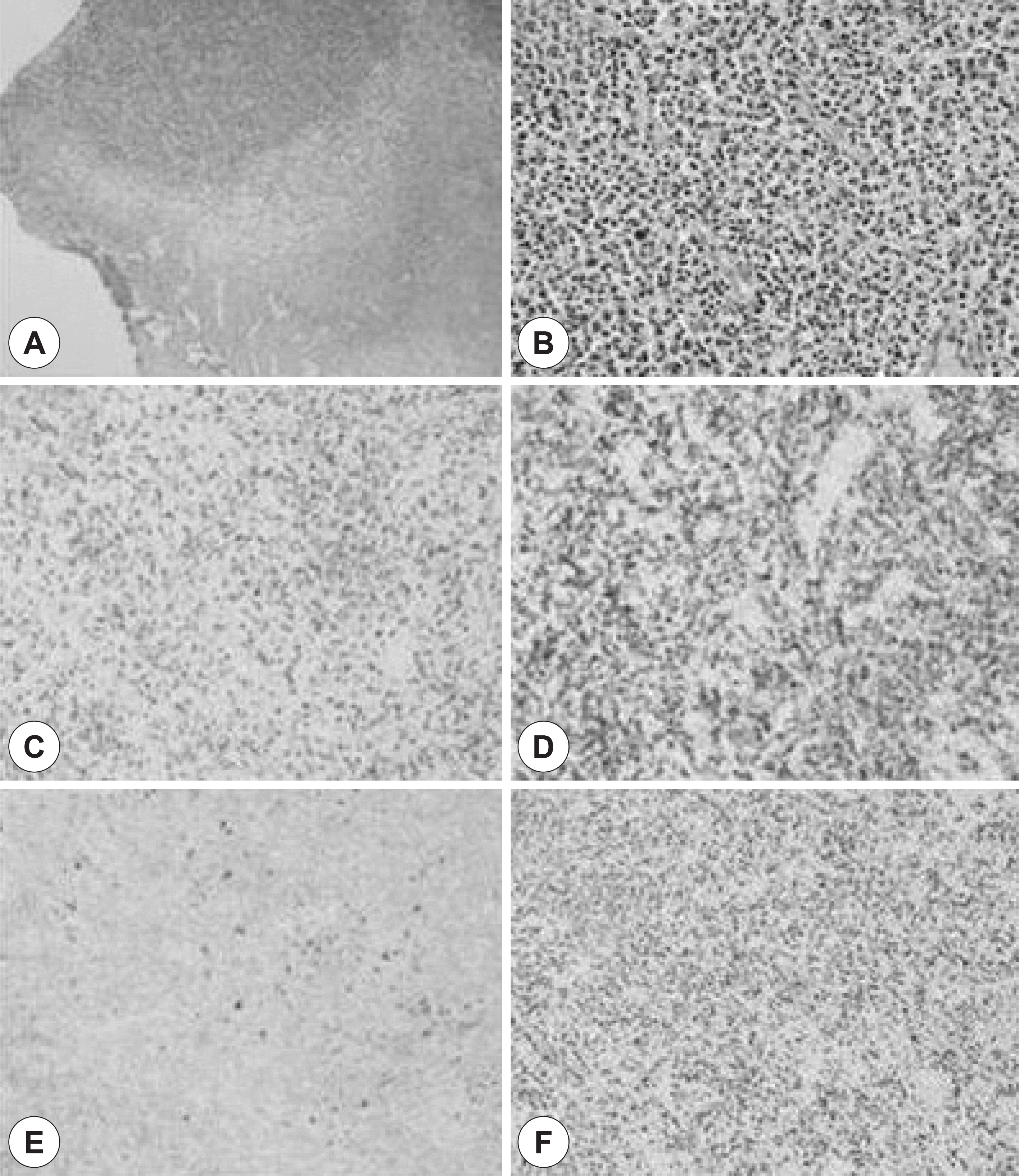
 | Fig. 4.Histologic and immunohistochemical findings. Small to medium sized atypical lymphoid cells with clear cytoplasm (HE ×100, ×400) (A and B). Tumor cells are positive for anti-CD3 and anti-CD56 antibody (×200)(C, D). Tumor cells are negative for anti-CD20 antibody (×200)(E). Tumor cells are positive for EBV-ISH (×200)(F). |


 PDF
PDF ePub
ePub Citation
 Print
Print


 XML Download