

 PDF
PDF ePub
ePub Citation
 Print
Print


INTRODUCTION
Cancer screening in medical imaging is one of the most important areas in computerized medical software. Especially, attempts to automate the early diagnosis of cancer using computer aided detection (CAD) algorithm on chest X-ray and mammography images were the most important research topic in the field of radiology [1]. However, the results of the clinical effects of CAD are still controversial. Even there was a research about screening performance of CAD reporting that sensitivity was significantly decreased for mammograms interpreted with vs without CAD in the subset of radiologists who interpreted both with and without CAD (odds ratio, 0.53; 95% CI, 0.29–0.97) [2]. But, deep learning technology, which has recently been greatly developed, is raising expectations for the possibility of computer software related to cancer screening again.
Deep learning is a kind of neural network. The neural network consists of an input layer, a hidden layer, and an output layer. Deep learning is a neural network with a large number of hidden layers. Over the past few years, deep learning has achieved tremendous performance improvements, especially in image classification [3] and speech recognition [4]. In recent years, deep learning has been used in a wide variety of areas. In particular, powerful image recognition and classification capabilities are being continuously adopted in high-tech devices such as autonomous vehicles and drones. The medical sector is no exception. Google has reported good results using deep learning for diagnosis of diabetic retinopathy [5], and Mount Sinai Hospital has introduced a system called “Deep Patient” that applies this technology throughout the electronic health records (EHRs) system, reporting improved accuracy in predicting the prognosis of severe diabetes, schizophrenia, and various cancers [6].
In this review, I would like to describe what the deep learning technology has done to cancer screening, what features of applied technology are, and what challenges to overcome in the future, focusing on the medical imaging field. As another review of this issue addresses CT, MRI and pathology applications, this review focuses on key cases that have been applied to optical imaging, chest X-ray, and mammography.
CONVOLUTIONAL NEURAL NETWORKS
Of the deep learning technologies, convolutional neural networks (CNNs) are particularly popular in the field of medical imaging. Most of the deep learning technologies related to cancer screening are also based on CNNs. CNN has been growing since the late 1970s and has been applied to medical imaging analysis since 1995 [7]. However, CNNs were able to be applied in the real world since Yann LeCun's LeNet was applied to hand-written digit recognition [8]. But, other than that, CNNs have become a technology with success not seen by many until 2012. The event that broke the downturn of CNNs was the December 2012 ImageNet Challenge. Krizhevsky et al, using CNNs called AlexNet, became the winners of the competition in a remarkable gap [9]. AlexNet contains eight layers with weights. The first five are convolutional and the remaining three are fully connected layers. Since the success of AlexNet, numerous CNNs architectures have emerged and continue to boost performance. Finally, the 2015 ImageNet Challenge comes with ResNet, a CNN that achieves an error rate of 3.57%, which is below the human average of 5% [10]. ResNet makes use of the residual function to enable deep learning of up to 152 layers, and since then there have been many similar CNNs with very good performances. From this point on, deep learning research using CNNs has started to increase rapidly in the field of medical imaging.
CANCER SCREENING IN OPTICAL IMAGING
Among cancer screening using optical imaging, melanoma detection is the biggest concern. According to The American Cancer Society, 87,110 new melanomas are diagnosed in the United States in 2017 and 9,730 are estimated to die. In addition, the 5-year survival rate of melanoma diagnosed as Stage IA and IB was 97% and 92%, respectively [11]. This means that although the problem is serious, early diagnosis can save many people. Therefore, early diagnosis of melanomas using optical imaging is very important. One of the most remarkable achievements of cancer screening in medical imaging using deep learning has come from melanoma detection. Stanford University researchers used CNNs to classify skin lesions. They used 129,450 clinical images to train classifying 2,032 different diseases and compared the test results with the results read by 21 board-certified dermatologists. The tasks tested in this study were to differentiate malignant carcinomas versus benign seborrheic keratoses and malignant melanomas versus benign nevi. In this experiment, CNN performed better than all dermatologists in all tasks [12]. This shows the potential to distinguish malignant skin cancer early from a photograph taken through a smart phone, and it has shown that cancer screening using optical imaging can be an inexpensive and efficient medical service.
In addition to dermatological diseases, cancer screening using optical imaging is expected to be useful for cervical cancer diagnosis. In 2016, several conference papers published improved results on cervical dysplasia diagnosis and cervical cancer classification [13]. Because cervical cancer ranks as the second most common type of cancer in women aged 15 to 44 years worldwide, these results have very important implications for women's health. To facilitate this research, Intel and MobileODT are collaborating on a cervical cancer screening challenge through Kaggle, a data platform. This challenge began on March 16, 2017 and ended on June 21, 2017, with 848 teams from 22 countries around the world participating in a hot competition [14]. Because the result of such a challenge is made with data only, it is necessary to carry out rigorous clinical experiments in order to become a practical applicable technology. However, the pace of development of this technology is expected to be very fast, because already top teams are releasing and discussing their technology.
CANCER SCREENING IN CHEST X-RAY
Finding pulmonary nodules on chest X-ray has the longest history in cancer screening using medical imaging. Neural network technology began to be applied before deep learning technology matured as it is seen now [7]. The reason for this early adoption of the new technology is that early diagnosis of lung cancer using chest X-ray has such an important clinical significance. According to the 2017 statistics of the American Cancer Society, there are about 222,500 cases estimated to be newly diagnosed with 155,870 deaths in 2017. This is the number one cause of cancer deaths regardless of gender, and it reaches one-quarter of all cancer deaths [15].
However, studies using deep learning techniques, including CNNs, for pulmonary nodule detection have begun to emerge after 2015. But, the applications were mainly focused on screening in CT images. There is relatively few research on pulmonary nodule detection using deep learning in chest X-rays. Bar et al used CNNs to identify different types of pathologies in chest X-ray images. They tested a dataset of 93 images trained with ImageNet, a well-known large scale nonmedical image database and try various combinations of features extracted from the CNN and a set of low-level features. They got an area under the curve (AUC) of 0.79 for classification between healthy and abnormal chest X-rays, where all pathologies are combined into one large class [16]. Bobadilla et al tried a family of CNNs known as DeepCNets composed of alternating convolutional and max-pooling layers with a linearly increasing number of filters. They trained and tested the Japanese Society of Radiological Technology (JSRT) database [17] composed of 154 nodules and 93 non-nodule chest radiographs. Their results show that CNNs can operate effectively on lung nodule classification through data augmentation and dropout regularization and achieve comparable results with state-of-the-art performance [18].
There are far fewer deep learning studies using chest X-rays compared to CT because there are not enough public datasets to accelerate this research. Conversely, in the case of CT screening, challenges such as LUNA16 or Kaggle Data Science Bowl 2017, which provide rich public datasets and large prize pools, are nurturing deep learning research. However, it is clear that the chest X-ray is the simplest and cheapest modality. Therefore, deep learning studies for lung cancer screening using chest X-rays will still be very important.
CANCER SCREENING IN MAMMOGRAPHY
The application of neural network technology for breast cancer screening in mammography has already begun in 1996 [19]. Also, in 2012, Jamieson et al. attempted mass classification of mammography and ultrasound using 4-layer AND [20]. In this way, compared with other cancers, breast cancer screening was applied relatively early. This is probably due to the importance of breast cancer as the number one female cancer mortality rate and commonly used cancer screening imaging modality such as mammography. However, deep learning has been widely applied in this field since 2015. Many studies have shown that tumor detection using CNNs is useful in breast cancer screening. Most of the results are from mammography [21222324], but studies using tomosynthesis [2526], ultrasound [27], and MRI [28] have also been published.
Although the results so far have shown promise, there are still no results showing state-of-the-art results that are well beyond radiologists. The main reason is that, like other medical imaging deep learning studies, it is difficult to acquire a large amount of data. Therefore, various studies are under way to overcome these limitations.
FUTURE CHALLENGE AND PERSPECTIVES
So far, I have reviewed the current state of deep learning technology used in cancer screening focusing on optical imaging, chest X-ray, and mammography. Although imaging modality and target cancer are different, we can see that there are similar kinds of future challenges.
First, it is not easy to acquire a large amount of medical image data required for deep learning. Therefore, most of the research results utilize the deep neural network used for other image recognition or additional training after transfer learning from other networks. To solve this problem, it is important to have a large number of public medical image datasets like ImageNet. Challenges such as Intel&MobileODT Cervical Cancer Screening and LUNA that target public competition through Kaggle and lung nodule detection are some of them, but more fundamental efforts to solve the problems should be done. Recently, Stanford University has announced plans that will build Medical ImageNet and activate research on medical imaging deep learning based on this plan. This effort should have global consensus and be able to collaborate with multiple medical institutions.
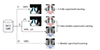
Second, it is difficult to learn if there are many medical image data but they are not properly labeled and annotated. Deep learning technology, which is applied to most medical images to date, is highly dependent on radiologists' labels and annotations. This is a fundamental problem with supervised learning technology. And, it seems that it may not be solved by unsupervised learning. Therefore, deep learning technology, which can provide good performance with a small number of correct answers, is actually very important. Hwang and Kim proposed a technique called weakly supervised learning to overcome this problem. They used class activation maps from CNNs for weakly-supervised learning with good localization and screening performance [22]. We can use traditional fully-supervised learning if we can obtain medical image data with enough labels and annotation, and weakly-supervised learning if we have no annotations. Semi-weakly-supervised learning can be applied in between. There is a difference between these approaches (Fig. 1). Depending on the data acquisition and performance of the training and test, we need to reasonably determine α (percentage of annotated medical images).
Finally, there is a need for technologies that can use different imaging modalities at the same time, link with EHR, and use genetic information for more comprehensive screening. This is a very difficult task, technically and clinically, but this goal must be accomplished through common effort. For this goal, among the deep learning technologies, CNNs that are mainly used in medical imaging, RNNs (Recurrent Neural Networks) capable of natural language processing, and reinforcement learning that can be continuously learned in various situations will be necessary together.
CONCLUSION
Cancer screening in medical imaging is a field that can achieve many good results with deep learning technology. As briefly reviewed here, deep learning technologies applied to optical imaging, chest X-ray, and mammography have flourished since 2015 and many studies have been published. Reviewed papers are summarized in Table 1. So far, the results are not very satisfactory, but the development of deep learning technology is very fast, the supply of researchable medical image data is getting bigger, and the research funds are getting rich, so the future is bright. In the future, it will be easier and more accurate to diagnose not only medical images but also EHR and genetic information with the help of deep learning technology. The development of deep learning technologies is important for this, but the role of physicians who understand and use these technologies becomes increasingly important. Therefore, I expect more physicians to study these deep learning technologies with interest.
 XML Download