

 PDF
PDF ePub
ePub Citation
 Print
Print


INTRODUCTION
Autism spectrum disorder (ASD) is a life-long neurodevelopmental condition characterized by persistent deficits in social communication and social interaction and restricted, repetitive patterns of behavior, interests, or activities [1]. The prevalence of ASD has increased markedly in the recent three decades and the median of prevalence estimates of ASD was 62/10,000 [2]. In 2014, the Centers for Disease Control and Prevention (CDC) estimated that ASD affects approximately 1 in 68 children [3]. Although increases in public awareness and research funding have led to scientific advances in understanding ASD and its treatment, there is a huge unmet demand for mechanism-driven successful interventions of core symptoms of ASD. Pharmacological treatments such as risperidone and aripiprazole can be beneficial in relieving some symptoms of ASD, but drugs for improvement of the core symptoms are yet to be developed [4].
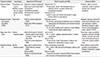
Brain stimulation and modulation has long been implemented in the treatment of psychiatric disorders and guidelines were suggested for several brain stimulation methods, such as electroconvulsive therapy (ECT), single-pulse or repeated transcranial magnetic stimulation (rTMS), vagus nerve stimulation (VNS), deep brain stimulation (DBS), et cetera [5]. Brain stimulation, unlike systemic pharmacological treatment delivered orally or parenterally, involves electrical mechanisms of the brain, which in turn induces localized neurochemical changes [5]. Applications of neurostimulation or neuromodulation by a variety of new and old techniques might be able to improve or correct underlying dysfunctions. Although brain stimulation therapies have been regarded as highly invasive treatment and reserved for patients with treatment-resistant disorders, several new stimulation methods are less invasive and can be used in less severe patients. Therapeutic applications of these tools in ASD have been tried in some cases but clinical experience and scientific evidence are limited (Table 1). Here we review the currently available techniques for neurostimulation and neuromodulation to provide useful information for researchers and clinicians.
ELECTROCONVULSIVE THERAPY
ECT applies an electric stimulus to the surface of the head and induces a widespread seizure with release of many neurotransmitters [5]. When it is adjusted for each patient, the parameters of stimulus can vary widely according to one's seizure threshold, clinical efficacy and adverse effects. Induced seizures with camphor and later electric shocks were introduced as convulsive therapy since 1930s for schizophrenia and depression [6]. ECT became spread all over the world because it was safer, more effective, and more convenient than camphor or metrazol therapy. Italian psychiatrists, Ugo Cerletti and his colleague Lucio Bini, were nominated for a Nobel Prize but did not become winners. In the past, ECT was applied as 'unmodified' form, without muscle relaxants, and the seizure resulted in a grand-mal convulsion. Fracture or dislocation of the bones resulting from the procedure was rare but a serious complication of unmodified ECT. To avoid that complication, 'modified' ECT with a short-acting anesthetic agent in addition to the muscle relaxant was developed. ECT devices have applied for psychiatric patients since 1938 prior to medical devices being regulated by the US FDA. However, the US FDA today considers ECT machinery to be experimental ones and has classified the ECT machinery as Class III medical devices, high risk devices except for patients suffering from catatonia [7].
In ASD, catatonia is not uncommon as 12-18% of young people with ASD also have catatonic symptoms [89]. However, diagnosis of catatonia in ASD is difficult to be made because there are common features in these two conditions such as mutism, echolalia, and repetitive behaviors. Specific criteria for 'autistic catatonia' or 'catatonia-like deterioration' have been suggested, including slowness in movement and verbal responses, difficulty in initiating and completing actions, increased reliance on physical or verbal prompting from others, increased passivity and apparent lack of motivation [8]. The fifth edition of Diagnostic Statistical Manual (DSM-5) defines catatonia as being characterized by the presence of at least three of the following: catalepsy, waxy flexibility, stupor, agitation, mutism, negativism, posturing, mannerisms, stereotypies, grimacing, echolalia and echopraxia and a diagnostic category Catatonia Not-Otherwise-Specified can be applied to catatonia in ASD [1].
It has been known that treatment of autistic catatonia is challenging [10]. ECT is an effective second line treatment option for coexisting acute phase of catatonia in ASD when first line benzodiazepine treatment is insufficient [1011]. Clinical guidelines for treatment of autistic catatonia suggest that bilateral ECT is one of the treatments of choice together with psychological approaches and high doses of lorazepam [12]. A systematic review of interventions used to treat catatonic symptoms in ASD indicated that 11 relevant papers on ECT treatment were rated as low quality reports and majority of cases presented with comorbid diagnoses, including depression, anxiety disorder, obsessive-compulsive disorder, psychosis, and tics and Tourette syndrome. Almost all cases reported a marked improvement with ECT such as increased speech, reduced posturing, improved social interaction and increased activity, but a few papers reported a more mixed or partial response to ECT [13]. There are some concerns about ECT, including rapid recurrence of symptoms, mild delirium, aggravation of symptoms, and prolonged seizure [1011].
Underlying neural mechanisms are still unknown. Recent neuroimaging studies suggested that ECT attenuates electroencephalography (EEG) pattern of activation and deactivation associated with cognitive task performance and alters cortical functional connectivity [14]. Multiscale entropy analysis of EEG showed that EEG complexity decreased in the frontocentral region and increased in the occipital region [15]. Abnormalities in gamma aminobutyric acid (GABA) has been hypothesized to play a role in pathophysiology of ASD and catatonia and the therapeutic effect of ECT might be related to enhancement of GABA function [16].
REPEATED TRANSMAGNETIC STIMULATION
rTMS is a widely used, safe and non-invasive tool for neurostimulation and modulation using series of pulsed magnetic stimuli delivered to the brain [17]. Stimulating coil generates magnetic fields which pass through scalp and skull and induces an electrical current in the brain. rTMS involves repetitive delivery of pulses (>1 Hz) to modulate cortical activity for research and therapy [18]. It is believed that rTMS can modulate activity of the targeted region immediately and also alter neuroplasticity mechanisms resulting in long-term effects. To assess neuroplasticity, two types of rTMS stimulation protocols have been used, that is, theta-burst stimulation (TBS) [19] and paired associative stimulation (PAS) [20]. TBS delivers a burst of three pulses at 50 Hz repeated at intervals of 200 ms in either continuous or intermittent mode (cTBS or iTBS, respectively). iTBS enhances cortical activity associated with long-term potentiation (LTP), whereas cTBS suppresses cortical activity associated with long-term depression (LTD). PAS delivers two paired stimulations of an electrical peripheral nerve stimulation of the right median nerve followed by a TMS pulse to the contralateral motor cortex in 25 ms, inducing LTP-like neuroplasticity.
In recent decades, pathophysiology of ASD has been studied extensively to find macro- and micro-structural, neurochemical, functional, anatomic, and genetic abnormalities, but the exact etiology of ASD is still unknown. Among many theories, the aberrant neuroplasticity hypothesis in ASD has been supported by research in multiple domains such as genetics, animal model, neuroimaging, and brain stimulation [21].
Neuroplasticity is defined as neuron's ability to reorganize and alter anatomical and functional connectivity in response to the environmental input. The most prominent morphological findings in neuroimaging studies in ASD is brain overgrowth in the early life [22]. Genetic mutations studied in patients with ASD are found in several genes linked to synaptogenesis and neuroplasticity, including neuroligin 3 and 4, c3orf58, NHE9, PCDH19, CNTNAP2, and SHANK3 [2324]. Excitation/inhibition (E/I) imbalance, caused by a deficit in the inhibitory and an excess in excitatory neurotransmission, might be a critical determinant of neuroplasticity in ASD [25].
Clinical and research use of rTMS has been done in Parkinson's disease, depression, schizophrenia, and migraine headache [26]. Diagnostic and therapeutic application of rTMS in ASD is emerging and its potential is being realized [26]. In the comparison between 27 individuals with ASD treated with 18 session long course of 1 Hz rTMS applied over the dorsolateral prefrontal cortex and 27 age-matched subjects with ASD as control, rTMS improves executive functioning and clinical symptoms such as irritability, hyperactivity, and stereotypic behaviors in ASD [27]. It was proposed that extended dosing (i.e., 6,000 pulses) of high-frequency (i.e., 20 Hz) rTMS in ASD might stabilize aberrant neuroplasticity and improve the impairment of social and cognitive performance [21]. A double blind randomized trial conducted in 28 adults with high-functioning autistic disorder or Asperger's disorder showed that deep rTMS to bilateral dorsomedial prefrontal cortex (PFC) resulted in a significant reduction in social relating impairment and socially-related anxiety than sham treatment [28].
VAGUS NERVE STIMULATION
VNS has been performed by intermittent repeated stimulation of the left vagus nerve with a small electrical pulse from an implanted neurostimulator to a bipolar lead wrapped around the nerve in the neck for patients with psychiatric disorders [29]. The left vagus nerve is stimulated rather than the right vagus nerve because the right one plays a role in cardiac function, the alteration of which may be potentially harmful [29]. This intervention requires surgical implantation of a neurostimulator that usually can be done as an outpatient procedure. Briefly, an incision is made in the upper left chest and the generator is implanted into a little "pouch" on the left chest under the clavicle. A second incision is made in the left side of neck, so that the surgeon can access the vagus nerve and then wrap the leads around the left branch of the vagus nerve, and connects the electrodes to the generator under the skin. Once successfully implanted, the generator sends electric impulses to the vagus nerve at regular intervals [29]. Non-invasive VNS through the skin does not have sufficient evidence for clinical application [30].
VNS was initially approved for treatment-resistant epilepsy [31]. In addition to typical autistic symptoms like difficulty in communication and repetitive behaviors, persons with ASD sometimes present mental retardation and seizures. It was reported that ASD is more common in children who experienced seizures during their first year of life than the general population and a third of adult patients with ASD had experienced seizures in their childhood and adolescence [31]. Since VNS was approved in 1997 by the US Food and Drug Administration for medically refractory partial-onset seizures in patients 12 years of age and older, a number of reports demonstrated the positive quality of life in patients with ASD and epilepsy following placement of a VNS device [32]. Interestingly, in some case reports, VNS in ASD was associated with modest behavioral improvement as well as seizure control in both child and adult [33].
Neural mechanisms of antiepileptic effects of VNS might be related with the locus coeruleus and dorsal raphe nucleus, because they have widespread projections to the brain and spinal cord, release neuromodulators with robust antiepileptic effects, are known to be activated by acute and chronic VNS, and abolish VNS-induced seizure suppression when lesioned [33]. However, how VNS could suppress and improve behavioral control, possibly cognition, is unclear. In consideration of the facts that interictal spikes can disrupt cognitive function and it is common that the patients with ASD have epileptiform abnormalities, therapeutic effect might be the result of reduced seizure and epileptiform activity [33]. Another plausible explanation is VNS might rectify the E/I imbalance in ASD, as was shown in a recent preclinical report that inflammatory stress-induced decrease in ratio between synaptic inhibition and excitation could be blocked by VNS as a result of the "anti-inflammatory reflex" [34].
DEEP BRAIN STIMULATION
DBS is a surgical procedure under stereotactic technique to implant electrodes at target regions in the brain [35]. An electrical stimulus of various intensity and frequency is delivered via implanted electrodes and induces an electrical field that modulates patterns of neuronal firing and thus modifies activity in the neuronal circuits. DBS was initially used in the treatment of movement disorders and the therapeutic indications are expanding including depression and obsessive-compulsive disorder [3637].
While patients with ASD at one end show high intelligence and high function such as living on their own with a professional career, those at the other end show lower function and critical symptoms such as self-injurious behaviors (SIB), and aggressive behaviors that are potentially life-threatening to themselves and their family members [38]. SIB is prevalent and very often refractory: 35-50% of people with ASD have SIB and 32% of 135 consecutive patients with ASD had refractory SIB [38]. Even though clinical application of DBS for severe autism is very limited, there are some case reports with successful results [3940]. A patient with severe stereotypies of self-injuring "picking" movements underwent DBS with electrode implantation in bilateral globus pallidus interna (GPi) and showed a remarkable improvement in her motor stereotypies of 91.3% in the John's Hopkins motor stereotypy rating scale (JHMRS) [39]. The other patient presenting prominent self-injurious stereotypies of severe body rocking and repetitive punches of his legs and arms together with biting his care givers underwent DBS in bilateral anterior limb of the internal capsule (ALIC) and GPi and showed an initial improvement of 71.6% in the JHMRS [39]. The third patient with life-threatening SIB underwent DBS in bilateral basolateral amygdaloid (BLA) nucleus and showed a significant decrease in SIB with a self-rating score of 1-2 by an ordinal scale from 6 (worst case) to 1 (best case) and improvement in social cognition [40].
Socioemotional processing, also termed as social cognition, refers to the processing and storage of information necessary to navigate social contexts [41] and the brain regions mediating socioemotional processing, which includes the limbic system, facial processing system and the mirror neuron network [42]. A revised limbic system model includes hippocampal-diencephalic, parahippocampal-retrosplenial, temporo-orbitofrontal, and default mode networks. The facial processing system consists of the amygdala, fusiform gyrus, and superior temporal sulcus [42]. The mirror neuron network is located at the temporoparietal junction and communicates anteriorly with the medial PFC. In ASD, functional and neuroimaging studies have shown abnormalities in these networks and their white matter tracts, including cortical and subcortical gray matter overgrowth early in brain development, especially in the PFC and amygdala, decreased activation in networks for socioemotional processing, and alterations of both resting-state and stimulus-induced oscillatory activities, notably in the gamma range [35]. Among these structures, the amygdala has a central role in social cognition and its abnormality is closely linked to social deficit and SIB in ASD, although it is not the only structure implicated in ASD. Especially, BLA connects between central amygdala and superficial nuclei and reciprocally communicates with orbitofrontal, anterior cingulate cortex, and medial PFC. These results suggest that DBS of the BLA might be a potential therapeutic intervention for refractory ASD with life-threatening SIB [35].
FUTURE DIRECTION: POTENTIAL APPLICATION OF OPTOGENETICS
Optogenetics is a technology that allows targeted fast control of precisely defined events in biological systems from cells to freely moving mammals [43]. To achieve such goal, two key characteristics are used in optogenetics: 1) light and light-sensitive proteins or opsins (e.g., channelrhodopsin and halorhodopsin); 2) cell-type specific expression of opsins using molecular biological techniques such as genetic engineering of animals, viral vectors to express opsins in a specific type of cells in living tissue. Such combination of genetic and optical methods enables gain or loss of function of well-defined events in specific cells of living tissue [43]. Although methods of neurostimulation and neuromodulation for ASD have been developed and are being used, they have inherent limitation in temporal and spatial resolution that hinders us from understanding exact etiology and mechanism of therapeutic effects of psychiatric illnesses in spite of their practical usefulness in clinical settings [44]. Optogenetics might be able to shed light on research and treatment of ASD.
BLA, discussed in the previous section as a potential target for DBS, can also be stimulated using optogenetic technique [45]. BLA has multiple projections to various brain regions, including centrolateral amygdala (CeL), the bed nucleus of the stria terminalis (BNST), and ventral hippocampus (vHPC) [45]. Because the BLA-vHPC circuit specifically facilitates anxiety-related behaviors and impairs social interaction simultaneously, axon terminals of glutamatergic neurons of BLA in vHPC might be a potential target for treatment of ASD [45].
One of the mostly cited models of ASD is the increased ratio of E/I in the brain. It is believed that a deficit in the inhibitory neuro-transmission could develop during neuronal maturation and such E/I imbalance could be one explanation in the pathophysiology of ASD [46]. Parvalbumin (PV) interneurons plays a critical role in the E/I balance and generating gamma band oscillations [47]. Recently E/I imbalance hypothesis has been proven by carefully performed optogenetic experiments [48]. The authors generated mice expressing both CaMKIIα::SSFO on pyramidal neurons and DIO-PV::C1V1 on PV interneurons in the medial PFC, which are activated by different wavelengths of light, i.e., 473 nm and 590 nm, respectively. When only pyramidal neurons were activated, social behavior of the animals significantly decreased; however, when pyramidal neurons and PV interneurons were activated simultaneously, social behavior increased to the normal level. These results suggest that enhancement of the activity of PV interneurons in the medial PFC might be an effective strategy to improve the social deficit in ASD. Another recent mouse model with abnormal PV interneurons (Dlx5/6+/- mice) showed cognitive inflexibility and deficient task-evoked gamma oscillations that could be restored by optogenetic stimulation of PV interneurons in the PFC [49]. However, the increased E/I balance might not be limited to the PFC, hence it should be more desirable to enhance PV interneuron activity in the broader area of the cortex.
PV neurons in the basal forebrain have been known to make cortical projection [50]. It was discovered that basal forebrain PV neurons could control cortical gamma band oscillations by their direct projection to cortical PV interneurons [50]. Therefore, these neurons might be an ideal target to increase the activity of cortical inhibitory neuronal activity and rectify the E/I imbalance. Based on recent research on the mechanism of ASD, optogenetic stimulation of basal forebrain PV neurons deserves careful study of its potential as a therapeutic intervention for ASD.
CONCLUSION
The core symptoms of ASD are not treatable with pharmacotherapy. Brain stimulation and modulation is getting attention as new therapy tools for ASD. Noninvasive brain stimulation techniques such as ECT and rTMS and invasive intervention methods like VNS and DBS have been applied to a variety of symptoms associated with neurological and psychiatric conditions, including Parkinson's disease, depression, tic disorder, epilepsy, and autism. In ASD, there have been limited supporting data for the efficacy and safety in humans, especially in pediatric patients. However, after reviewing the literature, we suggest that brain stimulation and modulation methods are potentially effective and viable options for treatment of ASD. For future direction, investigation of mechanism-driven treatment of core symptoms of ASD, using novel neuroscientific methodology such as optogenetics, is warranted.
 XML Download