

 PDF
PDF ePub
ePub Citation
 Print
Print


INTRODUCTION
Minimum invasive surgery (MIS) has been a key issue in medical surgery since it could not only minimize the operation scars (or other aftermath) but also significantly reduce the recovery time of the patients after the surgery. In fact, robotic technology has shown its effectiveness in many medical surgery areas such as neurologic surgery, orthopedic surgery, percutaneous surgery, radiosurgery, laparoscopic surgery, etc. [123456789101112]. More effort have been devoted to further extend its potential to other medical areas.
In particular, very high precision and safety are extremely important in neurosurgery since the fine and accurate operations either inside the brain or close to neural cords should be conducted. Slight mistakes or errors during the surgery could result in serious and unrecoverable damage to the patients. Thus, as efforts to reduce or to replace the role and the burden of the surgeons in neurosurgical operations such as spinal fusions, stereotactic operations, etc., many different types of advanced neurosurgical robot systems have been introduced [123456789101112].
Among those neurosurgical operations, the robotic system conducting stereotactic neurosurgical interventions such as Deep Brain Stimulation (DBS) will be mainly discussed in this paper. In DBS operation, a multi-contact electrode is inserted into the designated location of the brain where it provides high frequency electrical current pulses to a subthaelamic necleus. The operation is known to be very effective in the treatment for epilepsy, Alzheimer's and other diseases but requires extremely high precision and demands carefulness.
So far, many different forms of commercialized traditional stereotactic devices have been introduced. The joint structure of the most conventional stereotactic devices are either the PPPRR type or the PPRPR type where P and R denotes the prismatic joint and the revolute joint, respectively. The needle type inserting device or the micro-drive unit attached to the end of those stereotactic devices carries the electrode at its endpoint and inserts it into the designated region. For example, Leksell Stereotactic System® manufactured by Elekta Medical Systems [13], Zamurano Duchovny (ZD) stereotactic frame by Adhoc Medical [14], have the PPPRR structure. But Cosman-Roberts-Wells (CRW) Steoreotactic Arc system by Integra LifeSciences Corporation has the PPRPR structure [15]. In the past, the position accuracy of the manual stereotactic surgery was reported around 1mm~2mm [161718]. Recently, it is reported that the Leksell frame plus arc has a mean position accuracy of 0.7mm [19], the CRW Precision Arc and Phantom Base is contended to have an position accuracy of less than 0.5 mm [15], and Zamorano frame is reported to have a position error of 1.17±0.25mm [20].
In general, for this manual stereotactic neurosurgery, the CT images or MRI images obtained in the preoperative planning stage are employed both for the planning the operation and for registering the operation area of the patient and the instruments in the operating theater. Then intraoperative fluoroscopy images are employed, if needed, to provide the planar views on the brain of the patient during the needle insertion process. In this process, patients and surgeons are inevitably exposed directly and indirectly to the X-rays. In addition, surgeons should be able to properly align and position the needle to the target position, based on those planar fluoroscopy images of the brain of the patient. In fact, it is mandatorily recommended that allowable position error to the target position in DBS surgery should be less than 2mm for satisfactory results [21]. However, despite of its inconveniences and other uncertainties such as its long set-up procedure, its long operation time, exposure to the radio-active fluoroscopy, surgeon dependent position accuracy, registration errors, etc., majority of the surgeons still prefer to employ the manual stereotactic device.
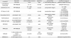
So far, there have been many commercialized neurosurgical robots and many neurosurgical robots developed in the laboratory that have already been successfully employed and verified in the role of assisting the surgeon in neurosurgery such as accurate positioning of the instrument, the electron placements, with the help of the preoperative and/or intra-operative images obtained from the CT, the MRI, the fluoroscopy, etc. [1234567891011122021222324252627282930]. Table 1 shows the summary of both the commercialized conventional stereotactic frames and the neurosurgical robotic systems in aspects of the kinematic structure, robot position accuracy, operation mode of the robot, data available in operation, etc.
STRUCTURE OF THE NEUROSURGICAL ROBOTS
The structure of the neurosurgical robot could be classified into two different types: the single type with either serial or hybrid structure and the macro-micro type. Note that the parallel robot alone may not be adequate to undertake an assisting role of the neurosurgery due to its intrinsically smaller workspace even though it has advantages of high accuracy and high stiffness than the serial type robot. Thus, most of commercialized neurosurgical robots have the serial structure such as the PathFinder, the NeuroMate, and the ROSA, which are reported to have a mean position accuracy of 0.44mm [22], 0.79± 0.82mm [23], and 0.1mm [24], respectively. As addressed, most of conventional stereotactic frames have the PPP-RR type structure, i.e., three translational degrees-of-freedom (DOFs) and two rotational DOFs having a remote center of motion(RCM). Note that this structure is very easy to control since the translational motion and the rotational motion are decoupled. Thus, many neurosurgical robots having the similar structure with the conventional PPP-RR type stereotactic frame, such as the MARS (Motor-Assisted Robotic Stereotaxy system) and the hybrid type robot employing parallel modules (the RCM mechanism and Scott-Rusell vertical motion linkages) to enhance the stiffness, have been introduced. The position accuracy of the MARS and the hybrid robot is reported as 0.6mm [30] with a maximal deviation of 1.05mm and 1.38±0.45mm [12], respectively.
As efforts to take advantages of distributing the burden of the requirements of the single robot into two different modules (the macro module and the micro module), the macro-micro type robots have been adopted in neurosurgery. In the operation of the macro-micro robot, the main task of the macro module could be i) to move the micro module to the neighborhood of the target position(global positioning) and ii) to stay fixed to firmly support the micro module. The role of the micro module could be to accurately move its end-effector to the target position (find positioning).
The EVOLUTION 1 robot consists of a serial type macro module and the hexapod type micro module (M-850, manufactured by Physik Instrumente [31]). The precision of the micro module is in the level of sub-micro millimeters but its bulky dimension and heavy weight tends to limit its wide applications.
The ROBOCAST is another macro-micro type stereotactic robotic system [2728]. The PathFinder which has an articulated 6 DOF serial structure and the MAZOR [32] which has the 6-UPS GSP (Gough-Stewart Platform) type is adopted as the macro and the micro module, respectively. The position accuracy of the macro module and the micro module is 0.5mm and less than 0.1mm, respectively. Note that the workspace size of the micro module is 40×40×10mm3.
The NISS robot [25] is composed of a 5-DOF macro module which has 2 active joints and 3 passive joints and a 6-DOF micro module which is a hexapod (Physical Instruments, M-850). The position accuracy of the macro-micro robot indicating the needle-to-target deviation is reported as 0.3±0.2mm. Note that the three passive axes of the macro module are moved manually to position the micro module to the suboptimal position close to the target position in the operation of the NISS system.
EFFECTS OF THE IMAGE TECHNOLOGY IN NEUROSURGERY
In neurosurgical operations, accurate image information on the operation area inside the brain of the patient could be extremely useful in all three stages, in the preoperative planning stage, in the intraoperative surgical operation stage, and in the postoperative evaluation stage. Thus, many different forms of available data such as the fluoroscopy image, the CT image, the MRI image, etc., have been employed in neurosurgical operations [923252627343536]. However, those fluoroscopy, CT and MRI image data may not be accurate or their long processing time may not be adequate to meet the requirements of the neurosurgical operations yet. Further, in neurosurgical operations, there are many uncertainties such as brain shift (up to 20mm) of the patient, registration errors of the instruments, and unexpected motions of the patients and instruments, etc., Thus, the importance of the intraoperative information such as CT images, MRI images, other real-time position tracking data have been recognized in the operating theater. Thus, more and more efforts to employ those intraoperative image information into neurosurgical operations in real time have been devoted and produced some promising results [10243637]. With the further advancement of the CT and the MRI technologies, the intraoperative image-guided neurosurgical robot system is expected to prevail over the other types of neurosurgical robot systems without intraoperative image-guiding. In such situations, the neurosurgical robot should be developed to be compatible to the CT and the MRI devices in aspects of its material, size, functions, etc.
DESIGN ASPECTS OF THE NEUROSURGICAL ROBOT
One of the most compatible structures to those constraints of the intraoperative image-guided neurosurgical robot (or image device compatible robot) could be the macro-micro type structure. As addressed before, the role of the macro module is to move the micro module to the neighborhood of the target position and stay fixed to firmly support the micro robot with minimal deviation against external loads and disturbances including the weight of the micro module. Thus, its high stiffness is extremely important but its high position accuracy may not be important as long as it achieves its role of gross positioning of the micro module.
Thus, the macro module could be designed to be either fully active, or partially active, or fully passive. Further, the macro module could have less restrictions to be designed to have a simple structure to minimize the hindrance to the surgeons or the other staffs in the operation theater. Note that due to the actuators required, the fully active macro module may be bulky and not cost-effective, compared to the other two types, partially active or fully passive.
The conventional stereotactic frames or the other PPP-RR type robot structures which is similar to the typical conventional stereotactic frames could be good examples of the fully passive macro robots. On the arc of RCM link of the macro module, the micro module could be attached to form a macro-micro robot. This type of the macro-micro robot would be acceptable only under the assumption that the sufficiently compact and light weight micro module is available.
Other forms of the macro robot employing the serial or the hybrid type structure could be considered. In general, the gravitational effects (weight of the floating links) of those structures are significantly large to give burden to the surgeon. Thus, two typical methods employed to minimize the gravitational effects are the counter weight balancing method and the counter balance spring method. For the structure employing the counter weight balancing method, the inertia load to the surgeon by the increased mass is increased [38]. For the structure employing counter balance spring method, it may not be easy to find the appropriate set of springs to achieve the perfect counter balancing throughout the whole workspace of the general multi-DOF robots [39].
On the other hand, the partially active macro module could be somewhat cost-effective since it is designed such that the floating link weights of the robot could be controlled by the minimal active joints along with additional counter weights. One example of this structure is implemented in [40] and more details will be discussed in the following section.
The role of the micro robot is to move its end-effector to the target point but with very high precision. The most of the existing candidates for the micro module in the macro-micro type neurosurgical robot seem to have over-qualified specifications in aspects of its workspace size, its payload, its weight, its costs, etc. especially for stereotactic surgery applications which requires relatively small operation space.
For example, the commercial hexapod M-850 (Physik instrumente [31]) is rather too bulky and heavy even though it has high payload and high accuracy of sub-millimeter. And the micro robot M-810 (Physik instrumente) has a compact size of 100mmD×118mmH, but its weight is slightly heavy as 1.7kgf. Note that the M-810 employs piezo-motors which may not be cost-effective but provides very high position accuracy (repeatability of 0.5µm) and payload capacity of 5kgf.
On the other hand, the micro robot, MARS (MiniAture Robot for Surgery), has a compact size of 50mm×50mm×70mm, a very light weight of 200g, and a lateral payload of 1kgf. Its translational and rotational workspace size is 40mm×40mm×10mm and 12°×12°×12°, respectively, and with very high position accuracy of less than 0.1mm.[32] The module was employed in the ROBOCAST system and showed promising results in [33]. In fact, this module has excellent specifications for the micro module in the stereotactic neurosurgery macro-micro robot over the other existing modules. However, there seems to be room for better modules in aspects of its DOFs, costs and workspace size, as will be discussed in the following section.
DESIRABLE DESIGN ASPECTS FOR THE MACRO-MICRO NEUROSURGICAL ROBOT
The desired design aspects for the macro-micro robot would be summarized as follows:
1. Passive joints could be employed in the macro module. The macro robot could be either fully passive or partially passive to be cost-effective,
2. No high position accuracy is required for the macro module. Under the assumption that the position data of the macro module are provided, if necessary, through external sensors such as CT, MRI, or motion tracking data, etc., the position accuracy of the macro module may not be important as long as it could move the micro robot to the neighborhood of the target position and be locked still at the position.
3. Minimization of the inertial, gravitational, friction effect of the macro module is required. For the fully passive or the partially active robots, they should be designed to minimize the inertial, friction, and gravitational effect so that the operator handles the macro-micro robot easily.
4. The micro robot should be light, compact, and cost-effective. Also, its small workspace could be acceptable, but it should be large enough to cover the region to manually reach to the target position by the macro module from its initial position.
5. The minimum DOF for the micro module is 4 DOFs, not 6 DOFs, for the stereotactic neurosurgery operation. Thus, all candidates of the 4-DOF, 5-DOF, and 6-DOF micro modules need to be considered.
6. Minimal workspace size of the micro module could be considered. In particular, the translational workspace size of from 10mm×10mm×5mm to 20mm×20mm×10mm and the range of the tile angle of 3° to 5° of the micro robot could be enough in stereotactic operations. Note that there is no restriction imposed on the range of the torsion angle since it is not required in the stereotactic operation. For the development of the macro-micro robot for the neurosurgery, the above design criteria needs to be considered.
EXAMPLE OF THE MACRO-MICRO SURGICAL ROBOT BASED ON THE DESIRABLE DESIGN ASPECTS
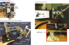
Recently, a macro-micro robot test bed is implemented to test the above design concept as shown in Fig. 1[40]. The micro module in Fig. 1(a) has a parallel structure and is equipped with commercial actuators which could be cost-effective. In fact, the module has 5-dof without the roll motion about z axis. Its translational and rotational workspace size is designed to have 10mm×10mm×5mm of space and an allowed tilt angle of 3° with no torsion angle, respectively. Its size and weight is 113mm×113mm×150mm and 1.38kgf, respectively. Its position accuracy is measured as 0.11mm when the tool endpoint is located 150mm away from the top bracket frame which is attached to the micro robot moving plate. And its payload capacity is greater than 1kgf.
The macro module has the PRR(RRR) type structure. Only the first prismatic joint along the vertical axis except all the other joints is selected to be active to support the weight of all the floating links. Note that the axes of the second and the third revolute joints are also vertical and thus are not subject to the gravitational load. The other three resolute joints (RRR) whose axes has a common intersection point (called a wrist point) are selected as passive since the weight of the wrist including the weight of the micro module could be counter-balanced by an additional counter weight to the other end of the wrist as shown in Fig. 1(b). Note that all joints are equipped with the encoder to trace the joint angles and that all passive joint are equipped with the brake such that all joints could be fixed at designated configuration.
PRELIMINARY EXPERIMENTS FOR THE MACRO-MICRO SURGICAL ROBOT
Preliminary experiments are conducted to see what size of the workspace of the micro module would be acceptable for neurosurgical surgery applications. To support the experiment, the motion simulator as shown in Fig. 1(c) is developed, which provides the visual information. The effectiveness of the robot is tested by checking the completion time to move the micro module by manually moving the macro module to within the designated neighborhood of the target position from the given position, as shown in Fig. 1(c). In the experiment, the subjects are asked to put the needle endpoint of the micro module into the target hole, whose diameter is 1 mm inside of the straw of diameter of 6mm, by moving the macro module manually. The preliminary results showed that the subjects feel comfortable to move the macro robot to within the neighborhood of the target whose size is defined as 10mm×10mm×5mm, Similarly, the subjects also feel comfortable for the size of the orientation workspace defined as the tilt angles of 3°~5°, As expected, for smaller size of the rotational workspace defined by the tilt angle of 3°, longer completion time is required than for the case of the tilt angle 5°, since more delicate adjustments are required to accomplish the given task. In particular, for the tilt angle of 3°, the completion time turns out to be less than 40 secs. Based on this preliminary test results, the acceptable size of the translational and the rotational workspace of the micro robot module could be roughly confirmed. In the next stage, the effect of both the additional visual and position information computed from the position sensors of the macro robot, the effective procedure protocol, and the locking actions need to be investigated, to identify the more reliable minimal workspace size for the micro robot module.
CONCLUSIONS
In this paper, the position accuracy of the existing stereotactic neurosurgery robots and their structures are briefly reviewed. And it is briefly addressed that the macro-micro type robot has more adequate and more promising features than other structures in aspects of its roles and its costs since the strict specification requirements for the stereotactic surgery could be distributed into the macro module and the micro module, in the stereotactic surgery. Desirable design aspects for the macro-micro neurosurgical robots for stereotactic surgery are suggested along with the introduction of an exemplary system which has been developed based on the suggested design aspects. Then, to show the effectiveness of the macro-micro robot in which the macro module is partially active, preliminary experiments are conducted to identify the minimal but acceptable size of the workspace of the micro module of the stereotactic neurosurgery macro-micro robot through its completion time for the specified tasks. In near future, in abreast with the drastic advancement of the position and image sensing technologies, the more cost-effective and reliable neurosurgical robots should be developed. And the appearance of a more cost-effective and more reliable macro-micro type stereotactic neurosurgery robot would be one of them which could be more widely accepted among surgeons.
 XML Download