

 PDF
PDF ePub
ePub Citation
 Print
Print


INTRODUCTION
Augmented reality (AR) is used in many fields including medicine, education, manufacturing, and entertainment. With advances in optics, computer systems, and surgical instruments, AR application to medicine is being vigorously researched. Particularly, as surgery using laparoscopy, endoscopy, or catheterized intervention have increased, AR takes an important role in many medical applications [1234].
AR denotes a technique to combine a real world and virtual objects which are artificially generated digital content by a computer [5]. As another aspect of AR is a registration between the real world and virtual objects, it aims to estimate threedimensional (3D) position of virtual objects related to the real world. Therefore, AR can allow the user to see 3D virtual objects superimposed upon the real world. With the help of AR in medicine, a surgeon can see hidden organs inside a body and improve the perception of treatment procedure by interacting with the real world. After a brief description of three components of the medical AR, its applications will be presented.
TECHNOLOGY FOR AUGMENTED REALITY
AR in medicine mainly comprises three technical parts such as camera calibration, patient registration, and object tracking [567].
1. Camera Calibration
Generally, real world objects are captured by a camera and reproduced on a display. AR merges virtual objects with the real world, which requires transformation between the camera and real world coordinates. Before estimating the transformation, the characteristics of the camera must be defined. Pinhole model is a simple camera model that maps the 3D real world onto two-dimensional (2D) coordinates called the image plane. 3D points are mapped onto the 2D image plane by translating the point on a straight line towards the camera center until it intersects the image plane [89]. This mapping is called perspective projection, and the transformation between the image and real world coordinates can be represented as a projection matrix. Thus, camera calibration is the estimation of the projection matrix parameters for a pinhole model [101112].
2. Patient Registration
Patient data for preoperative planning is 3D volume data taken from computed tomography (CT) or magnetic resonance imaging (MRI). Since it provides a view of the internal anatomy and target points for the surgeon, patient data should be registered with respect to a patient of the real world coordinates, which is called patient registration [5]. Point based registration is a reliable solution, where registration is performed with fiducials affixed on the patient. One set, consisting of more than four fiducial points, is registered to another set of corresponding points using a rigid transformation. However, the accuracy of fiducial based registration varies depending on the number of fiducials and measurement quality of each fiducial position, as well as their spatial arrangement [2]. To improve registration accuracy, iterative closest point (ICP) based surface matching is often used in combination with point based registration [131415]. However, careful selection and collection of 3D surface data is critical for final accuracy, usually expressed in terms of target registration error (TRE).
3. Object Tracking
Object tracking is to estimate the spatial position of the camera or marker on surgical instruments, and is an essential component of a medical AR system. In AR tracking, the relative position of an object on the basis of the camera position is generally calculated. When given a calibrated camera with known intrinsic parameters, the relative position can be determined as a set of three or more paired points between the 3D and projected 2D coordinates [1112161718].
Combining these technologies, we can implement an AR system that overlays virtual objects on the endoscope or surgical microscope view.
MEDICAL APPLICATIONS OF AUGMENTED REALITY
This section reviews four AR systems in medical applications: cardiac, bone tumor resection, sinus, and spinal surgery. These AR systems are major components of surgical navigation systems proposed by the authors' laboratory. We briefly explain the characteristics of each surgery, then the AR system configuration and results.
1. Cardiac Intervention
A surgical navigation system was proposed to guide chronic total occlusion intervention. Conventional intervention for chronic total occlusion of the coronary artery depends highly on 2D X-ray images and the surgeon's experience. Therefore, large displacements between the surgeon's hand-eye coordination may be generated by discrepancies in position or orientation between the patient and the acquired images [2]. This can lead to misidentification of the coronary artery or incorrect positioning of the stenosis on the coronary artery [5]. The proposed system merged the 3D CT angiography model with X-ray images to provide 3D anatomical information to the surgeon [19].
(1) AR system configuration
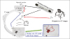
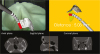
Fig. 1 shows the cardiac intervention AR system components. A commercial optical tracking system was used to track the location of markers attached to the patient and C-arm device. The different system coordinates were unified using a transformation matrix.
(2) Results
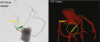
Fig. 2 shows the prototype software for the proposed system, combining AR and virtual reality (VR). The CT angiograph is overlaid onto the X-ray image and the VR images are positioned beside it. We expect that surgeons can easily understand anatomical information that is occluded in the original X-ray image, as well as the vascular anatomy and relative instrument location using the prototype proposed system. The system can also minimize X-ray exposure and injection of contrast medium, since fluoroscopy is less required than in conventional surgery.
Although several challenges still remain to apply the proposed navigation system to clinical use, the system is a promising alternative to fluoroscopy guided chronic total occlusion intervention.
2. Bone Tumor Resection Surgery
An AR navigation system for bone tumor resection was proposed. Resection of a pelvic tumor is a major surgical challenge because blood vessels are complexly intertwined with nerves. Safety resection margins should be confirmed intraoperatively during the surgery. An AR navigation system would be helpful, providing intuitive visualized information of resection margins [20].
(1) AR system configuration
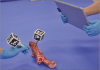
The AR system for bone tumor resection surgery included an embedded camera on a tablet PC to track the patient and tools. AR visualization was also displayed on the tablet PC. Fig. 3 shows the proposed AR system configuration, which includes just a tablet PC and multi-faced reference markers with no external optical tracking system. The tablet camera tracks and realizes patient and tool reference marker positions. Registration between patient and 2D camera image was achieved using the paired point registration method. The transformation to align two point sets was calculated after matching four to six anatomical and artificial markers. Their relationships between the camera and markers are then defined based on the registration using the perspective n-points algorithm.
(2) Results
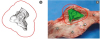
Fig. 4 shows a resection margin calculated from a reconstructed 3D tumor model and a captured scene for bone tumor AR navigation. To build a resection margin, dilation operation in image processing is applied to a reconstructed 3D tumor model and resection margin is represented as red contours. Fig. 4(b) shows the 3D tumor which is represented as green is overlaid on the image from a tablet PC embedded camera with its resection margin. In the experiment, the bone tumor resection was simulated with 10 mm resection margins to an artificial bone tumor made of a cement. The mean safety margin was 12.28 and 10.26 mm for the conventional and proposed AR method, respectively. The safety margin sizes were significantly different (t-test, p < 0.05), and the AR method was therefore significantly is closer to the desired 10 mm than the conventional method.
3. Sinus Surgery
Sinus surgery is also endoscopic surgery. The main problem is difficulty locating a surgical instrument to a specific object seen through the endoscope [6]. Since the access route to paranasal sinuses is complex, complications such as blindness and cerebrospinal fluid leak can occur due to damage of the orbit and skull base. To solve these problems, an integrated system was developed consisting of an AR based surgical navigation system and endoscope holder [21].
(1) AR system configuration
The proposed AR navigation system for sinus surgery was similar to those used for conventional surgery. The proposed system comprises three processes: patient image registration, camera calibration, and camera based tracking, which were provided by paired point registration, pinhole model based calibration, and perspective n-points algorithm, respectively.
The endoscope holder system consists of a 3 degrees of freedom (DOF) stackable parallel mechanism and 2-DOF end-effector. The 3-DOF stackable parallel mechanism combined a five-bar with two parallelograms, and the 2-DOF end-effector controlled the endoscope position. The system also included a brake, to hold the endoscope at any location the surgeon desired [21].
(2) Results
Fig. 5, which is the proposed AR navigation system, shows 2D multi-planar reconstruction (MPR) images (axial, coronal, and sagittal plane), along with the AR, and VR views. Warning and automatic transparency adjustment functions were also implemented. If the tip of the surgical instrument gets too close to a target, an alert sound is generated. The transparency of augmented objects is automatically changed according to the distance to the surgical instrument.
4. Spinal Surgery
For spinal surgery, the most important consideration is to correctly localize the surgical instrument inside the patient's anatomy. Therefore, AR based surgical navigation systems, which assist the surgeon to recognize patient anatomical structures, are widely accepted and have become a very an important research topic in this field [22]. However, although AR provides more intuitive visual information, inaccurate depth perception is a major issue. To improve the depth perception, an integrated VR and AR system was proposed that displayed in a single window with aligned view axes and provided the distance between surgical instruments and target organs [18].
(1) AR system configuration
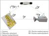
The proposed VR and AR switchable surgical navigation system consists of position tracking and visualization sections. In the position tracking section as shown in fig. 6, the transformation (TOC ) between the camera and patient is calculated by an optical tracker, and is updated with tracking data in real time. The visualization section used an open source visualization library and graphic processing unit(GPU) based depth peeling technique to display translucent objects [18].
) between the camera and patient is calculated by an optical tracker, and is updated with tracking data in real time. The visualization section used an open source visualization library and graphic processing unit(GPU) based depth peeling technique to display translucent objects [18].(2) Results
The user can switch from AR to VR by rotating the virtual camera around target objects, providing visualization of patient anatomy depth. Fig. 7 shows the proposed VR and AR switchable surgical navigation system. When the virtual camera is positioned within the range of a camera image, surgical navigation system is operated in AR mode. Otherwise, it is operated in VR mode. In addition, the depth which means the minimum distance between the tip of a surgical instrument and the nearest point of the target is also displayed on the screen.
CONCLUSION
Surgery is changing from open procedures to minimally invasive approaches. AR technology has a great potential to assist this change, and is becoming more important. The largest advantage is to visualize the region of interest, such as tumors, blood vessels, and nerves which are often invisible or obscured to direct vision. We believe AR will only become more ubiquitous in future medicine. Research on precise camera calibration and patient to image registration will help provide robust AR for clinical applications.

 XML Download