

 PDF
PDF ePub
ePub Citation
 Print
Print


INTRODUCTION
The use of computers and robots in the medical field is no longer a research interest but already a part of clinical routine. In otolaryngology/head and neck surgery, image-guided surgery (IGS) and robotic surgery are becoming more common. IGS provides the computerized real-time feedback to the surgeon about the location and orientation of the surgical devices along with the anatomical information of surrounding structures. The IGS foresees the hidden anatomical structures before they are exposed. IGS is now widely used in endoscopic sinus surgery where the effectiveness of surgical navigation has been reported [1]. The robotic surgery enhances the fine movement of the surgeon's hands to achieve the surgical goal with minimal damage to surrounding structures. The Da Vinci series (Intuitive Surgical Inc, USA), the only surgical robots currently available, are employed in laryngeal and pharyngeal surgery through the patient's mouth, which is now termed the transoral robotic surgery (TORS), with promising clinical outcomes [2]. On the other hand, IGS or robots in the otological field are much less popular and most of the otological procedures are exclusively performed manually, which have basically remained unchanged for decades. The otologists' concern about IGS and robots has been the balance between the required accuracy and the additional invasiveness. Many otologists demand the registration error of no more than 0.5 mm in otological procedures. This requirement almost reaches the inherent limit of accuracy defined by the physical resolution of the CT dataset, i.e. the pixel size, which is typically 0.2-0.5 mm. To achieve this high degree of accuracy, invasive procedures have often been justified, such as invasive fiducial marking, head clamping, or additional radioexposure by intraoperative CT scanning. This invasiveness, however, has restricted the use of IGS to unusually difficult cases that we rarely encounter. Otological cases that justify the robotic surgery with its underlying invasiveness are even rarer. Thus, research on reducing the invasiveness of IGS and robotic surgery is indispensable; otherwise, they will remain a scarcely applied technology that can only serve for courageous patients selected by technologically gifted surgeons [3]. Thus, IGS and robots for otological procedures must achieve high degree of accuracy and noninvasiveness, however, it is often difficult to achieve both objectives simultaneously. The research needs to overcome the dilemma of balancing the noninvasiveness and accuracy, and also has to overcome the otologists' skepticism that such goal is impossible to achieve.
The author herein reviews a series of projects aimed at developing computer- and robot-assisted otological surgery. Computers and robots did not come to the operating room instantly. Instead, the research required the accumulation of multiple projects. First, we replaced invasive procedures to noninvasive, but equally effective, procedures for the IGS. Specifically, we developed a noninvasive reference frame and a noninvasive registration method. Then we performed otological procedures under IGS and developed the interface for otologists. Finally, a surgical robot for otological procedures was developed based on the information provided by surgeons who used IGS during otological surgery. This user-oriented approach resulted in developing a unique robotic system of what we now express as human-robot collaborative control.
NONINVASIVE REFERENCE FRAME
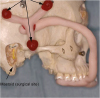
The reference marker tells the computer the location and orientation of the patient's temporal bone. The reference frame is one of the critical factors that contribute to the accuracy of the surgical navigation. The invasive reference frame that can be screwed on the skull is more accurate and stable than less invasive referencing [4]. However, noninvasive reference sticker to be attached on the skin of the forehead is usually the only justifiable choice, although it can be easily slid along with the skin and thus compromise the accuracy. We collaborated with dentists and developed a custom-made reference frame that fits on each patient's upper teeth (Fig. 1) [5]. This reference frame contributed to the stable referencing because upper teeth are connected to the temporal bone without any mobile joints. In addition, reference frame on the upper teeth is useful in terms of sterility because it does not interfere with the surgical field.
NONINVASIVE REGISTRATION
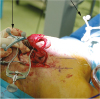
Most of the currently available surgical navigation systems use either paired-point matching or surface matching registration protocols. Surface matching registration is more popular because of its noninvasiveness and the fact that no additional CT scan is necessary. However, surface matching registration usually results in larger registration error than paired point matching method. To overcome the problem of limited accuracy, we developed the surface template-assisted marker positioning (STAMP) method (Fig. 2) [36]. The STAMP method uses a 3D-printed template of the bony surface, which was designed based on the preoperative CT. This method improved the accuracy of the IGS while maintaining the noninvasiveness of the surface matching registration method.
WARNING INTERFACE FOR THE PROTECTION OF IMPORTANT ANATOMICAL STRUCTURES
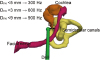
There has been a large dilemma when using IGS in the otological procedures, specifically speaking, a surgeon who is focusing on the microscope cannot look at the navigation screen. Therefore, we developed a warning sound system to deliver the most important information, e.g. distance from the facial nerve, to the surgeon. Anatomical structures were segmented with a predetermined amount of safety margin. When a surgical device reaches the segmented structures, the warning sound notified the surgeon of the proximity to the structure without necessitating the surgeon to look at the navigation screen (Fig. 3). We tested this function to protect the facial nerve in the temporal bone model and found that an inexperienced surgeon could drill the temporal bone with greater confidence [7]. This development had a significant impact on our series of research projects because it gave us insights of what robots should do in the operating room to help surgeons: to protect important structures from unnecessary injury.
ROBOTS FOR MASTOID SURGERY
Robots for otological procedures have been reported from a few institutes. The Da Vinci, the only approved surgical robot currently available, was used to perform mastoidectomy in cadavers for feasibility study [8]. Automatic mastoidectomy using an industrial robot was also performed in a cadaver study [9]. For a more specific purpose, a robot designed for cochlear implantation has been reported [10].
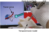
The ultimate goal of the computer assisted temporal bone surgery is to develop a fully computerized method which only allows minimal room for human errors. However, we learned several points from our previous research projects. First, surgeons still prefer to control the surgery with minimal automation or robotic restriction. Second, surgeons hoped that computers and robots could help protect the inner ear and the facial nerve. Third, the Da Vinci-style, master-slave robot was not perceived as an attractive device because otological procedures deal with shallow region of the bone. Based on these information, we proposed a robot for otological procedures (Fig. 4) [11]. A robot developed in Hanyang University has a function of human-robot collaborative control. This control usually does nothing while the important structures are not in danger. The surgeon can manipulate the robot's arms almost freely within the robot's working area. However, once the tip of the drill reaches a certain proximity to a predefined structure, the actuators of the robot will gradually restrict the movement of the drill so that the surgeon feels as if the drill hit a wall. In our temporal bone model study, an engineer, who had no background of surgical training, had performed mastoidectomy under the robotic assistance on five temporal bone models, and completed the procedure while never injuring the facial nerve of the protected region [11].
DISCUSSION – THE ROLE OF COMPUTERS AND ROBOTS
The currently established surgical procedure of the temporal bone is performed in a "landmark to landmark" fashion. The surgeon finds the first landmark and uses it to reach the second landmark. This process, of using available landmarks as guides to the next landmark, is repeated until the surgeon reaches the surgical target. Therefore, the landmarks must be readily accessible in the normal temporal bones. These classical landmarks should now be regarded as a part of "average anatomy" or "textbook anatomy". Surgery based on average or textbook anatomy is an important first step for a surgeon who is new to the field. However, there are many situations where the landmarks of average anatomy are absent, modified, or destroyed. Even in typical temporal bones, the landmarks have individual variation in the location and its size. Classical solution to overcome the missing landmarks was to acquire experience. The situation is now gradually changing; we now often use patient-specific shapes of the bone, old fracture lines, or marks of previous surgery that can be found only in the particular patient, along with classic landmarks. As computers and software programs for 3D reconstruction of the CT/MRI images became affordable, using the patient-specific anatomy in the surgery has become very easy for surgeons. In the author's institute, the electronic medical chart system has the function for 3D reconstruction of CT or MRI images and surgeons can easily find small anatomical features that has been difficult to detect in planar images. Thus, in the current standard, knowing the average anatomy should be regarded necessary but no longer sufficient for safe surgery. In future temporal bone surgery, surgeons must deal with the anatomy that is specific to the patient in addition to the average anatomy that can be learned in textbooks. The use of computers and robots would enhance this process by routinely providing patient-specific anatomy before and during surgery.
In this review, the author has introduced a potential model of future temporal bone surgery. The author believes that the role of computers and robots in future surgery lies in their objectivity. In unusually difficult cases, there are situations that abnormal anatomy is so peculiar that a surgeon can be misled by his/her own experience. In these cases, a surgeon must be objective; i.e., liberate him/herself from experience or textbook anatomy, and focus totally on the patient's anatomy. The assistance from computers or robots has clear strength in these cases because of its inherent nature of objectiveness. In temporal bone surgery, facial nerve monitoring is another example of surgical assistance with absolute objectivity. The facial nerve monitor is free from experience or anatomy, but it simply tells whether the facial muscles are stimulated. Interestingly, even the effectiveness of facial nerve monitoring once received some level of skepticism from experienced surgeons. The author thinks that the computers and robots may also go through a similar path of skepticism. However, after this step is passed, the collaboration between surgeons and computer/robots should contribute to safer and more complete surgery by utilizing patient-specific anatomy and image-guided robotic control.
 XML Download