

 PDF
PDF ePub
ePub Citation
 Print
Print


INTRODUCTION
Corticosteroid administration has been widely used as the empirical treatment for various inner ear diseases such as sudden sensorineural hearing loss (SNHL), Meniere disease, and autoimmune-induced hearing loss for a long time. After Sakata et al. [1] first tried intratympanic (IT) steroid injection to control Meniere disease, IT steroid injection treatment has been used as an alternative option to systemic steroid treatment over the last two decades. Two major lines of evidence support the feasibility of IT steroid injection treatment and explaining its mechanism of action. First, injected steroid into the middle ear cavity can penetrate the round window membrane and diffuse into the inner ear fluid [23]. Second, many glucocorticoid receptors and mineralocorticoid receptors have been found in the inner ear structures [456]. Moreover, there is a theoretical advantage that IT steroid injection could increase the concentration into the target organ while it could also reduce the systemic steroid exposure; therefore the use of IT steroid injection has become widespread in a short time. Animal studies have demonstrted markedly higher concentration of corticosteroids in the endolymph and perilymph of the cochlea when delivered via the intratympanic route in comparison with systemic administration [3].
After these studies, a large-scale prospective study demonstrated IT-steroid injection treatment was not inferior to oral steroid treatment on sudden SNHL in humans [7]. Recent literature recommends IT-steroid injection not only as an alternative to oral steroid in vulnerable subjects such as diabetic patients, but also for a salvage therapy after failure of initial therapy [89].
THE RATIONALE FOR IT-STEROID INJECTION TREATMENT FOR TINNITUS
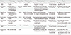
IT steroid injection for the treatment of tinnitus has been tried for over fifteen years. Sakata et al. [10] reported that tinnitus improved in 75% of 3,978 ears immediately after four IT-steroid injections and in 68% after 6 months. Cesarani et al. [11] reported 13.5% cure rate and 24% improvement rate 8 weeks after nine injections of dexamethasone. However, those studies were single arm studies without controls. Although a few controlled studies using IT saline as a placebo were recently conducted, no one has shown significantly better improvement than controls (Table 1) [12131415]. However, most of the studies of IT steroid injection for the treatment of tinnitus did not consider the duration of symptoms and the majority of enrolled patients had tinnitus lasting for more than 1 year before treatment. Given that the cochlea is the target organ of administered steroid, and that damage to the cochlea is not reversible after 3 months as shown in many animal studies, it might be unreasonable to expect a therapeutic effect of IT steroid injection for chronic tinnitus lasting over 1 year. In the acute stage, tinnitus is usually heard intermittently and makes little discomfort, so patients tend not to visit a clinic at that time, and even doctors might not realize a need to treat. Indeed, we could find terminology describing early onset tinnitus such as "acute tinnitus" or "recent onset tinnitus" in only a few recent reports [16171819]. The distinction between acute and chronic tinnitus is arbitrary and varies between 3 and 6 months. Recently published guidelines for tinnitus management suggested just waiting or minimal intervention without expensive or time-consuming evaluations and treatments because of the potential for resolution of the acute tinnitus [1819], however they did not show any statistical data concerning the spontaneous recovery rate. Only a meta-analysis of 314 wait-list subjects with tinnitus as control groups in 11 randomized controlled studies revealed a significant decrease in scores on tinnitus-specific measures of 3% to 8% [20].
However, what would happen if 'acute tinnitus' did not resolve naturally? Every chronic long-standing tinnitus has been, in the beginning, an acute one.
The majority of clinicians might agree with using systemic oral steroid or IT steroid within a short therapeutic window when acute tinnitus was combined with sudden SNHL, even though the argument of beneficial effect vs potential harm still remains [2122]. Although improvement of the hearing threshold in patients with sudden SNHL is usually correlated with positive outcomes on tinnitus, there is no consensus of treatment in cases of acute tinnitus combined with subtle hearing loss confined within a narrow frequency range or no decrease in hearing thresholds compared with the other side. Although the pathophysiological mechanism of tinnitus in the central auditory system is still unclear, the majority of tinnitus is believed to be triggered by cochlear damage. When the combined hearing loss is minimal and confined in narrow frequency range, tinnitus patients often recognize hardly any change in hearing. We can surmise that in many cases, the etiology and pathology of the cochlea in acute idiopathic tinnitus are common to those of sudden SNHL, such as viral infection and microvascular obstruction. Because the damage would be milder and more restricted in the early stage of tinnitus than that in sudden SNHL, we suggest that an application of the treatment given for sudden SNHL would produce better results for acute tinnitus than for sudden SNHL.
Tinnitus subjects with normal audiograms do not necessarily indicate the absence of cochlear damage. Several studies have demonstrated that tinnitus subjects with normal audiograms show increased hearing thresholds at extended high frequencies above 8 kHz compared to normal-hearing subjects without tinnitus [2324]. Some investigators found subtle damage to the outer hair cells that alters otoacoustic emissions can cause tinnitus [252627]. Other investigators have suggested that there could be central deafferentation in spite of normal audiogram and a marked reduction in the amplitude of the wave I potential originating from the auditory nerve [2829]. These studies support the rationale of treatment for cochlear damage in early stage in most cases of tinnitus whether they are combined with recognizable hearing loss or not.
In a recent randomized controlled study, the IT steroid showed meaningful therapeutic effect on acute idiopathic tinnitus developed within 3 months and unrelated to sudden SNHL. The improvement rate of alprazolam plus ITD group was greater than that of alprazolam only group (75.8% vs 40.3%), and the cure rate of alprazolam plus ITD group was also greater than that of alprazolam only ITD group (25.8% vs 9.8%) [16]. Considering that maximal improvement rates from the placebo effect are reported to be 30% to 40% [3031], the improvement rate of alprazolam only group might likely be attributed to the placebo effect, and the cure rate of 9.8% likely due to spontaneous resolution. Although there is no other controlled study supporting therapeutic effect of IT steroid injection on tinnitus, if the subjects were selected with strict criteria in terms of symptom duration, then IT steroid injection might be shown as a potential treatment modality for tinnitus.
THE ACTION MECHANISM OF STEROID
It has been shown that glucocorticoid receptor and mineralocorticoid receptors exist in the inner ear, and glucocorticoid activates both receptors [45]. Cochlear damage from numerous causes such as noise, ototoxic agents, endolymphatic hydrops, viral infection, and vascular ischemia are commonly related to inflammatory cytokines and production of reactive oxygen species. Anti-inflammatory and immunosuppressive actions of glucocorticoid could play a key role to both prevent and recover from the cochlear damage [32]. Mineralocorticoid receptor binding steroid stimulates ion-homeostasis action. The stria vascularis pumps Kccc over the blood-labyrinth barrier into the endolymph and then this K+ is transported from the Henson cell and Claudius cell through the spiral ligament fibrocyte to the stria vascularis for recycling. This mechanism called K+ cycling maintains the endolymphatic potential of +80 mV for cochlear activity. Cochlear damage from various causes involves not only hair cell but also stria vascularis which can impair ion-homeostasis. Mineralocorticoid maintains ion-homeostasis helping to prevent cochlear damage by activating Na+K+-ATPase, epithelial sodium channels, calcium channels, and the Na+/Cl- cotransporter of the stria vascularis [63233].
DELIVERY ROUTE AND PHARMACOKINETICS OF INJECTED STEROID INTO THE MIDDLE EAR CAVITY
Steroid administered to the middle ear enters into the scala tympani through the round window membrane, spreads to the scala vestibuli via the spiral ligament or Rosenthal's canal, and finally reaches the endolymph of the scala media [343536]. In this delivery route, all of the cochlear inner structures (hair cell, spiral ganglion, stria vascularis, etc.) can be exposed to steroid. Parnes et al. [3] administered methylprednisolone to guinea pigs via oral, intravenous, and intratympanic routes and measured the drug concentration in cochlear fluid. The maximum concentrations of methylprednisolone in perilymph was significantly higher via the intratympanic route (50.37 mg/L after 1 hour) vs oral administration (0.06 mg/L after 6 hours) or intravenous administration (0.31 mg/L after 1 hour). Comparison of absolute concentrations in endolymph and perilymph by the type of steroid showed hydrocortisone >methylprednisolone >dexamethasone, however, relative concentration corrected by anti-inflammatory equivalent factor revealed methylprednisolone >dexamethasone ≈ hydrocortisone. Regardless of the type of steroid, all three drugs fall as low as zero in concentration before 24 hours.
The thickness of the round window membrane is known to be about 70 µm in humans and consists of 3 layers of an outer epithelial layer, middle connective tissue layer, and inner mesothelial layer. Drug penetration through the round window membrane is mainly affected by drug factors such as molecular weight, concentration, electrical charge, and lipid solubility. Among these, the molecular weight is the most important factor. The lower the molecular weight, the easier it would penetrate the membrane. According to animal studies, substances with a molecular weight less than 1,000 such as steroid, aminoglycoside, etc. can easily permeate by simple diffusion [3738]. Another factor influencing drug delivery is the anatomy of the round window membrane such as thickness of membrane, presence of false membrane (21% in 202 temporal bone) [39], presence of tissue plug around round window niche, or bony obliteration. Silverstein et al. [40] examined the round window of 41 patients by microscope, and revealed that the round window in 5 patients was obstructed. According to this study, there might be a potential risk of steroid not reaching the lymph even though intratympanic injection was performed.
PROGNOSTIC FACTORS FOR THE OUTCOMES OF IT STEROID IN THE TREATMENT OF ACUTE IDIOPATHIC TINNITUS
A retrospective study looking at a large number of case reviews was conducted to determine possible prognostic factors for the outcomes of treating acute idiopathic tinnitus with IT dexamethasone [41]. This study was conducted in the institute to which the author belongs, and included 114 subjects treated with IT dexamethasone for acute tinnitus with symptom lasting for 3 months or less, and who subsequently completed questionnaires 3 months after treatment. Of the 114 patients, tinnitus was cured in 43 patients (37.7%), partially recovered in 42 patients, and did not improve in 29 patients. The subjective improvement rate, including cure and partial recovery, was 74.6%. The treatment outcomes of IT steroid were analyzed according to the duration of symptoms, unilaterality of tinnitus, and pure-tone asymmetric hearing loss. The cure rate was significantly greater in patients with symptoms for 2 weeks or less than in patients with symptoms for more than 1 month (64.7% vs 15.7%), and the correlation analysis reconfirmed that onset time of treatment is the most important factor correlating with the cure rate for acute idiopathic tinnitus. Even though the mean global improvement index (satisfaction questionnaire) was higher in the unilateral group than in the bilateral group, the cure and improvement rates were not significantly different between patients with unilateral tinnitus and patients with bilateral tinnitus. Among 95 patients with acute unilateral tinnitus, initial pure-tone audiometry revealed that 58 had asymmetric hearing threshold and 37 had symmetric hearing threshold. Although the audiometric response (>15 dB at any frequency) rate was significantly greater in patients with asymmetric hearing loss than in patients with symmetric hearing loss (48.8% vs 4.8%), there were no significant differences in the mean global improvement index, improvement rate, or the cure rate.
When all the analyses were combined, the best candidates for ITD are patients with acute unilateral tinnitus presenting 2 weeks or less after symptom onset with ipsilateral deteriorations on an audiogram. However, it is difficult to establish a definitive indication for ITD in patients with acute tinnitus and patients with bilateral tinnitus or symmetric hearing thresholds should not be excluded in IT steroid injection treatment. The reason might be that the occurrence of tinnitus does not depend upon the degree of damage to the OHCs, but rather upon different plastic changes in the central auditory system after cochlear damage.
There could be many reasons for the non-responsive cases of IT-steroid for even acute tinnitus. While cochlear damage may have been accumulating continuously, tinnitus may not be recognized until the damage reaches a threshold level, after which there is prolonged and increasing hyperactivity of the auditory neurons. In this case, IT steroid would not work because the cochlear lesion has been present for a long time even though the symptoms of tinnitus were only recently perceived.
SELECTION OF DRUG AND TREATMENT PROTOCOL
Methylprednisolone produces the highest relative concentration in lymphatic fluid, but it often provokes a burning sensation during injection. Hydrocortisone is reported to cause an inflammatory reaction of the round window membrane in a morphologic study with rat [42]. Dexamethasone is most widely used due to lack of the adverse reactions in other drugs as noted above. A high concentration of dexamethasone (10–25 mg/mL) is generally used in many research papers, but in Korea, only dexamethasone of 5 mg/mL is available domestically. The quantity of drug injected into the middle ear cavity is reported to range from 0.3–0.8 mL but it can depend on various factors such as the air-pressure in the middle ear or the leakage through the Eustachian tube, etc. Judged from the author's clinical experience, the counter-puncture of the tympanic membrane could be helpful to administer a greater amount of drug by air-pressure drop in the middle ear. Nevertheless the quantity of drug injected into the middle ear cavity in practice, usually seems less than 0.5 mL and the excess often drains out thr-ough the Eustachian tube. Physicians should encourage patients to spit out any drug drained to the pharynx so as to decrease potential systemic risk. There is no consensus about the length of the injection period or the number of injections which can give the best results. In many previous studies, 3–5 injections given as 2 injections per week were the most generally used protocols [1213141516]. The author's protocol is daily IT dexamethasone injection for 4 consecutive days, based on the pharmacokinetic profiles investigated by Parnes et al. [3], which has shown that the concentration of dexamethasone in lymphatic fluid may drop to zero within 24 hours after injection.
ADVERSE REACTIONS AND PRECAUTIONS
Transient pain, bitter taste, bleeding from tympanic membrane and vertigo could be provoked by IT steroid injection [43]. The temperature of the injected drug should be similar to body temperature to prevent caloric vertigo, but there is still a potential risk of vertigo broght on even with the drug at near body temperature. When using topical anesthesia such as lidocaine spray, enough suction to remove the lidocaine completely before injection should be ensured to prevent severe vertigo. An increase of tinnitus or development of ear fullness right after injection is common due to the presence of fluid in middle ear cavity, but it recovers in several hours. The worst complications are permanent tympanic membrane perforation or chronic otitis media, both of which occur very rarely. Generally, a systemic adverse reaction due to the IT steroid has rarely been reported. However, in the author's own experience, in a few cases where excess IT drug was released through the Eustachian tube, a slight increase of blood sugar and facial flushing attributed to the swallowed drug has been observed.
COST-BENEFIT
According to the latest information from the Centers for Medicare & Medicaid Services and the Korean National Health Insurance Service, intratympanic treatment is reimbursed at a rate of $172 per injection in the United States [7] compared with $16.50 per injection in Korea. For the 4 injections of IT steroid, it minimally costs $688 in US vs $66 in Korea, which is about a tenfold difference. Considering the potential therapeutic effects of ITS injection and the low rate of side effects, the cost-benefit might be acceptable especially in Korea.
CONCLUSIONS
Currently, IT steroid injection treatment of tinnitus requires more evidence provided by randomized controlled trials, but when the therapeutic target is limited to tinnitus in the acute phase, IT steroid injection may be a treatment option worth considering justified on the rational knowledge of mechanism, on the minimal potential for side effects, and on the result of a prospective study which showed a better clinical outcome with IT sterioid administration than with the alprazolam only treated group. Early intervention appears to be most important to relieve the acute tinnitus and to prevent the development of chronic symptoms. Although asymmetric hearing loss and unilateral tinnitus seems to be favorable factors for the outcome with IT steroid injection, patients with bilateral tinnitus or symmetric decreases in hearing thresholds should not be excluded in IT-steroid injection treatment.
 XML Download