

 PDF
PDF ePub
ePub Citation
 Print
Print


INTRODUCTION
About 50 years ago, the satellite cell was discovered through electron microscopic inspection of skeletal muscle. Satellite cells are mono-nucleated and located in the edge of the skeletal myofibers. These cells were imposed for their association with muscle regeneration. Satellite cells maintain dormant state in the mature muscle and are activated mitotically during muscle injuries, and finally differentiated to myoblasts. It contributes to the myofibrils resulting in muscle recovery from the injury [1]. Collins proved that satellite cells have both differentiation and self-renewal ability [2]. Sequentially, Kuang et al. established that the muscle stem cells achieve self-renewal through asymmetrical divisions. Also, satellite cells did not consist of only adult stem cells, rather both adult stem cells and committed progenitor cells [3].
Muscular dystrophies include many diseases, which have skeletal muscle wasting and limit mobility of patients. Among that, Du-chenne muscular dystrophy (DMD), fragility of respiratory muscles and absence of dystrophin protein in the cardiac muscle results in respiratory or cardiac failure and early death [4]. The underlying pathogenesis is already investigated, but the treatment has not been established yet. However, research has accumulated these days and clinical trials are also being performed now.
There are roughly four methods to research DMD treatments, 1) gene therapy replacing the mutated gene, 2) cell therapy which replace affected cells or using bone marrow-derived stem cells and mesoangioblasts, 3) repairing the endogenous gene using exon skipping, skipping premature termination, and 4) drug therapy which compensates for lack of dystrophin, promotes function in dystrophic muscle, improves muscle hypertrophy and minimizes muscle wasting, uses histone deacetylase inhibitors or NO-releasing anti-inflammatory drugs [5]. In this article, cell therapies in DMD are focused in a review and current clinical trials using cell therapy are introduced.
SATELLITE CELL IDENTIFICATION AND CHARACTERISTICS
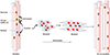
In 1961, Mauro first observed the presence of the satellite cells (SCs) in frogs through electron microscopy [6]. The mononucleated SCs were located in the periphery of the skeletal myofibers. Later, this cell type was also detected in humans, and their functions in muscle regeneration were established [7]. Location of SCs, which is adjacent to the myofibrils, implicated their involvement in tissue regeneration (Fig. 1). In muscle recovery, SCs enlarge, begin to proliferate and fuse to the multinucleated myofibrils [8]. More specifically, amount of undifferentiated cells are increased and the cells are lined up at the periphery of damaged fibers. During regeneration, precocious myogenic progenitors are substituted as mature myoblasts. Afterwards, undifferentiated cells start to join with the regenerated fibers. After the regeneration, the SCs go through self-renewing mitosis for maintaining their amount in the tissue. Many studies using H3-thymidine proved both the satellite cell section and differentiated nuclei in growing muscle [1]. Therefore, the abilities of SCs which can be asymmetrically divided and its self-renewal traits were confirmed. Fig. 1 shows the self-renewal and differentiation capacity of satellite cells.
In the adult skeletal muscle, satellite cells keep quiescence with mitosis and are localized between the basal lamina and the sarcolemma of muscle fibers. At dormant state, SCs expressed limited amount of gene and protein translation but they can be activated in response to stress, which is induced by injury, weight bearing, or the myo-degenerative disease [9]. What triggers the activation of SCs is still largely unknown but recent studies suggest some factors. For example, if there is inhibition of synthesis of sphingosine-1-phosphate, which is needed for the satellite cell to enter the cell cycle, muscle regeneration is surprisingly ceased [10]. Moreover, extrinsic mechanical stretch which leads to nitric oxide (NO) synthesis induces hepatocyte growth factor (HGF) release and SCs activation [11]. NO gas also encourages expression of Follistatin, which antagonizes Myostatin, an inhibitor of myogenesis express-ed by dormant SCs [12]. In result, SCs can be activated from quiescence. Microenvironment-secreted growth factors such as fibroblasts growth factors and p38α/β MAPK also can activate SCs. FGFs can induce pro-myogenic MAPK signaling pathways and if p38α/β is abrogated, cell-cycle exit is delayed and expression of cell-cycle regulators are adjusted [13].
Studies with radioisotope labeled SCs in growing muscle demonstrated that half of the daughter cells differentiate into myonuclei while the other half stay as constant dividing SCs [14]. This result suggests SCs have the ability of self-renewal. This capability of SCs has been clearly proved. Moreover, their myogenic differentiation and self-renewal functions are evidence of SCs as adult stem cells.
REGULATION OF SATELLITE CELL FUNCTION
1. Niche regulation
Stem cell niche means the microenvironment where stem cells are localized and it controls the fate and function of stem cells. As to satellite cells, the stem cell niche can regulate the asymmetric division and commitment of daughter cells without disturbing the stem cell homeostasis in the niche [15]. Because satellite cells are located along the myofibers below the basal lamina, the muscle fiber is one of the most important elements of the stem cell niche. All signals such as chemical, mechanical and electrical from the host fiber have been proved to be associated with the regulation of SCs function. The basal lamina also plays as an important role in the niche. It accounts for a major part of the extracellular matrix and is mainly made up of laminin, collagen, and proteoglycans [9]. Therefore, adhesion to the basal lamina is essential for the maintenance of stem cell characters. Another element of the niche is the microvasculature that supplies SCs and interstitial cells which interact with SCs [16]. In humans, 68% of satellite cells are localized within 5 µm from capillaries or vascular endothelial cells at both dormant and activated conditions [17]. Meanwhile, asymmetric division of the stem cell depends on cell polarity, which is achieved through cell to cell or cell to ECM interactions within the niche. Each side of SCs expresses different molecules - integrin α7β1 receptors on basal lamina side, M-cadherin on apical side - and this asymmetric allocation lets SCs form a structural basis for polarity and leads to the cell fate differences. This asymmetric cell division would also promote the fusion of the differentiating daughter cells [16].
Also, cytokines such as Il-1α, IL-13, TNF-α, and IFN-γ which is secreted by T cells are promoting factors of proliferation of muscle stem cells during muscle regeneration. In the study by Fu et al., these 4 cytokines were injected together into regenerating muscle of Rag1-/- mice and they helped in both decreasing muscle stem cell proliferation and correcting the impaired regenerative responses. In addition, muscle stem cells with these cytokines comparably participated more in muscle recovery than freshly isolated muscle stem cells. These results implicate that the muscle stem cells that expanded with the cytokines in vitro could achieve properties of undifferentiated progenitor cells [18].
2. Signaling pathway
In several decades, many signaling pathways such as Wnt, Notch, Bone morphogenetic protein (BMP), TGF-β are proved to be involved in activation of satellite cells. As well as activation, modulating pathways specific to muscle stem cells have also been demonstrated. Maintenance of dormant state, reversible quiescence and self-renewal, asymmetric destination, symmetric proliferation of stem cells are defined [19].
Notch signaling is needed to preserve the dormant state of satellite cells, suggesting that niche-derived Notch ligand should bind to a Notch receptor on the dormant satellite cell [20]. In this study, Rbp-j which is the downstream transcripton factor in the Notch pathways was eliminated from adult stem cells in normal muscle. This caused activation and ectopic differentiation of stem cells even if it did not enter the cell cycle and bypassed the transient amplifying progenitor stage [20]. Consequently, RBP-J plays as a restrictor of cell cycle entry and a mediator of Notch signaling [21].
Referred from in vitro experiments, it is supposed that multiple signaling pathways are associated with quiescence of the satellite cell. So far, Ang1/Tie2 [22], P38/MAPK [13], myostatin [23], Notch-3 [24], Spry1 [25], are proved to be involved in satellite cell cycles.
Although many signaling pathways that regulate stem cell function of satellite cells are revealed, there might be more unknown pathways [19]. During the cell cycle, many signaling molecules are repeatedly used. Representatively, Numb is used both to decide asymmetric fate and maintain progenitor cells [26]. Therefore, classical Wnt, notch and BMP signals might also be used in another way to regulate stem cell functions [19].
3. Epigenetic regulation of satellite cell function
Regulation of the SC's function is focused at the epigenetic level, which has cellular and physiological phenotypic trait variations caused by external or environmental factors that alter gene expression without modifications in the DNA sequences. It involves DNA methylation, histone modification, etc. Right after the identification of SCs, differences in chromatin organization between adult muscle and growing muscle accord with their shift from a dormant to an activated state [27]. The regulators of epigenetics are mediators of DNA methylation and demethylation, histone acetylases, methylases, miRNAs and so on [19]. These factors would contribute many changes in aged satellite cells that describe age-related declines in function and rejuvenation through exposure to the systemic environment [28]. So far, Pax7 functions with the Wdr5-Ash2L-MLL2 histone methyltransferase complex to methylate histone H3 lysine 4 at the Myf5 locus [29], and regulation of the repressive PRC2, EZH2 to control Pax7 expressions are established.
BIOMARKERS INVOLVED IN MUSCLE RECOVERY BY SATELLITE CELLS
Many biomarkers have been investigated so far. The most important molecule is Pax7, which regulates self-renewal in satellite cells and maintains myogenic potential. Pax7 is a transcription factor involved in the embryonic development of muscle stem cells and is expressed in both dormant and activated state of satellite cells [30]. It also regulates the expression of Myf5, which is associated with the embryonic myogenesis and stem cell differentiation [31]. Pax3 controls proliferation of the satellite cells along with Pax7 [32]. In addition to those, Barx2 [33], M-cadherin [34], c-Met [35], α7-integrin [36], CD34 [37], CXCR4 [38], syndecan-3, syndecan-4 [39], caveolin-1 [40], calcitonin receptor [41], lamins A and C, and emerin [42] are also expressed biomarkers in stem cells.
Above all, Pax7 is a uniform marker of muscle stem cells. When muscle injury occurs, Pax7+ satellite cells enter into the cell cycle and start to differentiate. Some of those cells return back to the dormant state to supply the cell pool. At that state, the satellite cell lacks MyoD. But during activation and progression, satellite cells begin to expresss MyoD and Myf5 protein, and finally have Myogenin which is the marker of differentiaton [19].
CURRENT STEM CELL-BASED THERAPY IN DMD PATIENTS
In recent studies, muscle stem cells have been proved to have self-renewable ability at the single-cell level and stem cell functions [19]. Therefore, satellite cells are being examined to treat muscle diseases. Among many skeletal muscle diseases, some muscular dystrophies which repeat degeneration and regeneration may consume pre-existed precursors.
Especially, DMD is a result of frame shift mutations of dystrophin gene located in the locus Xp21 and about 1/3,500 male birth is affected [4]. If mutations of dystrophin genes occurred, it causes the loss of functional protein on muscle fibers and consequently, the fibers become fragile and necrotize to death. Satellite cells can regenerate the deteriorated fibers but the new muscle fibers still have no dystrophin and will be degenerated once more (Fig. 2). As degeneration-regeneration cycles repeated, satellite cells will be senescent and lose their ability of proliferation, differentiation and muscle regeneration [43]. It leads to muscle weakness progressively and eventually the patients could not even use respiratory muscles. Cardiac defects that develop are the most frequent causes of death. Because current treatments of DMD is limited to management of inflammation [44], many studies focus at the regenerative function of satellite cells.
Cell therapy is infusing normal myoblasts and strengthening dystrophin expression on myofibers, so it is considered as one of the possible treatments of DMD. There are two methods in cell therapy, autologous graft and heterologous graft. Autologous graft is using patient's own muscle progenitor cells, but the mutations should be corrected before re-engraftment. The implanted progenitor cells can be involved in the muscle regeneration process and provide the corrected gene to tissue. The advantage of autologous graft is lack of immune response against dystrophin. But the proliferation capacities of cells are limited, so these confines should be considered. Whereas heterologous graft using progenitors of normal donors does not require gene correction, thus, continuous immunosuppression is needed. Consequently, neither of these me-thods is perfect for DMD treatment [45].
Besides the graft methods, the insufficient migration and death of the myoblasts are also reasons for the negative results seen in studies [46]. Some countermeasures such as increasing in number of myoblasts injected to the muscle or effect of radiation to defect muscle for boosting the release of myogenic factors are suggested but are still in doubt [47].
Many other cell therapies are currently undergoing. Substituting mutated cells is a process that involves injecting into dystrophic muscle and leads to the expression of dystrophin by myofibers. The limitation of this method is delivering myoblasts systemically by local injection [48]. Normal bone marrow-derived stem cells also can be used to regenerate skeletal muscle fibers. With this, bone marrow mesenchymal stem cells have little myogenic differentiation ability but could be altered to be more myogenic with high levels of intracellular Notch protein [49]. Donor mesoangioblasts which are intra-arterial transplanted can improve dystrophic muscle. When mesoangioblasts are transplanted to dogs, expression of dystrophin increased up to 70% of the muscle fibers and they had normal contraction force and mobility [50].
PRESENT PROGRESSIVE CLINICAL TRIALS OF CELL THERAPY IN DMD PATIENTS
Recently, many clinical trials in DMD patients have progressed. Of that, some trials with cell therapy are introduced in this review.
1. NCT02241434, stem cell therapy in DMD
Neurogen Brain and Spine Institute have fulfilled clinical trials from January, 2009. This phase 1 trial is aimed to see the effect of autologous bone marrow mononuclear cell therapy in DMD patients and 500 patients are now enrolled. It has single group assignment, open label test and recipients are 3 to 25 year old males and females from India. Primary completion data is manual muscle testing, which will be collected in January, 2016.
2. NCT01834040, study safety and efficacy of BMMNC for the patient with DMD
Chaitanya hospital, India is now carrying out a phase 1, 2 clinical trial from September, 2014. 30 DMD patients who are 4 to 20 year old males/females whom have consented to bone marrow-derived autologous cell therapy are enrolled. This Study is performed as a single arm, single center trial to check the safety and efficacy of BMMNC (100 million per dose) for the patients with DMD. Intervention method is intralesional/intravenous injection of autologous stem cells and the primary outcome is improvement seen in daily living scales planned to September, 2016. Secondary outcome is improvement of muscular dystrophy seen in specific functional rating scales.
3. NCT02285673, efficacy of umbilical cord mesenchymal stem cells in DMD
From November, 2013, Acibadem university is carrying out phase 1/2 clinical trials which identify the efficacy of umbilical cord mesenchymal stem cells in DMD patients and whether the wild type gene can be transferred to patients. 10 patients who are 7 to 20 year old males are enrolled who need partial respiratory support during the day (less than or equal to stage 1 NIH, liver, renal and cardiac function). Primary outcome is seen by DMD gene expression that will be collected in February, 2015.
4. NCT02196467, transplantation of myoblasts to DMD patients
In Centre Hospitalier Universitaire de Québec, DMD patients who are males and older than 16 years are enrolled from May, 2014 to examine whether the transplantation of normal myoblasts throu-ghout one muscle (the extensor carpi radialis) of the patients is safe and whether it will improve the strength of that muscle. The ultimate aim of this study is to evaluate the safety of a procedure of high-density injections of donor myoblasts throughout a muscle (under immunosuppression by tacrolimus). This study will be implemented by single group assessment in a double blind manner and the estimated primary completion date is January, 2018.
5. NCT01834066, study safety and efficacy of bone marrow derived autologous cells for the treatment of muscular dystrophy
In Chaitanya hospital, 25 patients who have consented to bone marrow derived autologous cell therapy and who are 6 to 25 years old are enrolled to this trial from September, 2014. The intervention method is intralesional transfer of autologous stem cell (MNCs) per dose, 6 doses in 3 months. Anticipated primary outcome is significant improvement in muscle strength by using kinetics muscle testing or by using MMT score. Secondary outcome is seen by improvement of daily living scales and baselines in EMG, which will be collected in November, 2016.
CONCLUSION
Therapeutic research for DMD has been rapidly developed in recent years. Many clinical trials have been started now and results will be available soon. However, there are still some limitations remaining. First, strategies to repair the dystrophin gene are available only for some mutations and cell and gene therapies have cost problems. Also, local delivery of the therapeutic agent is needed to minimize side effects and maximize the effect of agents, but clinical benefit can only be performed using systemic delivery [5]. Satellite cells still have a long way to go, but so far, numerous and brilliant research are being implemented even now.

 XML Download