

 PDF
PDF ePub
ePub Citation
 Print
Print


INTRODUCTION
The generation of desired functional cell types is a long-standing goal of regenerative medicine, and one that holds great promise for biomedical applications, notably in transplantation procedures. For example, liver transplantation is a successful treatment but because of a lack of donor livers it is calculated that 18% of adults in the UK listed for liver transplantation will die before livers become available, and artificially induced liver cells could be used for such therapeutic transplantation. Conventional strategies, particularly the directed differentiation of pluripotent stem cells, have been widely studied for this purpose, and effective procedures have been developed [1]. Embryonic stem cells are derived from blastocysts and so are pluripotent. They are non-transformed cells and can proliferate extensively when cultured on irradiated or mitomycin-treated fibroblast feeder layers together with leukemia inhibitory factor (LIF) or bFGF. They can be differentiated into derivatives of all three germ layers. However, ethical problems and teratoma formation hamper their widespread clinical application. Induced pluripotent stem cells (iPSCs) are another type of pluripotent stem cell derived from mouse and human somatic cells by overexpressing combinations of transcription factors (e.g. Oct-4, SOX2, Klf4, and c-Myc) [2]. They can also be differentiated into any cell type originating in the three embryonic germ layers. Although there are little or no ethical problems associated with their use, they, like embryonic stem cells they can produce tumors [3]. Compared to such pluripotent stem cells, directly reprogrammed cells have a low potential for tumor formation and can be reprogrammed into the desired cell types rapidly and efficiently.
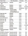
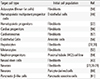
A strategy for reprogramming using transcription factor was first described in the case of MyoD [4]. The innovated discovery of iPSCs proved that combinations of cell specific transcription factors could alter cell fate [2]. This strategy has been applied to lineage reprogramming, and direct reprogramming has been used to obtain neural cells, cardiomyocytes, hepatocytes, and pancreatic cells [5678]. In this review we will outline the history of direct reprogramming and discuss recent progress (Tables 1,2). We will also consider future challenges and therapeutic applications.
HISTORY OF REPROGRAMMING TECHNOLOGY
In 1962, John Gurdon and his team showed that transfer of the nucleus of a fully differentiated cell into an enucleated frog egg could reprogram the cell and lead to the regeneration of an entire frog [9]. This research led to the cloning of mammals like Dolly the Sheep by somatic nuclear transfer [10]. In 1987, Weintraub and collaborators demonstrated that a single cell-type-specific transcription factor, MyoD, could act as a master switch inducing fibroblasts to form skeletal muscle cells [4]. This finding suggested that MyoD played a key role in myogenesis and muscle development [11]. Subsequent studies demonstrated that transcription factor-driven cell fate conversion could be achieved between related lineages such as blood, endoderm, and nervous system cells [121314]. In 2006, Takahashi and Yamanaka reported that overexpressing four transcription factors (Oct-4, Sox2, Klf4, and c-Myc) which are specific to embryonic stem cells converted mice fibroblasts into pluripotent stem cells [2]. They first compiled a list of 24 candidate genes for reprogramming cells by incorporating neomycin resistance and β-galactosidase reporter genes into Fbx15, a gene specifically expressed in pluripotent stem cells. The combination of these 24 factors activated Fbx15 and induced the formation of drug-resistant colonies with characteristic embryonic stem cell (ESC) morphology. By eliminating factors one by one, the 24 genes were narrowed down to only four transcription factors required to generate iPSCs. These genes were Oct-4, Sox2, Klf4, and c-Myc, currently known as "Yamanaka factors." Ongoing studies have demonstrated that the Yamanaka factors can induce iPSCs in other mammalian and human cells [151617]. Likewise, other cell sources such as neural stem cells, liver and stomach cells, and terminally differentiated blood cells, can be reprogrammed into iPSCs by the same factors [181920] and iPSCs can be induced by other combinations of transcriptional factors such as Nanog, Lin28, ESRRB, NR5A2 that play core roles in development [1721]. The breakthrough production of iPSCs attracted interest in the further challenge of converting mature cells directly into other types by overexpressing lineage specific transcriptional factors and bypassing the stem cell state [5722]. Recently, many studies have described direct reprogramming generating cell types such as neural cells, cardiomyocytes, hepatocytes and pancreatic β cell [5678].
DIRECT REPROGRAMMING TO FORM NEURAL CELLS
It seems quite a challenge to induce one cell type directly from one of another lineage. Direct reprogramming of fibroblasts into neurons was first achieved using 19 candidate genes [7]. After continuous elimination of genes, three genes, Ascl1, Brn2, and Myt1l, were found to be the minimal neuron-specific factors for lineage reprogramming of fibroblasts into neural cells. The induced neuronal cells produced spontaneous action potentials, expressed multiple neuron-specific proteins and were able to form functional synapses. Subsequent work demonstrated that mouse fibroblasts could be reprogrammed into dopaminergic neurons by Ascl1, Lmx1a, and Nurr1 [23], and that striatal astrocytes could directly transdifferentiate into neural cells in vivo in the presence of Asxl1, Brn2, and Myt1l [24]. NeuroD was also shown to induce reactive glial cells to form functional neural cells in a brain injury model [25]. Several studies demonstrated that human fibroblasts could be converted into glutamatergic neurons by combinations of factors such as Ascl1, Brn2, Myt1l, and NeuroD1 [26], Brn2, Myt1l and miR-124 [27], and Axl1, Myt1l, neuroD2, miR-9/9 and mmiR-124 [28]. Dopaminergic neurons could be induced to form from human fibroblasts by the combination of Asxl1, Lmx1a, and Nurr1 [29]. Similarly, human fibroblasts could be converted into motor neurons by Asc1l, Brn2, Myt1l, Lhx2, Hb9, Isl1, and Ngn2 [23]. These studies were the first to demonstrate that direct lineage reprogramming is not limited to the same germ layer or lineage system, since fibroblasts, which are derived from mesoderm, could be converted into neurons, which are of ectodermal origin.
DIRECT REPROGRAMMING INTO CARDIOMYOCYTES
Although embryonic mesoderm cells could be induced to differentiate into cardiomyocytes, the master genes of cardiac differentiation had not been identified, despite much research influenc-ed by the identification of MyoD [430]. Fourteen candidate factors were initially selected as able to induce cardiomyocyte-like cells [31]. Then by knockout studies in mice, the requirements were narrowed down to the minimal set of Gata4, Mef2c, and Tbx5. However, the transcriptome of the induced cardiomyocyte-like cells differed from that of neonatal cardiomyocytes and only a small percentage of the cells were able to contract. Subsequently two groups reported that retroviral delivery of the combination of Gata4, Mef2c, and Tbx5 could convert fibroblasts into cardiomyocytes at sites of infarction and decrease injury size [32]. Another study demonstrated that direct conversion into cardiomyocytes was more efficient when the transcription factors Hand2 was added, in both an in vitro and in an in vivo myocardial infarction model [33]. A different group found that the combination of Tbx5, Mef3c, and myocardin could induce a wider spectrum of myocardial genes than previous combinations [34]. A subsequent study showed that fibroblasts could be converted into cardiomyocytes by micro-RNAs in the combination miR-1, miR-133, miR-208, miR-499, and JAK inhibitor both in vitro and in vivo [35]. Another group looked for robust calcium oscillations and spontaneous beating and concluded that the combination of Hand2, Nkx2-5, Gata4, Mef3x, and Tbx5 was the most effective inducer [36]. These results demonstrate that a variety of combinations of transcription factors and microRNAs can induce direct lineage reprogramming and are of promise in regenerative medicine. It will be important to understand more about the molecular mechanisms of cardiac cell induction in vitro and to enhance the efficiency of induction of cardiac cells in vivo.
DIRECT REPROGRAMMING TO HEPATOCYTE CELL TYPE
It is now accepted that terminally differentiated cell types can undergo direct transdifferentiated into cells of other lineages in response to overexpressing lineage-specific factors but it is not always clear whether the transdifferentiated cells can function in damaged organs when transplanted. Two groups have shown that hepatocytes induced from fibroblasts can improve the function of injured hepatic tissues after transplantation [56]. Hepatocytes induced by a combination of Gata4, Hnf1a, and Foxa3 and inactivation of p19Arf expressed hepatic genes and restored the function of damaged livers in mice [5]. The hepatocytes in another study were generated by Hnf4a plus Foxa1, Foxa2, or Foxa3 and also had several hepatocyte-specific features, and their transplantation rescued damaged hepatic tissues [6]. Two further studies demonstrated direct reprogramming of fibroblasts into human hepatocytes. The combination of HNF4 (instead of GATA4), HNF1A, and FOXA3 converted human fibroblasts into induced hepatocytes [37] and the same study showed that hepatocytes induced by overexpressing SV40 large T antigen could be grown in vitro. Another group used a combination of HNF1A, HNF4A, HNF6, CEBPA, ATF5, and PROX1 together with overexpression of c-Myc and knockdown of p53 for reprogramming [38]. This combination had the advantage of avoiding forced proliferation of mature cells, which could cause some damage. In both cases, the human induced hepatocytes displayed metabolic activities comparable to those of control hepatocytes. These studies demonstrate novel applications of induced hepatocytes to improve liver engineering and for use in regenerative medicine. Nevertheless, transplantation of the induc-ed hepatocyte-like cells was only partially effective, suggesting that these cells are not identical to normal hepatocytes. However, in vivo induced hepatocytes are proving to be able to substitute for normal hepatocytes in studies involving disease modelling, cell transplantation and tissue engineering. Further basic studies are required to clarify the mechanisms underlying the induction of hepatocytes and to address whether human fibroblasts can also be converted into functional hepatocytes.
DIRECT REPROGRAMMING TO FORM PANCREATIC β CELLS
Progress in cell reprogramming suggests the possibility of treating hyperglycemia in type 1 DM with induced insulin-producing β cells. Pairs of cell types such as hepatocytes and non-endocrine pancreatic cells that share a common lineage have a better chance of being reprogrammed into β cells. Although these cells cannot secrete insulin for lack of some key factors, they express some typical proteins. For instance, hepatocytes cannot transform proinsulin into insulin but, like β cells, express factors associated with stimulus-secretion coupling such as GLUT2 and glucokinase. Hence, fewer factors are necessary to induce insulin-producing β cells from these cells. Accordingly, transfer of Pdx1 into mice by adenovirus-mediated gene transfer were able to reprogram hepatocytes to form cells that can control blood sugar levels by producing insulin [39] and the ability to produce insulin was considerably enhanced when Pdx1 was fused with the VP16 transcriptional activation domain (Pdx1/VP16)[40]. As a result, adenoviral transfection of PDX1 reprogrammed hepatocytes into cells decreased blood glucose levels in DM animal models [41]. Likewise, delivery of Ngn3, MafA, and Pdx1 was able to reprogram pancreatic acinar cells into insulin-positive cells in an immunodeficient mice model [8]. Although the induced β cells were not very efficient because they could not form islet structures, this study underlines the potential of direct reprogramming by defined transcription factors in vivo. In another study, ectopic expression of Pax4 reprogrammed pancreatic progenitor cells expressing Pdx1 into glucagon-expressing α cells, which could then be reprogrammed into insulin-positive cells [42]. However, normoglycemia could not be restored in older animals, for reasons that are unclear. Nevertheless, this study suggests that alpha cells from DM patients could serve as a source of glucose-responsive, insulin-secreting β cells.
STRATEGIES FOR PRODUCING LARGE NUMBERS OF INDUCED CELLS FOR TRANSLATIONAL PURPOSES
Like iPSC technology, lineage reprogramming can be used in biomedical applications, including disease modeling, cell therapy and drug testing [3]. However the direct reprogramming strategy has the theoretical advantages of shortening the time needed for reprogramming and minimizing the risk of teratoma formation. An important limitation of direct reprogramming is that scaling-up is difficult because the reprogrammed cells proliferate poorly. Recently, new approaches have been developed for obtaining large numbers of cells by direct conversion. In one approach somatic cells are induced to form stem cells or progenitor/precursors and then further differentiated into terminally differentiated cells. The stem cells or progenitors/precursors can then be directly reprogrammed into neural stem cells or their progenitors [4344], oligodendrocyte precursor cells [45], hepatic stem cells [46], HSCs [47], and hematopoietic multipotent progenitors [48]. Another approach is to induce transient expandable intermediates during lineage differentiation. This strategy has been used for producing human hepatocytes [38].
TRANSLATION OF LINEAGE REPROGRAMMING FOR CELL THERAPY
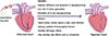
Undoubtedly the most exciting application of direct reprogramming is for cell replacement therapy. Compared with iPSC technology, lineage reprogramming has the obvious advantage that it can be conducted in vivo and could theoretically avoid the risks of teratoma formation and genetic alterations caused by long-term in vitro culture. Moreover, in vivo lineage reprogramming could also circumvent the transplantation process, which could be problematic when cells are induced in vitro. There have now been many reports demonstrating lineage reprogramming in vivo. The induction of cardiomyocytes from fibroblasts in vivo is currently the most interesting application of lineage reprogramming in regenerative therapy. In a coronary ligation model, functional cardiomyocytes induced from cardiac fibroblasts by local delivery of cardiac reprogramming factors in vivo decreased cardiac infarct size and reduced dysfunction (Fig. 1)[32]. In another study in a heart injury model, lineage reprogramming enhanced cardiac function by inducing cardiomyocyte-like cells from endogenous cardiac fibroblasts [33].
Despite the promising advances, there are significant obstacles to the clinical application of in vivo lineage reprogramming. In many cases, some transcripts characteristic of the original cell type persist after reprogramming, indicating that reprogramming is not complete [649]. Also more safe and efficient methods of in vivo delivery of transcription factors need to be developed, and the side effects of reprogramming in vivo must be investigated.
CONCLUSIONS
The technology of induced reprogramming may prove useful in therapeutic cell transplantation. The iPSCs strategy has the merit of scalability but carries a risk of tumorigenicity. On the other hand, the strategy of directly inducing changes of somatic cell type without passing through the pluripotent stage is less likely to generate teratomas and does not require the delivery of integrated genes. Another interesting clinical application of direct lineage reprogramming is in vivo reprogramming. However many obstacles remain, and further studies are needed to produce functional cells of higher purity and develop safer and more efficient delivery method. Studies of potential side effects are also called for.
 XML Download