

 PDF
PDF ePub
ePub Citation
 Print
Print


Abstract
Ferroportin-1 (FPN) is a transporter protein that is known to mediate iron export from macrophages. The purpose of this study was to investigate the effect of copper on the regulation of FPN gene expression in J774 mouse macrophage cells. J774 cells were treated with various concentrations of CuSO4 and RT-PCR analyses were performed to measure the steady-state levels of mRNAs for FPN and divalent metal transporter 1 (DMT1, an iron importer). Copper treatment significantly increased FPN mRNAs in a dose-dependent manner, but didn't change the levels of DMT1 mRNA. Experiments with transcriptional inhibitor actinomycin D (0.5 µg/mL) revealed that copper treatment did not affect the half-life of FPN mRNAs in J774 cells. On the other hand, results from luciferase reporter assays showed that copper directly stimulated the promoter activity of FPN. In summary, our data showed copper induced FPN mRNA of macrophages via a transcriptional rather than post-transcriptional mechanisms.
Figures and Tables
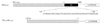
Fig. 2
Effect of copper on the levels of mRNA for ferroportin (FPN) and for divalent metal transporter 1 (DMT1) in J774 macrophage cells. RT-PCR analyses were performed to determine mRNA for FPN, DMT1, and β-actin (upper panel). Levels of FPN and DMT1 mRNA were normalized to β-actin mRNA levels (lower panel).
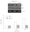
Fig. 3
Effects of varying doses of copper on the viability of J774 macrophage cells. Cell viability were measured by MTT assays. Results are the mean ± s.d. of three independent experiments.
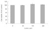
Fig. 4
Effects of copper on FPN mRNA stability in J774 macrophage cells. J774 cells were pretreated with 200 µM CuSO4 for 20 hr and throughly washed. Then actinomycin D (0.5 µg/mL) was added with CuSO4 (Cu+) or with medium alone (Cu-). Total RNA was isolated at 0, 2, 6, 9 hours after actinomycin D treatment, and RT-PCR analyses were performed (upper panel). The rate of FPN mRNA decay of untreated and copper-treated cells was determined after normalization with β-actin mRNA. Results are the mean ± s.d. of three independent experiments.
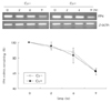
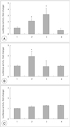
Fig. 5
Effects of copper on FPN promoter/5'-UTR driven luciferase reporter activity. HeLa cells were transiently transfected with A: FPN-Luc, B: FPNΔIRE-Luc, or C: pGL-control (empty) vector. Twelve hours later, cells were exposed to various agents for 20 h, and cell lysates were assayed for luciferase activity. Luciferase activities were normalized to protein concentraion of cell lysates and expressed as fold changes compared to untreated controls. 1, untreated; 2, copper; 3, iron; 4. iron chelator. Results are the mean ± s.d. of three independent experiments. *: p < 0.05 vs. untreated controls.
References
1. Conrad ME, Umbreit JN. Iron Absorption and Transport-an Update. Am J Hematol. 2000. 64(4):287–298.

2. Ganz T, Nemeth E. Regulation of Iron Acquisition and Iron Distribution in Mammals. Biochim Biophys Acta. 2006. 1763(7):690–699.
3. Chua AC, Graham RM, Trinder D, Olynyk JK. The Regulation of Cellular Iron Metabolism. Crit Rev Clin Lab Sci. 2007. 44(5-6):413–459.
4. Abboud S, Haile DJ. A Novel Mammalian Iron-Regulated Protein Involved in Intracellular Iron Metabolism. J Biol Chem. 2000. 275:19906–19912.
5. Muckenthaler MU, Galy B, Hentze MW. Systemic Iron Homeostasis and the Iron-Responsive element/iron-Regulatory Protein (IRE/IRP) Regulatory Network. Annu Rev Nutr. 2008. 28:197–213.
6. Knutson MD, Oukka M, Koss LM, Aydemir F, Wessling-Resnick M. Iron Release from Macrophages After Erythrophagocytosis is Up-Regulated by Ferroportin 1 Overexpression and Down-Regulated by Hepcidin. Proc Natl Acad Sci USA. 2005. 102(5):1324–1328.
7. Beutler E. Hemochromatosis: Genetics and Pathophysiology. Annu Rev Med. 2006. 57:331–347.
8. Pietrangelo A. Hemochromatosis: An Endocrine Liver Disease. Hepatology. 2007. 46(4):1291–1301.
9. Knutson MD, Vafa MR, Haile DJ, Wessling-Resnick M. Iron Loading and Erythrophagocytosis Increase Ferroportin 1 (FPN1) Expression in J774 Macrophages. Blood. 2003. 102(12):4191–4197.
10. Lymboussaki A, Pignatti E, Montosi G, Garuti C, Haile DJ, Pietrangelo A. The Role of the Iron Responsive Element in the Control of ferroportin1/IREG1/MTP1 Gene Expression. J Hepatol. 2003. 39(5):710–715.
11. McKie AT, Marciani P, Rolfs A, Brennan K, Wehr K, Barrow D, Miret S, Bomford A, Peters TJ, Farzaneh F, Hediger MA, Hentze MW, Simpson RJ. A Novel Duodenal Iron-Regulated Transporter, IREG1, Implicated in the Basolateral Transfer of Iron to the Circulation. Mol Cell. 2000. 5:299–309.
12. Liu XB, Hill P, Haile DJ. Role of the Ferroportin Iron-Responsive Element in Iron and Nitric Oxide Dependent Gene Regulation. Blood Cells Mol Dis. 2002. 29(3):315–326.
13. Ludwiczek S, Aigner E, Theurl I, Weiss G. Cytokine-Mediated Regulation of Iron Transport in Human Monocytic Cells. Blood. 2003. 101(10):4148–4154.
14. Yang F, Liu XB, Quinones M, Melby PC, Ghio A, Haile DJ. Regulation of Reticuloendothelial Iron Transporter MTP1 (Slc11a3) by Inflammation. J Biol Chem. 2002. 277(42):39786–39791.
15. Chung J, Haile DJ, Wessling-Resnick M. Copper-Induced Ferroportin-1 Expression in J774 Macrophages is Associated with Increased Iron Efflux. Proc Natl Acad Sci USA. 2004. 101(9):2700–2705.
16. Sareila O, Hämäläinen M, Nissinen E, Kankaanranta H, Moilanen E. Orazipone inhibits activation of inflammatory transcription factors nuclear factor-kappa B and signal transducer and activator of transcription 1 and decreases inducible nitric-oxide synthase expression and nitric oxide production in response to inflammatory stimuli. J Pharmacol Exp Ther. 2008. 324(2):858–866.
17. Gonzalez M, Reyes-Jara A, Suazo M, Jo WJ, Vulpe C. Expression of Copper-Related Genes in Response to Copper Load. Am J Clin Nutr. 2008. 88(3):830S–834S.
18. Muller P, van Bakel H, van de Sluis B, Holstege F, Wijmenga C, Klomp LW. Gene Expression Profiling of Liver Cells After Copper Overload in Vivo and in Vitro Reveals New Copper-Regulated Genes. J Biol Inorg Chem. 2007. 12(4):495–507.
19. Davis SR, Cousins RJ. Metallothionein Expression in Animals: A Physiological Perspective on Function. J Nutr. 2000. 130(5):1085–1088.
20. Jeney V, Itoh S, Wendt M, Gradek Q, Ushio-Fukai M, Harrison DG, Fukai T. Role of Antioxidant-1 in Extracellular Superoxide Dismutase Function and Expression. Circ Res. 2005. 96(7):723–729.
21. Itoh S, Ozumi K, Kim HW, Nakagawa O, McKinney RD, Folz RJ, Zelko IN, Ushio-Fukai M, Fukai T. Novel Mechanism for Regulation of Extracellular SOD Transcription and Activity by Copper: Role of Antioxidant-1. Free Radic Biol Med. 2009. 46(1):95–104.
22. Itoh S, Kim HW, Nakagawa O, Ozumi K, Lessner SM, Aoki H, Akram K, McKinney RD, Ushio-Fukai M, Fukai T. Novel Role of Antioxidant-1 (Atox1) as a Copper-Dependent Transcription Factor Involved in Cell Proliferation. J Biol Chem. 2008. 283(14):9157–9167.
23. Hart EB, Steenbock H, Waddell J, Elvehjem CA. Iron in Nutrition. VII. Copper as a Supplement to Iron for Hemoglobin Building in the Rat. 1928. J Biol Chem. 2002. 277(34):e22.
24. Owen CA Jr. Effects of Iron on Copper Metabolism and Copper on Iron Metabolism in Rats. Am J Physiol. 1973. 224(3):514–518.
25. Williams DM, Kennedy FS, Green BG. Hepatic Iron Accumulation in Copper-Deficient Rats. Br J Nutr. 1983. 50(3):653–660.
26. Harris ZL, Durley AP, Man TK, Gitlin JD. Targeted Gene Disruption Reveals an Essential Role for Ceruloplasmin in Cellular Iron Efflux. Proc Natl Acad Sci USA. 1999. 96(19):10812–10817.
27. Yamamoto K, Yoshida K, Miyagoe Y, Ishikawa A, Hanaoka K, Nomoto S, Kaneko K, Ikeda S, Takeda S. Quantitative Evaluation of Expression of Iron-Metabolism Genes in Ceruloplasmin-Deficient Mice. Biochim Biophys Acta. 2002. 1588(3):195–202.
28. Hellman NE, Gitlin JD. Ceruloplasmin Metabolism and Function. Annu Rev Nutr. 2002. 22:439–458.
29. Bates GW, Workman EF Jr, Schlabach MR. Does Transferrin Exhibit Ferroxidase Activity? Biochem Biophys Res Commun. 1973. 50(1):84–90.
30. Mukhopadhyay CK, Attieh ZK, Fox PL. Role of Ceruloplasmin in Cellular Iron Uptake. Science. 1998. 279(5351):714–717.
31. Attieh ZK, Mukhopadhyay CK, Seshadri V, Tripoulas NA, Fox PL. Ceruloplasmin Ferroxidase Activity Stimulates Cellular Iron Uptake by a Trivalent Cation-Specific Transport Mechanism. J Biol Chem. 1999. 274(2):1116–1123.
32. Sarkar J, Seshadri V, Tripoulas NA, Ketterer ME, Fox PL. Role of Ceruloplasmin in Macrophage Iron Efflux during Hypoxia. J Biol Chem. 2003. 278(45):44018–44024.
 XML Download