

 PDF
PDF ePub
ePub Citation
 Print
Print


Abstract
Objective
The aim of this study was to evaluate the biomechanical aspects of peri-implant bone upon root contact of orthodontic microimplant.
Methods
Axisymmetric finite element modeling scheme was used to analyze the compressive strength of the orthodontic microimplant (Absoanchor SH1312-7, Dentos Inc., Daegu, Korea) placed into inter-radicular bone covered by 1 mm thick cortical bone, with its apical tip contacting adjacent root surface. A stepwise analysis technique was adopted to simulate the response of peri-implant bone. Areas of the bone that were subject to higher stresses than the maximum compressive strength (in case of cancellous bone) or threshold stress of 54.8MPa, which was assumed to impair the physiological remodeling of cortical bone, were removed from the FE mesh in a stepwise manner. For comparison, a control model was analyzed which simulated normal orthodontic force of 5 N at the head of the microimplant.
Results
Stresses in cancellous bone were high enough to cause mechanical failure across its entire thickness. Stresses in cortical bone were more likely to cause resorptive bone remodeling than mechanical failure. The overloaded zone, initially located at the lower part of cortical plate, proliferated upward in a positive feedback mode, unaffected by stress redistribution, until the whole thickness was engaged.
Go to : 
REFERENCES
1.Chen YH., Chang HH., Chen YJ., Lee D., Chiang HH., Yao CC. Root contact during insertion of miniscrews for orthodontic anchorage increases the failure rate: an animal study. Clin Oral Implants Res. 2008. 19:99–106.

2.Asscherickx K., Vannet BV., Wehrbein H., Sabzevar MM. Root repair after injury from mini-screw. Clin Oral Implants Res. 2005. 16:575–8.
3.Bae SM. The repair of the root and pulp tissue after intentional root injury by the orthodontic microimplant in dog. PhD thesis. Daegu, Korea: Kyungpook National University;2005.
4.Asscherickx K., Vande Vannet B., Wehrbein H., Sabzevar MM. Success rate of miniscrews relative to their position to adjacent roots. Eur J Orthod. 2008. 30:330–5.
5.Kang YG., Kim JY., Lee YJ., Chung KR., Park YG. Stability of mini-screws invading the dental roots and their impact on the paradental tissues in beagles. Angle Orthod. 2009. 79:248–55.
6.Kuroda S., Yamada K., Deguchi T., Hashimoto T., Kyung HM., Takano-Yamamoto T. Root proximity is a major factor for screw failure in orthodontic anchorage. Am J Orthod Dentofacial Orthop. 2007. 131(4 Suppl):S68–73.
7.Park HS. An anatomical study using CT images for the implantation of micro-implants. Korean J Orthod. 2002. 32:435–41.
8.Bae SM., Park HS., Kyung HM., Kwon OW., Sung JH. Clinical application of micro-implant anchorage. J Clin Orthod. 2002. 36:298–302.
9.Bae SM., Kyung HM. Clinical application of micro-implant anchorage (MIA) in orthodontics (2) - Anatomic consideration and surgical procedures. Korean J Clin Orthod. 2002. 1:16–29.
10.Kim SH., Choi YS., Hwang EH., Chung KR., Kook YA., Nelson G. Surgical positioning of orthodontic mini-implants with guides fabricated on models replicated with cone-beam computed tomography. Am J Orthod Dentofacial Orthop. 2007. 131(4 Suppl):S82–9.
11.Poggio PM., Incorvati C., Velo S., Carano A. "Safe zones": a guide for miniscrew positioning in the maxillary and mandibular arch. Angle Orthod. 2006. 76:191–7.
12.Frost HM. Perspectives: bone's mechanical usage windows. Bone Miner. 1992. 19:257–71.
13.Gelgör IE., Büyükyilmaz T., Karaman AI., Dolanmaz D., Kalayci A. Intraosseous screw-supported upper molar distali-zation. Angle Orthod. 2004. 74:838–50.
14.Liou EJ., Pai BC., Lin JC. Do miniscrews remain stationary under orthodontic forces? Am J Orthod Dentofacial Orthop. 2004. 126:42–7.
15.Klineberg I., Jagger RG. Occlusion and clinical practice: an evidence-based approach. Edinburgh, New York, Wright: Elsevier;2004. 84.
16.NISA II/DISPLAY III User's Manual, Engineering Mechanics Research Corporation (EMRC).
17.Sevimay M., Turhan F., Kiliçarslan MA., Eskitascioglu G. Three-dimensional finite element analysis of the effect of different bone quality on stress distribution in an implant-supported crown. J Prosthet Dent. 2005. 93:227–34.
18.Papavasiliou G., Kamposiora P., Bayne SC., Felton DA. 3D-FEA of osseointegration percentages and patterns on implant-bone interfacial stresses. J Dent. 1997. 25:485–91.
19.Misch CE., Qu Z., Bidez MW. Mechanical properties of trabecular bone in the human mandible: implications for dental implant treatment planning and surgical placement. J Oral Maxillofac Surg. 1999. 57:700–6.
20.Meyer U., Vollmer D., Runte C., Bourauel C., Joos U. Bone loading pattern around implants in average and atrophic edentulous maxillae: a finite-element analysis. J Craniomaxillofac Surg. 2001. 29:100–5.
22.Yu W., Jang YJ., Kyung HM. Combined influence of implant diameter and alveolar ridge width on crestal bone stress: a quantitative approach. Int J Oral Maxillofac Implants. 2009. 24:88–95.
23.Wolff J. The law of bone remodelling. Berlin, Heidelberg, New York: Springer. 1986.
24.Jung ES., Jo KH., Lee CH. A finite element stress analysis of the bone around implant following cervical bone resorption. J Korean Acad Implant Dent. 2003. 22:38–48.
25.Kitamura E., Stegaroiu R., Nomura S., Miyakawa O. Influence of marginal bone resorption on stress around an implant - a three-dimensional finite element analysis. J Oral Rehabil. 2005. 32:279–86.
26.Hembree M., Buschang PH., Carrillo R., Spears R., Rossouw PE. Effects of intentional damage of the roots and surrounding structures with miniscrew implants. Am J Orthod Dentofacial Orthop. 2009. 135:280. .e1-9.
27.Brisceno CE., Rossouw PE., Carrillo R., Spears R., Buschang PH. Healing of the roots and surrounding structures after intentional damage with miniscrew implants. Am J Orthod Dentofacial Orthop. 2009. 135:292–301.
28.Dao V., Renjen R., Prasad HS., Rohrer MD., Maganzini AL., Kraut RA. Cementum, pulp, periodontal ligament, and bone response after direct injury with orthodontic anchorage screws: a histomorphologic study in an animal model. J Oral Maxillofac Surg. 2009. 67:2440–5.
29.Reina JM., García-Aznar JM., Domínguez J., Doblaré M. Numerical estimation of bone density and elastic constants distribution in a human mandible. J Biomech. 2007. 40:828–36.
Go to :
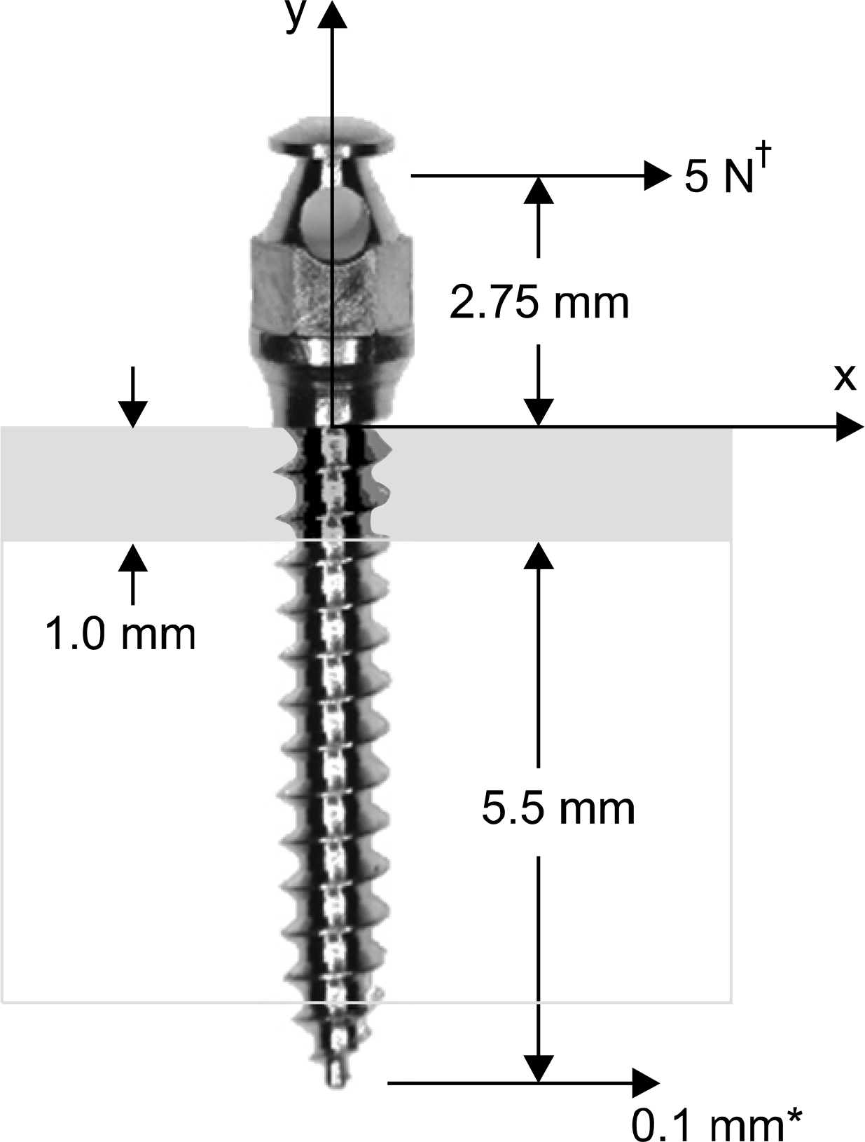 | Fig 1.Schematic diagram for the SH1312-7 (Dentos Inc., Daegu, Korea) microimplant with its apical part intruding the PDL space and contacting the root surface. The implant was assumed to be loaded either by an orthodontic force at its head, or apically by the adjacent root in contact with its tip (*a displacement load of 0.1 mm was assumed to be transmitted from the root surface to the microimplant, †5 N was assumed as an orthodontic force). |
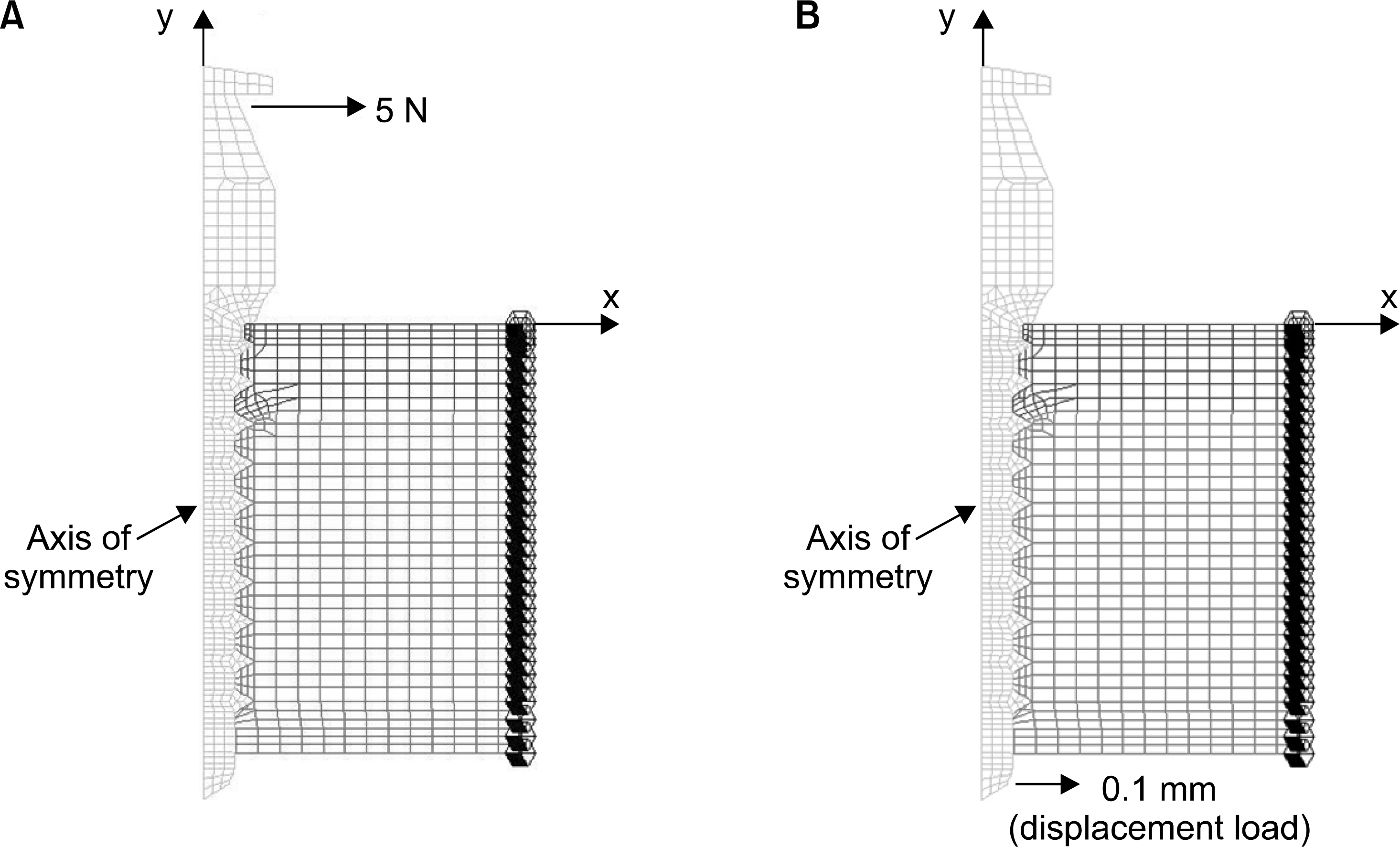 | Fig 2.Axisymmetric finite element (with non-axisymmetric loading) models simulating. A, Control model which presents a microimplant subject to orthodontic load of 5 N at the head, without root contact; B, a microimplant in contact with root surface, subject to apical excitation of 0.1 mm. Horizontal axis x represents the loading direction. Vertical axis y represents the axis of symmetry. |
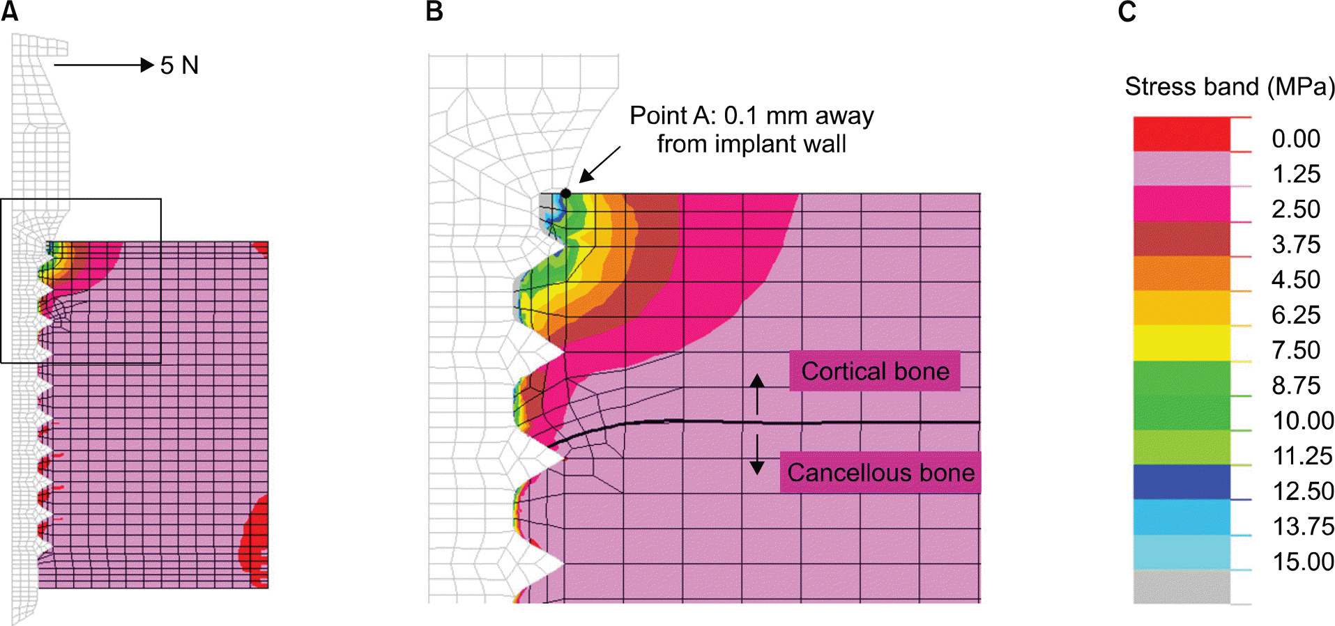 | Fig 3.Stresses (maximum compressive stress) in control model subject to orthodontic force of 5 N at the head. A, Overall stress distribution; B, magnified view of the cervical area; C, stress band (a cut off stress was specified at 15 MPa for clear observation of stress concentration occurring at the implant/cortical bone junction). |
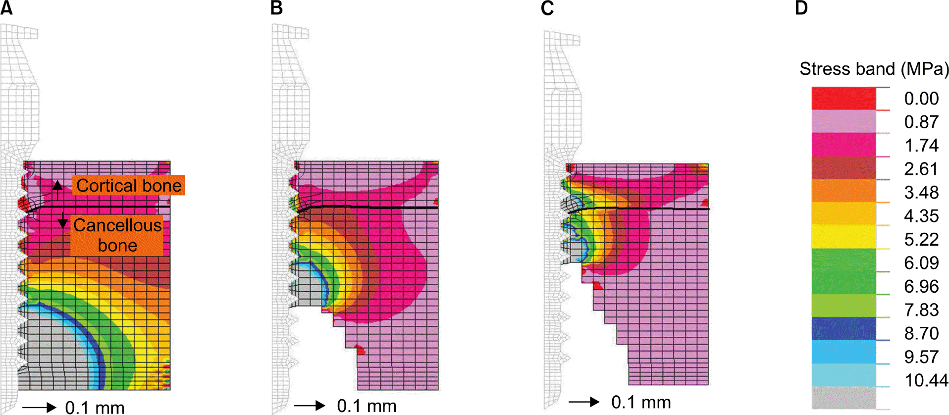 | Fig 4.Stress development in the cancellous bone. Calculation was performed in a stepwise manner by removing the cancellous bone elements subject to stresses of higher than ultimate compressive strength of 10.44 MPa (Table 1). A, Base model; B, Stage 1; C, Stage 2; D, stress band stress band with cut off stress at 10.44 MPa. |
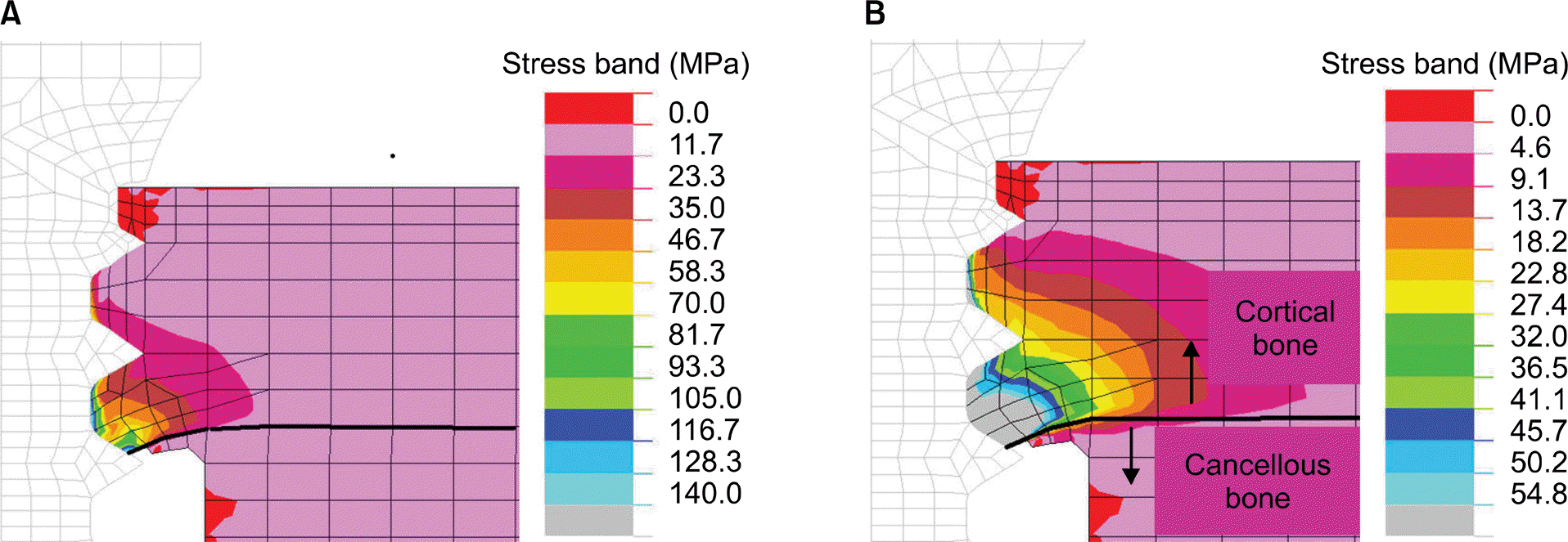 | Fig 5.Stresses in cortical bone after the entire peri-implant cancellous bone along the implant has lost structural integrity due to displacement of 1.0 mm at the apex. A, With the cut off stress at 140 MPa (maximum compressive strength of cortical bone, Table 1); B, with the cut off stress at 54.8 MPa (threshold for resorptive remodeling, Table 1). |
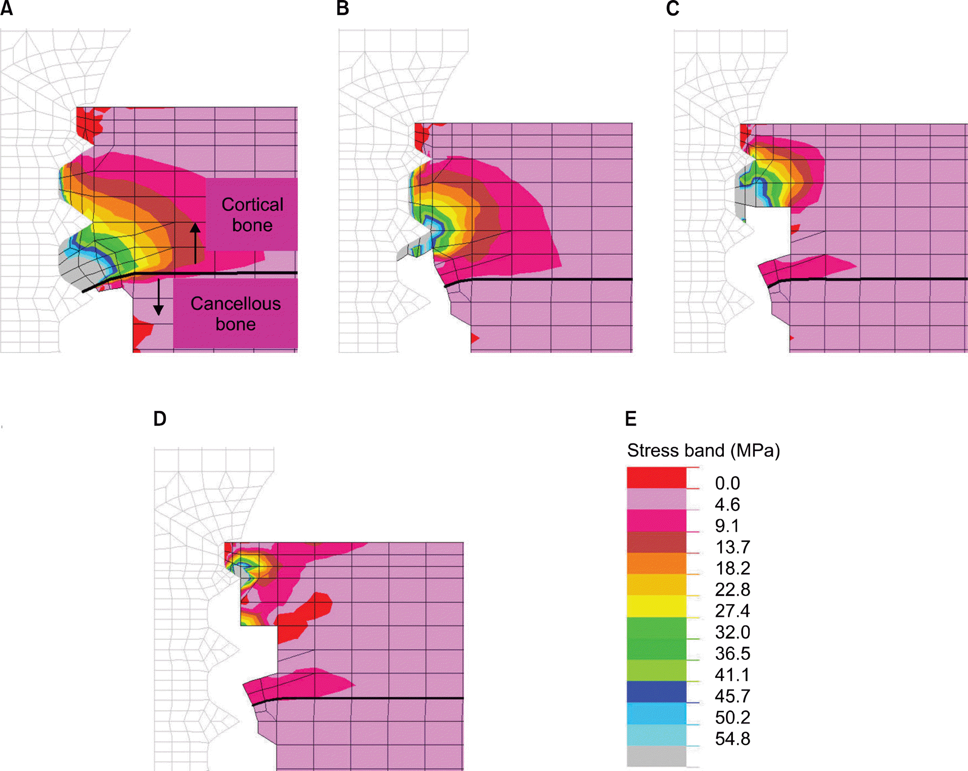 | Fig 6.Stress development in the cervical cortical bone (due to displacement of 1.0 mm at the apex) calculated in a successive manner by removing the elements from model where resorptive bone remodeling was predicted due to overload. A, Stage 3; B, Stage 4; C, Stage 5; D, Stage 6 models; E, stress band with cut off stress at 54.8 MPa (threshold for resorptive remodeling, Table 1). |
Table 1.
Mechanical properties used in this study
| Material | Young’s modulus (GPa) | Poisson ratio | Strength (MPa) | Threshold stress (MPa) |
|---|---|---|---|---|
| Titanium17 | 114.0 | 0.35 | - | - |
| Cortical bone18 | 13.7 | 0.3 | 72-76 (tensile) | 54.8* |
| 140-170 (compressive) | - | |||
| Cancellous bone18,19 | 1.37 | 0.3 | 0.22-10.44 (compressive) | - |
 XML Download