

 PDF
PDF ePub
ePub Citation
 Print
Print


INTRODUCTION
Direct bonded lingual retainers are the most commonly preferred retention devices1 as their usage result in less relapse in the long term evaluation.2 Besides various designs, their basic construction consists of a length of wire attached to the etched enamel with composite.1 These composites include either conventional restorative or specific orthodontic bonding resins.1 Photoactivated, resin-based composites (PARBCs) have become the material of choice for bonding lingual retainers today, as they offer ease of application and optimal handling characteristics to allow the clinician to shape and finish the adhesive around the lingual retainer wire for maximum patient comfort.3 For prolonged use in the oral cavity, the lingual retainer composites must possess certain physical properties to ensure clinical success.
These tooth-colored polymeric restorative materials have been extensively studied and improved throughout the last three decades. The main adverse effect of methacrylate polymerization reaction is volumetric shrinking, which contributes to stress formation along the bonded interfaces of restorations.4,5 For this reason, much research have mainly focused on dental adhesion improvements to achieve higher shear bond strength (SBS) and to improve the stability of the adhesive interface over time.6,7 Despite significant increases in immediate bond strength, the occurrence of gap formation mainly at the adhesive tooth interface has not been completely resolved.8-10 These gaps may cause seeping and leakage of oral fluids and bacteria between the tooth and restoration surface, an event that defines microleakage in dentistry.11 The amount of composite on the retainer wire may be inversely affected by potential gaps at the composite/enamel and composite/wire interfaces and may cause failure of flexible spiral wire retainers (FSWRs).
Recently, a new category of resin matrix for dental composite was developed based on ring-opening monomers.12 This hydrophobic composite derives from the combination of siloxane and oxirane, thus the name silorane.13 The major advantages of this innovative restorative material are its reduced shrinking and its mechanical properties comparable to those of the methacrylate based composites.13 Previous studies revealed higher marginal adaptation and reduced microleakage formation and lower material deflection when silorane-based materials were used compared to methacrylate composites.14,15
In the orthodontic literature, different band cements,16 light sources,17,18 composites and brackets19-23 have been evaluated for microleakage but these studies primarily focused on enamel demineralization. The FSWRs are intended to serve for a long time in the mouth and are exposed to various chemical and mechanical degradation.3
No studies in the literature appear to have evaluated Silorane-based material in orthodontics as lingual retainer composite. Therefore, the aim of this study was to evaluate the SBS, fracture mode, wire pull out (WPO) resistance and microleakage of low-shrinking composite for bonding orthodontic retainers.
MATERIAL AND METHODS
A total of 120 human mandibular incisor teeth, extracted for periodontal reasons, were collected. Teeth with hypoplastic areas, cracks, or irregularities of the enamel structure were excluded. The criteria for tooth selection dictated no pre-treatment with chemical agents such as alcohol, formalin, or hydrogen peroxide. The extracted teeth were stored in distilled water until use (maximum 1 month). The water was changed weekly to avoid bacterial growth. Calculus and debris were removed with a scaler and the teeth were pumiced.
All bonding procedures were performed according to the manufacturer's instructions by one author (T.U.). In group I (30 teeth), a 37% phosphoric acid gel (3M Dental Products, St Paul, Minnesota, USA) and Transbond XT Primer (3M Unitek, Monrovia, California, USA); in group II (30 teeth), Silorane System Adhesive (SSA) self-etch primer and SSA bond (3M Espe, Seefeld, Germany) were applied.
The materials were cured with a quartz tungsten halogen light source (Hilux 350, Express Dental Products, Toronto, Ontario, Canada). The curing times were set at 20 seconds for Transbond-LR and 40 seconds for low-shrinking composite, according to the manufacturers' instructions.
Bonding procedure
Sixty freshly extracted human mandibular incisor teeth were used in this part of the study. The teeth were moulded in square acrylic blocks with the long axis perpendicular to the upper surface of the blocks. Transbond-LR [group I; 3M Unitek; sample size (n): 30] and Silorane (group II; 3M-Espe; n = 30) were added to the lingual surface of the teeth by packing the material into cylindrical shaped plastic matrices with an internal diameter of 2.34 mm and a height of 3 mm (Ultradent, South Jordan, Utah, USA).
SBS testing
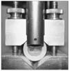
For SBS testing, the specimens were mounted in a universal testing machine (Hounsfield Test Equipment, Salford, Lancashire, UK). A notch-shaped apparatus (Ultradent) attached to a compression load cell at a crosshead speed of 0.5 mm/minute was applied in the vertical direction to each specimen at the interface between the tooth and composite until failure occurred (Fig 1). The maximum load (N) was divided by the cross-sectional area of the bonded adhesive posts to determine bond strength in megapascals (MPa).
Fracture mode
Fracture analysis was performed using an optical stereomicroscope at × 20 magnification (SZ 40, Olympus, Tokyo, Japan). The amount of adhesive remaining on the enamel surface was coded by one investigator (T.U.) who was blinded to group allocations. Failures were classified as cohesive if more than 80% of the resin remained on the tooth surface, and as adhesive if less than 20% of the resin remained on the tooth surface, or as mixed if certain areas exhibited cohesive fractures and others adhesive fractures.
WPO testing
In order to perform WPO testing, 40 acrylic blocks, with a diameter of 25 mm and a height of 10 mm, were prepared in moulds. In each block, a hole, 4 mm in diameter and 3 mm in height, was drilled and a slot 0.6 mm wide and 1 mm deep was cut. Inclusion of the hole resulted in, clinically similar composite thickness and width, while the slot permitted the application of a standard 1 mm composite thickness over the wire. Similar to SBS testing, group I was prepared with Transbond-LR and group II with low-shrinking composite. Multistranded PentaOne® wire (Masel Orthodontics, Bristol, Pennsylvania, USA) 0.0215 inches in diameter was used in both groups. The wires were cut into 10 mm lengths. After insertion of the wires into the prepared slots, different composites were placed in the hole and cured. The curing procedure was the same as SBS testing.
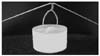
The free ends of the wire were drawn up and bent with an orthodontic plier (Fig 2). The attachment arm of the tensile load cell of the universal testing machine was secured and the force applied at a crosshead speed of 0.5 mm/minute through the long axis of each sample. Data were recorded when the wires were pulled out from the resin.
Microleakage evaluation
The remaining 60 incisor teeth were used in this part of the study. PentaOne® wire (Masel Orthodontics) of 0.0215 inch diameter was used in all groups. Wires were cut into 20 mm lengths to ensure standardization, and the wires were bent to fit the lingual curvature of the incisor teeth. Transbond-LR (n = 30) and Silorane (n = 30) composites were added to the lingual surface of the teeth and cured.
Prior to dye penetration, the apices were sealed with sticky wax, rinsed in tap water and air dried nail varnish was applied to the entire surface of the tooth except for approximately 1 mm away from the composite bulk. To minimize dehydration of the restorations, the teeth were replaced in water as soon as the nail polish dried. The teeth were immersed in 0.5% solution of basic fuchsine for 24 hours at room temperature. After being removed from the solution, the teeth were rinsed in tap water, and the superficial dye was removed with a brush and dried.
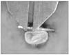
Each specimen was sectioned in a transverse plane (parallel to the lingual retainer wire) just above the wire with a low speed water-cooled diamond saw (Isomet, Buehler, Lake Bluff, IL, USA). The specimens were evaluated first under a stereomicroscope (×20 magnifications) (SZ 40) for dye penetration along the composite/enamel interface. Then lingual retainer wires were gently removed from the composite bulk and the dye penetration between the composite/wire interface in both the mesial and distal direction was also evaluated under a stereomicroscope. Microleakage was determined by direct measurement using an electronic digital caliper (Mitutoyo Co., Miyazaki, Japan) (Fig 3) and recording the data to the nearest value as a range 0.5 to 5 millimeters.
Statistical analysis
All statistical analyses were performed with the Statistical Package for Social Sciences, version 13.0 for Windows (SPSS, Chicago, IL, USA). When the p-value was less than 0.05, the statistical test was determined as significant.
Descriptive statistics, including the mean, standard deviation, minimum, and maximum values, were calculated for the two groups. The normality test of Shapiro-Wilks and Levene's variance homogeneity test were applied to the data. The data were found not normally distributed, and there was no homogeneity of variance between the groups. A non-parametric Mann-Whitney U-test for two independent variables was used to compare the SBS and WPO data of the two investigated composites. Fracture modes were analyzed using a Pearson chi-square test.
For each composite interface (composite/wire and composite/enamel), the microleakage scores were obtained by measuring the mesial and distal scores. After the statistical evaluation of mesial and distal leakage for each specimen, the score for each group was obtained by measuring the mean of mesial and distal microleakage scores. Microleakage comparisons were performed using Wilcoxon and Mann-Whitney U tests with Bonferroni correction.
RESULTS
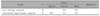
Descriptive statistics and the results of Mann Whitney-U test, comparing the SBS of two composites are presented in Table 1. The SBS differences for Silorane (mean 20.2 ± 8.5 MPa) and Transbond-LR (mean 23.5 ± 8.9 MPa) composites were not statistically significant.
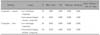
The fracture modes of the specimens are shown in Table 2. A greater percentage of fractures showed a mix type failure (85%) for Silorane and an adhesive type failure (60%) for Transbond-LR. There was a statistically significant difference between the groups (p < 0.001).
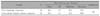
Descriptive statistics and the results of statistical tests comparing the WPO values of the two groups tested, are shown in Table 3. The WPO test results were not statistically significant between the groups.
Total microleakage comparisons between the composite-enamel and composite-wire interfaces for each of the two investigated composite groups are shown in Table 4. The values were statistically significant between the two groups (p < 0.001). The mean microleakage values at the composite-enamel interface for conventional lingual retainer composite and low-shrinking composite were 0.038 mm and 0.000 mm, respectively. At the composite-wire interface the mean microleakage values were 0.750 mm and 0.000 mm, respectively.
DISCUSSION
To our knowledge, this study is the first to evaluate the bonding properties of a low-shrinking composite in comparison with a conventional orthodontic composite for bonding lingual retainers. The use of bonded lingual retainers, particularly in the mandibular incisor area, has become increasingly popular among orthodontists. There is a general agreement on the necessity of fixed lingual retainers to prevent relapse after active orthodontic treatment.1 Different composites have been suggested for bonding of lingual retainers, including both restorative and orthodontic bonding materials; however the two major properties of these dental composites that still have to be improved are; their polymerization shrinking and the related polymerization stress.13
In the present study for SBS testing, the samples were prepared using a standardized cylindrical mold from Ultradent to build up composite cores on the etched enamel surfaces on which the composites were prepared directly. The WPO-testing methodology was adopted from the study of Bearn et al.24 This method was used to evaluate mean detachment forces for both composites and these forces were interpreted as resistance to failure. The total depth of the composite and wire was selected as 1 mm, because Bearn et al.24 showed that increasing the thickness to greater than 1.0 mm produced only a relatively small increase in force needed to detach the wire from the composite, so the increase was likely to give little clinical benefit. To determine the extent of microleakage on the bonded specimens, the dye penetration method was chosen. This is the most commonly used method to assess microleakage of dental materials.25 It is easy to perform, fast, and economical, but the shortcoming of the technique is the subjectivity involved in reading the specimens.26
According to Reynolds,27 adequate bond strength needed for clinical orthodontic bracket bonding varies between 5.9 and 7.8 MPa. In the current research, SBS values of both investigated composites were above the necessary values. Descriptive statistics and the results of statistical tests comparing the SBS of two groups showed that these values were similar and the findings were not statistically different.
It is stated in the literature that the most common failure type for lingual retainers is detachment at the wire-adhesive interface.1 It has been also reported that abrasion of composite over the retainer wire is also a reason for failure of lingual retainers.1 The mean detachment values for conventional lingual retainer composite (20.3 ± 5.1 N) were higher than for low-shrinking composite (20.1 ± 4.7 N) and the difference between the groups were not statistically significant. Bearn et al.24 compared six different composite resins, which were proposed as lingual retainer adhesives, via WPO tests, and reported scores of between 11.2 and 24.4 N. Transbond-LR in the present study showed higher detachment forces than those found by Bearn et al.24 Different from Bearn et al.24 PentaOne 0.0215 inch wire was used in this study. This wire is commonly used in orthodontics for lingual retainer fabrication and a study by Bearn et al.24 showed that an increased diameter from 0.0175 to 0.0215 inches significantly increased the force required to pull the wire from the composite. It can be assumed that samples prepared with low-shrinking composite could result in lower WPO forces if 0.0175 inch PentaOne wire had been used.
Most orthodontic bonding studies have shown mixed or cohesive-type failure.28,29 In those studies, after SBS testing, a part of the composite resin remained either on the enamel surface or the bracket base, causing cohesive rather than adhesive failure between enamel and composite resin. In the present study, more adhesive failures occurred for Transbond-LR (%60).
In restorative dentistry, microleakage is defined as seeping and leaking of fluids and bacteria between the tooth-composite interface.11 O'Reilly and Featherstone30 and Ogaard et al.31 have shown that visible white lesions can develop within 4 weeks, and according to Gladwin and Bagby11 microleakage increases the likelihood of recurrent caries and postoperative sensitivity. From an orthodontic perspective, it is possible to interpret this fact as the likelihood of formation of white spot lesions or caries at and under the enamel-composite interface. It is also likely that microleakage under the composite holding the retainer wire may result in failure of the fixed retainer. Thus, the investigation of microleakage between wire-composite interfaces might be an important topic for the clinical success of treatments and lingual retainers.
In the present study, the results of statistical tests, comparing the total microleakage values between the composite-enamel and composite-wire interfaces for each of the two investigated materials showed that there was no microleakage between the composite-enamel and composite-wire interfaces with low-shrinking composite. In the present study, more adhesive failures were observed in the Transbond-LR group. The difference between the two groups was statistically significant. In the present study, no microleakage found either at the composite-enamel or the composite-wire interfaces may be attributed to the low shrinking ability of the Silorane composite. However, clinical conditions may differ significantly in vivo. The present research was an in vitro study and the test conditions were not subjected to the rigors of the oral cavity.
The microleakage values found for low-shrinking composite in this study support the use of these composites in routine orthodontic practice. It is stated in the literature that the most common failure type for lingual retainers is detachment at the wire-adhesive interface.1 According to the results of the present study, with the shortcomings of an in vitro setting, it can be stated that low-shrinking composites are reliable for bonding orthodontic retainers.
CONCLUSION
Low-shrinking composite produced sufficient in vitro SBS and WPO values. These test results were not statistically different between the two composites.
There were statistically significant differences between the groups in terms of fracture mode. Greater percentages of the fractures showed mix type failure (85%) for Silorane and adhesive type failure (60%) for Transbond-LR group.
Total microleakage differences at the composite-enamel and composite-wire interfaces were statistically significant between the two groups. Microleakage values were lower in low-shrinking composite than the control.
 XML Download