

 PDF
PDF ePub
ePub Citation
 Print
Print


Abstract
Objective
The aim of this study was to evaluate the lip and perioral soft tissue changes after bracket bonding.
Methods
The soft tissue changes in 45 adult patients (age greater than 18 years and less than 29 years) without severe skeletal discrepancy were evaluated using three-dimensional images acquired with a laser scanner before and after bracket bonding was performed using 4 types of labial orthodontic brackets.
Results
Among the statistically significant changes in distance observed for the landmarks, the biggest change was observed in forward movement. The landmarks on the lateral sides also showed significant changes. While the landmarks on the upper lip showed significant upward movement, those on the lower lip showed significant downward movement. However, the changes were smaller for the landmarks on the upper lip (average, 0.87 mm) than for the landmarks on the lower lip (average, 1.21 mm). The type of bracket used did not significantly affect the soft tissue changes.
Figures and Tables
Fig. 2
A, VIVID 900 (Konica Minolta Sensing, Inc., Osaka, Japan) laser scanner; B, C, Procedure for orienting the patient's posture by using 2 spirit levels attached to a plastic hair band.
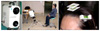
Fig. 3
Soft tissue landmarks. A, B, 3-D laser scanning images (texture mode); C, 3-D laser scanning image (vertex mode). N', Soft tissue nasion; Ex, exocanthion; En, endocanthion; Ala, nasal ala; Pn, pronasale; Sn, subnasale; ULP, upper lip point; Ls, labrale superius; Ls Rt, labrale superius right; Ls Lt, labrale superius left; Ch, cheilion; Stm, stomion; Li, labrale inferius; Li Rt, labrale inferius right; Li Lt, labrale inferius left; B', soft tissue B point; Pog', soft tissue pogonion; Tra, tragus; CK, cheek point.
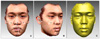
Fig. 4
Coordinate system. X-axis, Left (+) and right (-); Y-axis, superior (+) and inferior (-); Z-axis, anterior (+) and posterior (-).
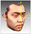
Fig. 5
A, Markings on the forehead and the 5 reference points; B, registered 3-D images with 1 coordinate system. N', Soft tissue nasion; Ex, exocanthionl; En, endocanthion.
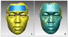
Fig. 6
Shell-to-shell deviation. A, Absolute color (absolute value of the deviation); B, signed color (anterior movement [+], posterior movement [-]).

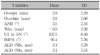
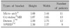
Table 3
Changes in the coordinate values (X, Y, and Z) and distance of landmarks after bracket bonding (mm)
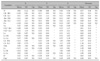
Table 4
Comparison of distance according to type of bracket (%)
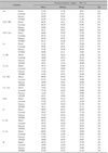
Sig, Significance; NS, not significant. The abbreviation is the same as Fig 3.
References
1. Kim KS, Kim YJ, Lee KH, Kim YH, Kook YA. Level of perception of changed lip protrusion and asymmetry of the lower facial height. Korean J Orthod. 2006. 36:434–441.
2. Lee JS, Choy K, Park YC, Kim KH. Changes in lip and perioral soft tissue after bracket removal. Korean J Orthod. 2007. 37:125–136.
3. Arnett GW, Jelic JS, Kim J, Cummings DR, Beress A, Worley CM Jr, et al. Soft tissue cephalometric analysis: diagnosis and treatment planning of dentofacial deformity. Am J Orthod Dentofacial Orthop. 1999. 116:239–253.
4. Baumrind S. Integrated three-dimensional craniofacial mapping: background, principles, and perspectives. Semin Orthod. 2001. 7:223–232.

5. Brown T, Abbott AH. Computer-assisted location of reference points in three dimensions for radiographic cephalometry. Am J Orthod Dentofacial Orthop. 1989. 95:490–498.
6. Arridge S, Moss JP, Linney AD, James DR. Three dimensional digitization of the face and skull. J Maxillofac Surg. 1985. 13:136–143.
7. Kanazawa E, Kamiishi H. Evaluation of facial osteotomy with the aid of Moiré contourography. J Maxillofac Surg. 1978. 6:233–238.
8. Ayoub A, Garrahy A, Hood C, White J, Bock M, Siebert JP, et al. Validation of a vision-based, three-dimensional facial imaging system. Cleft Palate Craniofac J. 2003. 40:523–529.
9. Han SY, Baik HS, Kim KD, Yu HS. Facial soft tissue measuring analysis of normal occlusion using three-dimensional CT imaging. Korean J Orthod. 2005. 35:409–419.
10. Park SH, Yu HS, Kim KD, Lee KJ, Baik HS. A proposal for a new analysis of craniofacial morphology by 3-dimensional computed tomography. Am J Orthod Dentofacial Orthop. 2006. 129:600.e23–600.e34.
11. Moss JP, Linney AD, Grindrod SR, Arridge SR, Clifton JS. Three-dimensional visualization of the face and skull using computerized tomography and laser scanning techniques. Eur J Orthod. 1987. 9:247–253.
12. Kusnoto B, Evans CA. Reliability of a 3D surface laser scanner for orthodontic applications. Am J Orthod Dentofacial Orthop. 2002. 122:342–348.
13. Baik HS, Lee HJ, Lee KJ. A proposal for soft tissue landmarks for craniofacial analysis using 3-dimensional laser scan imaging. World J Orthod. 2006. 7:7–14.
14. Baik HS, Jeon JM, Lee HJ. Facial soft-tissue analysis of Korean adults with normal occlusion using a 3-dimensional laser scanner. Am J Orthod Dentofacial Orthop. 2007. 131:759–766.
15. Baik HS, Kim SY. Facial soft-tissue changes in skeletal Class III orthognathic surgery patients analyzed with 3-dimensional laser scanning. Am J Orthod Dentofacial Orthop. 2010. 138:167–178.
16. Kau CH, Zhurov A, Richmond S, Bibb R, Sugar A, Knox J, et al. The 3-dimensional construction of the average 11-year-old child face: a clinical evaluation and application. J Oral Maxillofac Surg. 2006. 64:1086–1092.
17. Oliver BM. The influence of lip thickness and strain on upper lip response to incisor retraction. Am J Orthod. 1982. 82:141–149.
18. Erbay EF, Caniklioğlu CM, Erbay SK. Soft tissue profile in Anatolian Turkish adults: Part I. Evaluation of horizontal lip position using different soft tissue analyses. Am J Orthod Dentofacial Orthop. 2002. 121:57–64.
19. Burstone CJ. Lip posture and its significance in treatment planning. Am J Orthod. 1967. 53:262–284.
20. Hillesund E, Fjeld D, Zachrisson BU. Reliability of soft-tissue profile in cephalometrics. Am J Orthod. 1978. 74:537–550.
21. Day CJ, Robert T. Three-dimensional assessment of the facial soft tissue changes that occur postoperatively in orthognathic patients. World J Orthod. 2006. 7:15–26.
22. Soncul M, Bamber MA. Evaluation of facial soft tissue changes with optical surface scan after surgical correction of Class III deformities. J Oral Maxillofac Surg. 2004. 62:1331–1340.
23. McCance AM, Moss JP, Fright WR, Linney AD, James DR. Three-dimensional analysis techniques--Part 2: Laser scanning: a quantitative three-dimensional soft-tissue analysis using a color-coding system. Cleft Palate Craniofac J. 1997. 34:46–51.
 XML Download