

 PDF
PDF ePub
ePub Citation
 Print
Print


Abstract
Objective
The purpose of this study was to analyze the stress distribution and the displacement pattern of mandibular anterior teeth under various intrusive force vectors according to the position of orthodontic miniscrews and hooks, using three-dimensional finite element analysis.
Methods
A three-dimensional finite element model was constructed to simulate mandibular teeth, periodontal ligament, and alveolar bone. The displacement of individual tooth on three-dimensional planes and the von Mises stress distribution were compared when various intrusion force vectors were applied.
Results
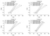
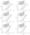
Intrusive forces applied to 4 mandibular anterior teeth largely resulted in remarkable labial tipping of the segment according to the miniscrew position. All 6 mandibular anterior teeth were labially tipped and the stress concentrated on the labiogingival area by intrusive force from miniscrews placed mesial to the canine. The distointrusive force vector led to pure intrusion and the stress was evenly distributed in the whole periodontal ligament when the hook was placed between the central and lateral incisors and the miniscrew was placed distal to the canine.
Figures and Tables
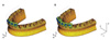 | Fig. 1Three dimensional finite element models and coordination system. A, Mandibular 4 anterior teeth model; B, mandibular 6 anterior teeth model. X, bucco-lingual (+) lingual, (-) buccal direction; Y, anterio-posterior (+) anterior, (-) posterior direction; Z, superio-inferior (+) superior, (-) inferior direction.
|
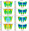 | Fig. 4Von Mises stress distribution in periodontal ligament of 4 anterior teeth by intrusion force (g/mm2) - Red color means high stress distribution and blue color means low stress distribution.
|
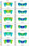 | Fig. 6Von Mises stress distribution in periodontal ligament of 6 anterior teeth by intrusion force (g/mm2) - Red color means high stress distribution and blue color means low stress distribution.
|
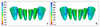 | Fig. 8Von Mises stress distribution in the periodontal ligament of Condition 2E before A and after B the application of large deflection theory. Red color indicates area of high stress and blue color, area of low stress. The difference is negligible.
|
References
1. Dermaut LR, Vanden Bulcke MM. Evaluation of intrusive mechanics of the type "segmented arch" on a macerated human skull using the laser reflection technique and holographic interferometry. Am J Orthod. 1986. 89:251–263.

2. Burstone CR. Deep overbite correction by intrusion. Am J Orthod. 1977. 72:1–22.
3. Woods MG. The mechanics of lower incisor intrusion: experiments in nongrowing baboons. Am J Orthod Dentofacial Orthop. 1988. 93:186–195.
4. Clifford PM, Orr JF, Burden DJ. The effects of increasing the reverse curve of Spee in a lower archwire examined using a dynamic photo-elastic gelatine model. Eur J Orthod. 1999. 21:213–222.
5. Dake ML, Sinclair PM. A comparison of the Ricketts and Tweed-type arch leveling techniques. Am J Orthod Dentofacial Orthop. 1989. 95:72–78.
6. Greig DG. Bioprogressive therapy: overbite reduction with the lower utility arch. Br J Orthod. 1983. 10:214–216.
7. Kanomi R. Mini-implant for orthodontic anchorage. J Clin Orthod. 1997. 31:763–767.
8. Melsen B, Verna C. Miniscrew implants: the Aarhus anchorage system. Semin Orthod. 2005. 11:24–31.
9. Park YC, Lee KJ. Nanda R, editor. Biomechanical principles in miniscrew-driven orthodontic. Temporary anchorage devices in orthodontics. 2009. St Louis: Mosby Elsevier;93–144.
10. Yared KF, Zenobio EG, Pacheco W. Periodontal status of mandibular central incisors after orthodontic proclination in adults. Am J Orthod Dentofacial Orthop. 2006. 130:6.e1–6.e8.
11. Rudolph DJ, Willes PMG, Sameshima GT. A finite element model of apical force distribution from orthodontic tooth movement. Angle Orthod. 2001. 71:127–131.
12. Tanne K, Sakuda M, Burstone CJ. Three-dimensional finite element analysis for stress in the periodontal tissue by orthodontic forces. Am J Orthod Dentofacial Orthop. 1987. 92:499–505.
13. Reimann S, Keilig L, Jäger A, Bourauel C. Biomechanical finite-element investigation of the position of the centre of resistance of the upper incisors. Eur J Orthod. 2007. 29:219–224.
14. Andrews LF. The six keys to normal occlusion. Am J Orthod. 1972. 62:296–309.
15. Yang SD. Evaluation and establishment of occlusal plane in clinical orthodontics. J Korean Found Gnatho-Orthod Res. 2001. 5:5–38.
16. Choi B, Baek SH, Yang WS, Kim S. Assessment of the relationships among posture, maxillomandibular denture complex, and soft-tissue profile of aesthetic adult Korean women. J Craniofac Surg. 2000. 11:586–594.
17. Arnett GW, Jelic JS, Kim J, Cummings DR, Beress A, Worley CM Jr, et al. Soft tissue cephalometric analysis: diagnosis and treatment planning of dentofacial deformity. Am J Orthod Dentofacial Orthop. 1999. 116:239–253.
18. Coolidge ED. The thickness of the human periodontal membrane. J Am Dent Assoc. 1937. 24:1260–1267.
19. Jeong GM, Sung SJ, Lee KJ, Chun YS, Mo SS. Finite-element investigation of the center of resistance of the maxillary dentition. Korean J Orthod. 2009. 39:83–94.
20. Sung SJ, Baik HS, Moon YS, Yu HS, Cho YS. A comparative evaluation of different compensating curves in the lingual and labial techniques using 3D FEM. Am J Orthod Dentofacial Orthop. 2003. 123:441–450.
21. Andrews LF. The straight-wire appliance, explained and compared. J Clin Orthod. 1976. 10:174–195.
22. Lee SK, Lim WH, Chun YS. Quantitative evaluation of cortical bone and soft tissue thickness in the mandible. Korean J Orthod. 2007. 37:212–219.
23. Park YC. New development in the segmented arch technique. J Korean Dent Assoc. 1986. 24:499–505.
24. Shroff B, Yoon WM, Lindauer SJ, Burstone CJ. Simultaneous intrusion and retraction using a three-piece base arch. Angle Orthod. 1997. 67:455–461.
25. Kim DW, Yang HC, Kim GT, Kim SS, Son WS. The effect of labial inclination on intrusion of the upper and lower incisors by three-dimensional finite element analysis. Korean J Orthod. 2003. 33:259–277.
26. Nauert K, Berg R. Evaluation of labio-lingual bony support of lower incisors in orthodontically untreated adults with the help of computed tomography. J Orofac Orthop. 1999. 60:321–334.
27. Wainwright WM. Faciolingual tooth movement: its influence on the root and cortical plate. Am J Orthod. 1973. 64:278–302.
28. Hixon EH, Aasen TO, Clark RA, Klosterman R, Miller SS, Odom WM. On force and tooth movement. Am J Orthod. 1970. 57:476–478.
29. Cobo J, Sicilia A, Argüelles J, Suárez D, Vijande M. Initial stress induced in periodontal tissue with diverse degrees of bone loss by an orthodontic force: tridimensional analysis by means of the finite element method. Am J Orthod Dentofacial Orthop. 1993. 104:448–454.
30. Matsui S, Caputo AA, Chaconas SJ, Kiyomura H. Center of resistance of anterior arch segment. Am J Orthod Dentofacial Orthop. 2000. 118:171–178.
31. Pedersen E, Isidor F, Gjessing P, Andersen K. Location of centres of resistance for maxillary anterior teeth measured on human autopsy material. Eur J Orthod. 1991. 13:452–458.
32. Vanden Bulcke MM, Burstone CJ, Sachdeva RC, Dermaut LR. Location of the centers of resistance for anterior teeth during retraction using the laser reflection technique. Am J Orthod Dentofacial Orthop. 1987. 91:375–384.
33. Burstone CJ. Biomechanics of deep overbite correction. Semin Orthod. 2001. 7:26–33.
34. Martinelli FL, Luiz RR, Faria M, Nojima LI. Anatomic variability in alveolar sites for skeletal anchorage. Am J Orthod Dentofacial Orthop. 2010. 138:252.e1–252.e9.
35. Choy K, Pae EK, Park Y, Kim KH, Burstone CJ. Effect of root and bone morphology on the stress distribution in the periodontal ligament. Am J Orthod Dentofacial Orthop. 2000. 117:98–105.
36. Tanne K, Nagataki T, Inoue Y, Sakuda M, Burstone CJ. Patterns of initial tooth displacements associated with various root lengths and alveolar bone heights. Am J Orthod Dentofacial Orthop. 1991. 100:66–71.
37. Koenig HA, Burstone CJ. Force systems from an ideal arch--large deflection considerations. Angle Orthod. 1989. 59:11–16.
38. Lee KJ, Cho YS, Kim SP, Lee KH. Prediction of the bending effect of an archwire for orthodontics. Journal of the Korean Society of Manufacturing Process Engineers. 2010. 9:95–100.
 XML Download