

 PDF
PDF ePub
ePub Citation
 Print
Print


Abstract
Objective
The purpose of this study was to investigate the prevalence of dental anomalies in outpatient clinics.
Methods
The subjects of this study were 3,133 patients who visited the clinic between January 2009 and June 2011. The dental records and panoramic films of the patients and detection of supernumerary, missing, and impacted teeth, transposition, and peg lateralis were reviewed. The results were analyzed according to gender and types and locations of dental anomalies.
Results
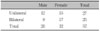
Among the patients, 362 had dental anomalies, with a prevalence rate of 11.55%. Congenital missing teeth (5.71%) ranked first in the categories, and impacted teeth (3.09%) ranked second. The percentage of patients having supernumerary teeth, peg lateralis, and dislocated teeth were 1.79%, 1.66%, and 0.45%, respectively.
Figures and Tables
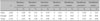
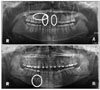 | Fig. 1Panoramic radiograph showing several dental anomalies. A, Impacted maxillary right canine, peg lateralis of both maxillary lateral incisors; B, impacted supernumerary tooth and impacted mandibular right first premolar.
|

References
1. Stafne EC, Gibilisco JA. Oral roentgenographic diagnosis. 1985. 5th ed. Philadelphia: WB Saunders;18–45.
2. Bhaskar SN. Orban's oral histology and embryology. 1976. 8th ed. St Louis: CV Mosby;23–205.
3. Schulze C. Gorlin RJ, Goldman HM, editors. Developmental abnormalities of the teeth and jaws. Thoma's oral pathology. 1970. Vol. I:6th ed. St Louis: CV Mosby;112–122.
4. Grahnen H. Hypodontia in the permanent dentition. A clinical and genetical investigation. Odont Revy. 1956. 7:Suppl 3. 1–100.
5. Silverman NE, Ackerman JL. Oligodontia: a study of its prevalence and variation in 4032 children. ASDC J Dent Child. 1979. 46:470–477.
6. Kotsomitis N, Dunne MP, Freer TJ. A genetic aetiology for some common dental anomalies: a pilot twin study. Aust Orthod J. 1996. 14:172–178.
7. Clayton JM. Congenital dental anomalies occurring in 3557 children. J Dent Child. 1956. 23:206–208.
8. Buenviaje TM, Rapp R. Dental anomalies in children: a clinical and radiographic survey. ASDC J Dent Child. 1984. 51:42–46.
9. Cha MH, Kim JT, Woo WS. Study on incidence of supernumerary and congenital missing teeth by Orthopantomography. J Korean Acad Pediatr Dent. 1975. 2:53–56.
10. Lee JM, Lee SR. A clinical and radiographic study of congenitally missing teeth. J Korean Acad Oral Maxillofac Radiol. 1991. 21:275–285.
11. Lee MS, Lee JG. The statistical study of prevalence of dental anomalies. J Korean Acad Pediatr Dent. 1985. 12:175–190.
12. Goaz PW, White SC. Oral radiology. 1987. 2nd ed. St Louis: CV Mosby;421–427.
13. Craig CE. Abnormalities in number and in the eruption path of teeth. Dent Clin North Am. 1968. 435–447.
14. Muller TP, Hill IN, Peterson AC, Blayney JR. A survey of congenitally missing permanent teeth. J Am Dent Assoc. 1970. 81:101–107.

15. Rosenzweig KA, Garbarski D. Numerical aberrations in the permanent teeth of grade school children in Jerusalem. Am J Phys Anthropol. 1965. 23:277–283.
16. Hunstadbraten K. Hypodontia in the permanent dentition. ASDC J Dent Child. 1973. 40:115–117.
17. Endo T, Ozoe R, Kubota M, Akiyama M, Shimooka S. A survey of hypodontia in Japanese orthodontic patients. Am J Orthod Dentofacial Orthop. 2006. 129:29–35.
18. Niswander JD, Sujaku C. Congenital anomalies of teeth in Japanese children. Am J Phys Anthropol. 1963. 21:569–574.
19. Lee JH, Shon HK, Jeon SJ, Choi HJ. A study on prevalence and pattern of dental anomalies. J Korean Acad Pediatr Dent. 1996. 23:429–449.
20. Ranta R, Tulensalo T. Symmetry and combination of hypodontia in non-cleft and cleft palate children. Scand J Dent Res. 1988. 96:1–8.
21. Glenn FB. A consecutive six-year study of the prevalence of congenitally missing teeth in private pedodontic practice of two geographically separated areas. J Dent Child. 1964. 31:264–270.
22. Primosch RE. Anterior supernumerary teeth--assessment and surgical intervention in children. Pediatr Dent. 1981. 3:204–215.
23. Ruhlman DC, Neely AR. Multiple impacted and erupted supernumerary teeth. Report of a case. Oral Surg Oral Med Oral Pathol. 1964. 17:199–203.
24. Stafne EC. Supernumerary teeth. Dental Cosmos. 1932. 74:653–659.
25. Kim YL, Hwang EH, Lee SR. A radiographic study of mesiodenses occurred in the maxillary central incisor region. J Korean Acad Oral Maxillofac Radiol. 1991. 21:367–375.
26. Jang YD, Hwang EH, Lee SR. A Roentgenocephalometric study of supernumerary teeth. J Korean Acad Oral Maxillofac Radiol. 1991. 21:393–403.
27. Rubenstein LK, Lindauer SJ, Isaacson RJ, Germane N. Development of supernumerary premolars in an orthodontic population. Oral Surg Oral Med Oral Pathol. 1991. 71:392–395.
28. Yang S, Kim JD. A study of dental anomalies. J Korean Acad Oral Maxillofac Radiol. 1993. 23:303–314.
29. Bolk L. Supernumerary teeth in the molar region in mandible. Dent Cosmos. 1914. 56:154–167.
30. Thilander B, Myrberg N. The prevalence of malocclusion in Swedish school children. Scand J Dent Res. 1973. 81:12–21.
31. Turner C, Hill CJ. Supernumerary mandibular premolar: the importance of radiographic interpretation. ASDC J Dent Child. 1986. 53:375–377.
32. Proffit WR. Proffit WR, editor. The development of orthodontic problems. Contemporary orthodontics. 1997. 2nd ed. St Louis: Mosby;110.
33. Langland OE, Langlais RP, Morris CR. Principles and practice of panoramic radiology. 1982. Philadelphia: WB Saunders Co.;157–204.
34. Altug-Atac AT, Erdem D. Prevalence and distribution of dental anomalies in orthodontic patients. Am J Orthod Dentofacial Orthop. 2007. 131:510–514.
35. Kwon TH, Lee JG, Sohn HK, Kim HT. The surgical exposure and application of direct traction of impacted maxillary canine: a case report. J Korean Acad Pediatr Dent. 1993. 20:407–414.
36. Uslu O, Akcam MO, Evirgen S, Cebeci I. Prevalence of dental anomalies in various malocclusions. Am J Orthod Dentofacial Orthop. 2009. 135:328–335.
37. Grover PS, Lorton L. The incidence of unerupted permanent teeth and related clinical cases. Oral Surg Oral Med Oral Pathol. 1985. 59:420–425.
38. Peck L, Peck S, Attia Y. Maxillary canine-first premolar transposition, associated dental anomalies and genetic basis. Angle Orthod. 1993. 63:99–109.
39. Mader C, Konzelman JL. Transposition of teeth. J Am Dent Assoc. 1979. 98:412–413.
40. Shapira Y, Kuftinec MM. Maxillary tooth transpositions: Characteristic features and accompanying dental anomalies. Am J Orthod Dentofacial Orthop. 2001. 119:127–134.
41. Sandham A, Harvie H. Ectopic eruption of the maxillary canine resulting in transposition with adjacent teeth. Tandlaegebladet. 1985. 89:9–11.
 XML Download