

 PDF
PDF ePub
ePub Citation
 Print
Print


INTRODUCTION
Understanding the relationship between the dental and basal arch forms is of diagnostic and therapeutic importance because the expansion of the dental arch is limited. Periodontal complications and increased risk of relapse may be expected if the teeth are moved beyond the apical base limit, especially in the mandibular arch.1-8 Lundström2 defined the apical base as the limit of the expansion of the dental arch that remains unaffected by orthodontic tooth movement or masticatory function. Previous reports have stressed the importance of the coordination between the upper and lower arches and claimed that the absence of this coordination may lead to functional and esthetic problems.9-12
Few studies have been performed on the shape of the basal arch,13-16 whereas several studies have examined the characteristics of the dental arch form in different ethnic groups.17-23 Some previous studies were aimed at finding mathematical relations to describe the dental arch.24-28 Most of these studies have analyzed the dental arch by employing two-dimensional (2D) methods.17-31 The reference points used for the measurements in these studies were incisal edges and cusp tips17,19,24-28 or indirect clinical bracket points derived from the contact points.18,20,23 However, these landmarks do not represent the clinical archwire forms. Recent studies have evaluated the arch dimensions on the basis of facial axis (FA) points.30,31
The dental arch has been previously evaluated in a three-dimensional (3D) study.32 Analysis using this method seems to be clinically relevant, especially for the application of preformed superelastic archwires, because digitizing the FA points in a 2D manner is not reliable. Kook et al.30 assessed the horizontal relationship between the upper and lower dentition with respect to the FA points in a 3D study. They found that the anterior and posterior overjets were homogeneous. In recent times, several studies14-16 have evaluated the relationship between the dental and basal arches on the basis of the WALA ridge (named after Will Andrews and Larry Andrews), which connects the most convex points on the mucogingival junction.13 However, none of these studies analyzed the maxillary arch, and therefore, the horizontal relationship between the upper and lower dentition has not yet been assessed. Further, the differences among the arch forms in terms of the basal arch dimensions has not yet been evaluated.
We hypothesized that the overjet and difference in the widths of the maxillomandibular basal arches have a significant positive correlation. The purposes of this study were to evaluate the relationship between the dental and basal arches; to analyze their differences in the tapered, ovoid, and square arch forms of the upper and lower dentitions in normal occlusion, by using 3D virtual models; and to test the postulated hypothesis.
MATERIAL AND METHODS
The samples comprised 77 maxillary and mandibular plaster casts obtained from subjects with normal occlusion. The ages of the subjects ranged from 20.4 to 25 years, with the mean being 23.2 years. The subjects meeting the following criteria were included:
Angle's Class I molar and canine relationships
0° < ANB angle < 4°
Normal overbite and overjet (> 0 mm, < 4 mm)
Minor arch length discrepancy (< 3 mm of crowding, < 1 mm of spacing)
Flat or slight curve of Spee (< 2 mm)
Absence of deviations in the dental midline and crossbite
Permanent dentition with normal tooth size and shape, except in the case of the third molars
The exclusion criteria included the presence of gingival defects or unidentifiable mucogingival junction on the model; history of previous orthodontic treatment; and restorations extending to contact areas, cusp tips, incisal edge, or facial surface.
The maxillary and mandibular casts were placed in the occluded relationship and scanned with an Orapix KOD-300 3D laser scanner (Orapix Co., Ltd, Seoul, Korea), at a resolution of 20µm.
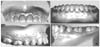
The FA point33 and WALA point13 were digitized for each tooth from the right first molar to the left first molar on each virtual model by using the software Rapidform 2006 (INUS technology, Inc., Seoul, Korea) to represent the dental and basal arches, respectively (Fig 1). The WALA point was measured directly below the FA of each tooth perpendicular to the occlusal plane. The occlusogingival position of this point varied from tooth to tooth. Digitization of all points was performed by an investigator (M.B.) with considerable experience in 3D technology.
The transverse direction was represented by the X-axis; the antero-posterior direction, the Y-axis; and the line perpendicular to the X and Y planes, the Z-axis. The FA point on the upper right second molar was set as the origin of the X and Y axes, and the Z-axis values were nullified.
Four linear and 2 ratio variables were measured and calculated for each arch form (Table 1).
The amounts of overjet at the anterior and posterior segments in each arch form were measured as follows:
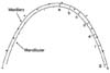
The X and Y coordinates for the FA points of each case were inputted into a mathematical software (MATLAB® 7.5 (R2007b), The MathWorks Inc., Natick, MA, USA) to generate the best fitting curve that represent the arch, by using the fourth degree polynomial equation (f(x) = ax4 + bx3 + cx2 + dx + e).27,28,34,35 The amount of overjet was measured as the shortest distance from each FA point on the mandibular arch to the maxillary one (Fig 2). To evaluate the intraexaminer reliability, 10 randomly selected scans were measured 2 weeks later. Intraclass correlation (ICC) test revealed high reliability between the 2 assessments (ICC > 0.8).
The best fitting curve representing each dental mandibular arch was matched to the arch form templates (Orthoform™, 3M Unitek, Monrovia, CA, USA), and the samples were classified into the following 3 groups according to the arch form: tapered (n = 20), ovoid (n = 20), and square (n = 37).32
Statistical analysis
Since analysis using the Chi square test did not reveal any significant association between gender and arch form, the data of the male and female subjects were combined before further analysis. Analysis of variance (ANOVA) was used for comparing the dental and basal arch dimensions independently among the 3 arch form types.
Independent sample t-test was performed to evaluate the difference in the overjet between the right and the left sides, and since there were no statistically significant differences, the data of both sides were combined. The Pearson correlation coefficients between the intercanine and the intermolar widths at the FA and WALA points were calculated. Pearson correlation coefficients were also calculated between at the amount of overjet at canine and the difference in intercanine width between maxillary and mandibular basal arches, and between the amount of overjet at the first molar and the difference in intermolar width between maxillary and mandibular basal arches. These analyses were performed to evaluate the relationship between the dental arch and the basal arch.
RESULTS
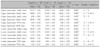
The 3 arch forms differed significantly with respect to all the dimensions of the dental arches, except the intermolar depths. For both the upper and lower arches, the square arch form had a larger intercanine width than the tapered ones and a larger intermolar width than both the tapered and ovoid arch forms (Table 2).
Unlike the case with the dental arch dimensions, the 3 arch forms differed significantly only in some dimensions of the basal arch. The upper and lower intermolar widths in the square arch forms were wider than those in the tapered and the ovoid arch forms. In the mandibular arch, the tapered arch form showed a larger intermolar depth (25.86 mm) than the ovoid (23.83 mm) and a smaller intermolar width/depth ratio (2.19) than both the ovoid (2.43) and square (2.44) arch forms (p = 0.001 and 0.0002, respectively) (Table 3).
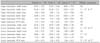
With regard to the horizontal relationship among the 3 arch forms, the tapered arch form had significantly larger overjet than the ovoid one from the canine to the second molar areas. It also had a greater overjet than the square one at the canine and premolar areas. Further, the 3 arch forms did not vary significantly with respect to the differences between the intercanine and intermolar widths of the maxillary and mandibular basal arches (Tables 4 and 5).
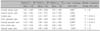
With regard to the relationship between the dental and the basal arches, strong correlations were found in the upper and lower arches with respect to the intermolar width (r = 0.83 and 0.85, respectively) and moderate correlations, with respect to the intercanine widths (r = 0.65 and 0.48, respectively). Nevertheless, in all the arch forms, no significant correlation was found between the amount of overjet at the canine area and the difference in intercanine width between maxillary and mandibular basal arches. Similarly, no significant correlation was found between the amount of overjet at first molar areas and the difference in intermolar width between maxillary and mandibular basal arches (p > 0.05).
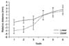
The average of the distances between the corresponding WALA and FA points, considering their relative position to each other, were similar in both the maxilla and mandible (Table 6). The WALA points were buccal to the FA points in the posterior area, but lingual, in the anterior. Further, the distance between the 2 points decreased in the canine direction, in both arches. However, the distances in the anteriors of the maxilla were larger than those of the mandible (Fig 3).
DISCUSSION
The purpose of this study was to evaluate the relationship between the dental and basal arches in an attempt to detemine the limit of expansion of the dental arch for stable treatment results.
Several methods have been used to evaluate the basal arch width from dental casts.36-39 Fujita et al.36 measured the basal arch between the innermost points at the buccal folds, while Hesby et al.39 measured it between points located 9.0 mm apical to the most occlusal point of the buccal developmental groove. Andrews and Andrews13 defined the WALA ridge as the ridge of the tissue at the mucogingival junction that represents the level of the basal bone. They suggested that it is similar to the archwire form of the dental arch. Identifying the WALA ridge is easier than estimating the root apex. In a recent study, Ronay et al.16 measured the basal arch width using the WALA points to evaluate the relationship between the dental and basal arch forms in Class I samples, while Ball et al.14 assessed it in Class I and Class II Division 1 malocclusions.
Virtual models have been confirmed to be accurate and reliable, and the measurements obtained using them have been shown to be reproducible; therefore, these models are considered a feasible alternative to plaster models.40-42 In this study, the FA points were digitized on 3D virtual models because the models show a direct relationship to the clinical archwire shape. The WALA points were also digitized because they represent the basal arch form.
The amount of overjet may be influenced by several factors, such as torque, bucco-lingual offset, marginal ridge relationships, and occlusal functions such as incisal guidance, and working and nonworking side interference.43,44 In this study, the tapered arch form showed a significantly larger overjet than the 2 other arch forms. These findings highlight the importance of selecting the archwire form that matches the patient's pretreatment arch from in order to achieve proper coordination between upper and lower arches.
This study revealed significant differences between the arch forms in only some dimensions of the basal arch; these include the upper and lower intermolar width, the lower intermolar depth, and the lower intermolar W/D ratio. However, the 3 arch forms differed significantly in several dimensions of the dental arch. This may suggest that the arch shape variation is mainly dental, and not skeletal. The moderate correlation between the dental and basal intercanine width may support this suggestion.
In this study, a strong correlation between the basal and dental intermolar widths was found in both maxillary and mandibular arches. This finding is consistent with those reported by Ronay et al.16 and supports Andrews' WALA ridge theory,13 which suggests that the dental arch form is related to the basal arch form in each patient. Changing the form of the dental arch during treatment without consideration of the basal arch form may result in relapse or give rise to periodontal problems. The strong correlation is also reflected in the significance of the difference of the basal intermolar width among the 3 arch forms.
The relation between the basal and dental arches seemed to have the same trend in the upper and lower arches. Consistent with the findings of Ronay et al.,16 the distances between the corresponding WALA and FA points were negative in the anterior area and positive in the posterior area because the WALA points were lingual and buccal in relation to the FA, respectively. However, the distance was larger in the maxilla (3.97 mm for the central incisor) than in the mandible (1.31 mm) because the upper incisors have a more labial inclination than the lower (Fig 3).
Although the overjet differed significantly among the 3 arch forms, it did not show any significant correlation to the basal transverse relation between the upper and lower arches at the canine and molar areas in each arch form. Therefore, the overjet cannot be used as an indicator of the discrepancy in the basal transverse relation.
Fujita et al.36 evaluated the basal arch widths between the innermost points at the buccal folds as part of a classification algorithm. Later, Hesby et al.39 reported that the upper basal width was 59.80 mm, while the lower was 55.63 mm. These values were comparable only to those obtained for the tapered group in our study. This discrepancy may be attributed to the ethnic differences.
Further research using cone-beam computed tomography to evaluate the dental and basal arch form relationship is necessary to confirm these findings based on the 3D models.
CONCLUSION
Individuals with the tapered, ovoid, and square dental arch showed only minimal differences in the skeletal arch dimensions. Further, although strong correlations were found between the basal and dental intermolar widths, only moderate correlations were noted for intercanine widths. These findings suggest that the basal arch may not be the principle factor in determining the dental arch form, especially in the anterior area.


 XML Download