

 PDF
PDF ePub
ePub Citation
 Print
Print


Abstract
Objective
The purpose of this study was to confirm the reliability of a cone beam computed tomography (CBCT)-generated panoramic view based on a CBCT 3D image and to find the most helpful 2D panoramic image compared with CBCT 3D image when examining the mesiodistal tooth axis.
Methods
A test model was constructed according to cephalometric norms. The test model was repeatedly repositioned for CBCT and panoramic radiographic imaging. Panoramic radiographs were acquired at each of the following 3 occlusal plane positions: -5°, 0°, and +5°. Measurements of mesiodistal tooth axis in CBCT 3D image, CBCT-generated panoramic view, and panoramic radiographs were compared.
Results
Compared with the CBCT-generated panoramic view, CBCT 3D image showed significant difference in the mesiodistal tooth axis in the premolars and no significant difference in the mesiodistal tooth axis in the incisors and canines. Mesiodistal tooth axis on the CBCT-generated panoramic view was significantly different from that on panoramic radiographs.
Figures and Tables
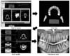
 | Fig. 3Angular measurement of tooth axis to reference line. A, CBCT 3D image; B, CBCT-generated panoramic view; C, panoramic radiograph. CBCT, Cone beam computed tomography.
|
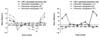 | Fig. 4Mean differences of tooth axis between CBCT 3D image and each panoramic image (CBCT-generated panoramic view and panoramic radiographs). CBCT, Cone beam computed tomography. Tooth number follows FDI system.
|
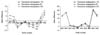 | Fig. 5Mean angular differences of the mesiodistal tooth axis between CBCT-generated panoramic view image and panoramic radiograph images. CBCT, Cone beam computed tomography.
|
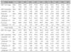
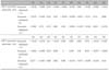
Table 1
Mesiodistal teeth axes measured on CBCT 3D image, CBCT generated panoramic view, and panoramic radiographs (unit: degree)
![]()
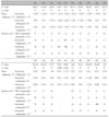
Table 2
Comparisons of mesiodistal tooth axis between CBCT 3D image and CBCT-generated panoramic view, and between CBCT 3D image and panoramic radiographs
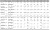
CBCT, Cone beam computed tomography; NS, no significance. Tooth number follows FDI system. Mean difference (°), differences of measured mesiodistal tooth axis between CBCT 3D image and CBCT-generated panoramic view, and between CBCT 3D image and panoramic radiographs. Negative (-) values of mean differences iindicate mesiodistal tooth axis on CBCT 3D image is smaller than that on other images. *p < 0.05.
![]()
References
1. Dewel BF. Clinical observations on the axial inclination of teeth. Am J Orthod. 1949. 35:98–115.

2. Mayoral G. Treatment results with light wires studied by panoramic radiography. Am J Orthod. 1982. 81:489–497.
3. Ursi WJ, Almeida RR, Tavano O, Henriques JF. Assessment of mesiodistal axial inclination through panoramic radiography. J Clin Orthod. 1990. 24:166–173.
4. Graber TM. Postmortems in posttreatment adjustment. Am J Orthod. 1966. 52:331–352.
5. Edwards JG. The prevention of relapse in extraction cases. Am J Orthod. 1971. 60:128–144.
6. Hatasaka HH. A radiographic study of roots in extraction sites. Angle Orthod. 1976. 46:64–68.
7. Keim RG, Gottlieb EL, Nelson AH, Vogels DS 3rd. 2002 JCO study of orthodontic diagnosis and treatment procedures. Part 3. More breakdowns of selected variables. J Clin Orthod. 2002. 36:690–699.
8. American Board of Orthodontics. Grading system for dental casts and panoramic radiographs [CD-ROM]. 2002. St Louis:
9. Xie Q, Soikkonen K, Wolf J, Mattila K, Gong M, Ainamo A. Effect of head positioning in panoramic radiography on vertical measurements: an in vitro study. Dentomaxillofac Radiol. 1996. 25:61–66.
10. Wyatt CC, Pharoah MJ. Imaging techniques and image interpretation for dental implant treatment. Int J Prosthodont. 1998. 11:442–452.
11. Lam EW, Ruprecht A, Yang J. Comparison of two-dimensional orthoradially reformatted computed tomography and panoramic radiography for dental implant treatment planning. J Prosthet Dent. 1995. 74:42–46.
12. Frederiksen NL. Diagnostic imaging in dental implantology. Oral Surg Oral Med Oral Pathol Oral Radiol Endod. 1995. 80:540–554.
13. Garcia-Figueroa MA, Raboud DW, Lam EW, Heo G, Major PW. Effect of buccolingual root angulation on the mesiodistal angulation shown on panoramic radiographs. Am J Orthod Dentofacial Orthop. 2008. 134:93–99.
14. Lucchesi MV, Wood RE, Nortjé CJ. Suitability of the panoramic radiograph for assessment of mesiodistal angulation of teeth in the buccal segments of the mandible. Am J Orthod Dentofacial Orthop. 1988. 94:303–310.
15. Mckee IW, Glover KE, Williamson PC, Lam EW, Heo G, Major PW. The effect of vertical and horizontal head positioning in panoramic radiography on mesiodistal tooth angulations. Angle Orthod. 2001. 71:442–451.
16. Jeon HS, Choi GL, Lim SH, Kim KW. Distortion of tooth axes on panoramic radiographs taken at various head positions. Korean J Orthod. 2008. 38:240–251.
17. Stramotas S, Geenty JP, Petocz P, Darendeliler MA. Accuracy of linear and angular measurements on panoramic radiographs taken at various positions in vitro. Eur J Orthod. 2002. 24:43–52.
18. Choi GL, Lim SH, Kim KW, Kim JD. Change in tooth length and angulation on panoramic radiographs taken at different labiolingual and buccolingual inclinations. Korean J Orthod. 2007. 37:114–124.
19. Lou L, Lagravere MO, Compton S, Major PW, Flores-Mir C. Accuracy of measurements and reliability of landmark identification with computed tomography (CT) techniques in the maxillofacial area: a systematic review. Oral Surg Oral Med Oral Pathol Oral Radiol Endod. 2007. 104:402–411.
20. Marmulla R, Wörtche R, Mühling J, Hassfeld S. Geometric accuracy of the NewTom 9000 Cone Beam CT. Dentomaxillofac Radiol. 2005. 34:28–31.
21. Peck JL, Sameshima GT, Miller A, Worth P, Hatcher DC. Mesiodistal root angulation using panoramic and cone beam CT. Angle Orthod. 2007. 77:206–213.
22. Van Elslande D, Heo G, Flores-Mir C, Carey J, Major PW. Accuracy of mesiodistal root angulation projected by conebeam computed tomographic panoramic-like images. Am J Orthod Dentofacial Orthop. 2010. 137:4 Suppl. S94–S99.
23. Mckee IW, Williamson PC, Lam EW, Heo G, Glover KE, Major PW. The accuracy of 4 panoramic units in the projection of mesiodistal tooth angulations. Am J Orthod Dentofacial Orthop. 2002. 121:166–175.
24. McNamara JA Jr. A method of cephalometric evaluation. Am J Orthod. 1984. 86:449–469.
25. Frykholm A, Malmgren O, Sämfors KA, Welander U. Angular measurements in orthopantomography. Dentomaxillofac Radiol. 1977. 6:77–81.
26. Philipp RG, Hurst RV. The cant of the occlusal plane and distortion in the panoramic radiograph. Angle Orthod. 1978. 48:317–323.
27. Baba R, Ueda K, Okabe M. Using a flat-panel detector in high resolution cone beam CT for dental imaging. Dentomaxillofac Radiol. 2004. 33:285–290.
28. Samawi SS, Burke PH. Angular distortion in the orthopantomogram. Br J Orthod. 1984. 11:100–107.
29. Mischkowski RA, Scherer P, Ritter L, Neugebauer J, Keeve E, Zöller JE. Diagnostic quality of multiplanar reformations obtained with a newly developed cone beam device for maxillofacial imaging. Dentomaxillofac Radiol. 2008. 37:1–9.
30. Scarfe WC, Nummikoski P, McDavid WD, Welander U, Tronje G. Radiographic interproximal angulations: implications for rotational panoramic radiography. Oral Surg Oral Med Oral Pathol. 1993. 76:664–672.
 XML Download