

 PDF
PDF ePub
ePub Citation
 Print
Print


INTRODUCTION
Several researchers have evaluated the relationship between dental occlusion and temporomandibular joint (TMJ) anatomy and health, and have emphasized the influence of malocclusion, vertical discrepancy, increased overjet, cross-bite, and tooth loss on TMJ functions and form.1-3 However, most of the previous research on jaw and joint movements was conducted in adults, who might have already developed degenerative changes in their TMJ anatomy due to existing malocclusions,4-6 given that the incidence of TMJ dysfunction or pathology caused by a malocclusion increases with age.7,8
TMJ function is generally assessed on the basis of morphometric data and functional analysis.9 Functional analysis can be performed by graphical methods, recording devices, interocclusal records, and electromyographic data.4,6,10,11 Axiographic devices can indicate mandibular movement capacity numerically and the borders and forms of axiographic tracings are helpful for diagnosing TMJ disorders.12
To discriminate between healthy and diseased TMJs and predict future TMJ dysfunction, physiologic differences in mandibular movement capacity and normative data among individuals with different occlusal relationships should be evaluated in childhood or adolescence, before they become affected by malocclusion. The aim of this study was to obtain normative data of malocclusion in adolescence and compare the opening and protrusive mandibular movements between Class II malocclusion, which is the most common malocclusion (incidence of 17.3%) requiring orthodontic treatment in Turkey,13 and Class I malocclusion in healthy adolescents by means of clinical and axiographic evaluations.
MATERIAL AND METHODS
Subjects
This research included 78 non-orthodontically treated volunteers aged 12 - 16 years. The inclusion criteria were a dentition minimally comprising 28 teeth and the absence of systemic or hormonal disorders, physical trauma, masticatory tenderness, psychologic problems, and dysfunctions or parafunctions that could cause TMJ dysfunction.
The Class I group comprised 38 adolescents (16 boys, 22 girls) with Class I molar and canine relationships, ideal overjet and overbite, mean chronologic age of 14 years 6 months (± 10 months), and mean skeletal age of 14 years 3 months (± 9 months). The Class II group comprised 40 adolescents (19 boys, 21 girls) with Class II molar and canine relationships, a minimal overjet of 5 mm, mean chronologic age of 13 years 7 months (± 9 months), and mean skeletal age of 13 years 9 months (± 8 months). The skeletal ages were estimated by hand-wrist radiographs.14 All subjects were informed about the experimental procedures and written consent was obtained from their parents. The Health Science Institute of Gazi University approved the research.
Clinical examination
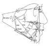
The same investigator performed all the clinical examinations. Overjet and overbite were measured with a boley gauge. A millimetric ruler (Staedtler, Nuernberg, Germany) was used to measure the maximum jaw opening by adding the overbite value to the distance between the upper and the lower incisal edges during mouth opening and the maximum protrusion by adding the overjet value to the distance between the incisal edges of the lower and upper incisors during mandibular protrusion. Detailed TMJ and masticatory-muscle examinations were conducted. The mandibular corpus and ramus lengths, mandibular plane inclination, gonial angle, and lower and upper incisal inclinations were evaluated from lateral cephalometric radiographs by the same investigator (Fig 1).
Axiographic examination
The right and left condylar paths were recorded during opening and protrusive mandibular movements with an axiographic recording device (AXO 516MK Axiograph III Kit; SAM Präzisionstechnik GmbH, Munich, Germany) by the same investigator. All condylar movements were recorded habitually, without any manipulation, in the upright sitting position.
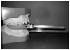
Paraocclusal clutches were prepared on lower dental casts and cemented to the labial and buccal surfaces of the mandibular teeth without covering their occlusal surfaces (Fig 2).
Upper and posterior frames, providing head support, were placed and a nasofrontal frame was adjusted at nasion. Vertical frames were immobilized at the tragus (Fig 3A). The infraorbital margin was considered the anterior reference point (infraorbital point). The lower recording frame was adjusted to the cemented clutch after its horizontal adjustment was calibrated to zero; later, the recording frame was attached to the clutch with two styli at the end bilaterally pointing to the condylar heads.
For the recordings, the subjects performed mandibular opening and closing movements up to 10 mm without any guidance, and the point at which the styli showed pure rotational movement was determined as the terminal hinge axis point (Fig 3B). From the center of the terminal hinge axis point, all movements were repeated four times without any guidance (Fig 3C), and the longest drawings were used for axiographic evaluation (Fig 4A). The terminal hinge axis and infraorbital points were plotted for both sides and the line joining these points was used as the reference plane. Linear (mm) and angular (°) values were measured on axiographic tracings by using a millimetric ruler. The linear values were calculated as the distance between the initial point and the finishing point of the tracing and the angular values were calculated as the acute angle between the line tangent to the initial curve and the reference plane (Fig 4B).
Statistical analysis
SPSS 10.0 for Windows software (IBM-SPSS Inc., Chicago, IL, USA) was used for the statistical analysis. All data are expressed as the mean ± standard deviation (SD). Intergroup and gender comparisons were evaluated by using the Student's t-test and intergroup gender comparisons were performed by using the chi-square test. Correlations of the skeletal and dental parameters with the axiographic measurements were examined by Pearson correlation analysis. p ≤ 0.05 was considered statistically significant.
To determine the method error, 50 randomly selected cephalograms from each group were retraced and axiographic records were recompleted 15 days after the first evaluation. The Bland-Altman method15 and intraclass correlation were utilized to evaluate the method error. All measurements declared sufficient reliability at the 95% level and agreement was excellent (ICC = 0.99).
RESULTS
No TMJ dysfunction symptoms and irregular axiographic tracings were detected in any of the subjects.
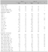
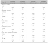
Table 1 shows the intergroup differences in the clinical, cephalometric, and axiographic data. In terms of opening mandibular movements, a significant difference was found in the maximum clinical mouth opening capacity between the Class I and the Class II groups (47.8 ± 4.7 mm vs. 45.4 ± 5.0 mm; p ≤ 0.05). The other initial opening axiographic tracings revealed no statistically significant difference between the groups. The opening condylar path inclinations were similar between the groups. On the other hand, significant differences in the protrusive mandibular movements were noted between the groups. The maximum clinical protrusion was 8.6 ± 1.5 mm in the Class I group and 11.2 ± 1.5 mm in the Class II group (p < 0.001). Further, the maximum protrusive length was 7.2 ± 1.4 mm in the Class I group and 8.0 ± 1.4 mm in the Class II group (p < 0.01). Again, there was no significant difference in the protrusive condylar path inclinations between the groups.
Table 2 presents the intragroup gender differences in the clinical, cephalometric, and axiographic measurements. The maximum clinical mouth opening capacity was significantly different between the boys and the girls in each group (Class 1 group: 50.4 ± 1.8 mm vs. 45.9 ± 5.2 mm, p < 0.01; Class II group: 47.5 ± 4.5 mm vs. 43.4 ± 4.8 mm, p ≤ 0.05). Further, the ramus and corpus lengths and effective mandibular length were significantly different between the genders in both groups.
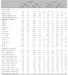
Tables 3 and 4 show correlations of the axiographic measurements with the skeletal and dental parameters in both the groups. In the Class I group, both the axiographic mouth opening angle (SNGoGn: r = -0.330, p = 0.041; CGoGn: r = -0.350, p = 0.029) and the axiographic protrusive angle (SNGoGn: r = -0.350, p = 0.028; CGoGn: r = -0.379, p = 0.019) were negatively correlated with the mandibular inclination. The mouth opening angle showed a positive correlation with the lower incisor angulation (IMPA: r = 0.460, p = 0.004; Table 3). None of the skeletal and dental parameters showed significant correlations with the clinical opening and protrusive measurements (data not shown).
In the Class II group, a negative correlation was noted between the axiographic protrusive angle and the mandibular inclination (SNGoGn: r = -0.442, p = 0.004; CGoGn: r = -0.360, p = 0.021). Further, the axiographic mouth opening length was negatively correlated with the lower incisor position (L1-NB: r = -0.460, p = 0.003) and positively correlated with the corpus length (GoGn: r = 0.440, p = 0.004). The axiographic protrusive length was negatively correlated with the lower incisor angulation (IMPA: r = -0.510, p = 0.001) and lower incisor position (L1-NB: r = -0.560, p = 0.001) and positively correlated with the overjet (r = 0.39, p = 0.004) and corpus length (GoGn: r = 0.330, p = 0.033; Table 4). In the clinical measurements, only the maximum clinical protrusion showed a positive correlation with the overjet (r = 0.44, p = 0.003; data not shown).
DISCUSSION
Solberg et al.16 investigated the relationships of the TMJ and facial morphology with occlusion by using skulls and found characteristic structural morphologies and greater anterior condylar displacement in skulls with Class II malocclusion. Further, the articular eminence morphology undergoes changes during growth and represents an adaptive capacity against dental functions.17 Although differences in the condylar and fossa morphology have been noted in skeletal Class I patterns, skeletal Class II patterns showed differences in the condylar position.18 These findings emphasize the importance of considering TMJ-related structures and functions in orthodontic treatment plans, especially for children who might be at risk of developing TMJ dysfunctions in the long term.8
The deciduous dentition has an unstable occlusal relation because of the non-sharp-edged tuberculum fossae and non-anatomically formed glenoid fossa and articular eminence, causing difficulty in repetition of centric occlusion.19 A large overjet with lack of incisor guidance can prevent maximum intercuspation in centric relation, causing less-than-optimal posterior interdigitation.11 For example, epidemiologic studies have demonstrated clinical symptoms of mandibular dysfunction and functional disorders in 35 - 80% of the Swedish children and adolescents.7,20 Considering that a subsisted dysfunction in childhood can lead to pathologic changes in the long term,7 it is essential to determine mandibular movement and understand functional evolution during the growth period.10
Adolescence, the period in which adaptive and degenerative changes are not developed, would be the most suitable time for determining the causative factors of TMJ disorders. In the present study, adolescents were examined to identify possible adaptive changes in the TMJ and obtain normative data for adolescence. Exclusion of the muscular, psychogenic, or parafunctional causes of TMJ dysfunction might be the reason for the lack of TMJ disorders and atypical axiographic tracings in this study.
In this study, the groups showed no significant difference in the axiographic opening and closing recordings but had significantly different protrusive movement ranges. Similarly, Zimmer et al.6 noted similar axiographic opening and closing angles between Angle's Class I and Angle's Class II occlusion in adults. In addition, studies to evaluate TMJ functions according to Angle's classification in adults showed significant differences in protrusive movements versus opening and closing movements.21 In this regard, protrusion should be considered as primarily caused by bodily translation of the condyles, whereas opening is a combined rotational-translational condylar movement.6 Therefore, the current results are mainly attributable to differences in the translational capacity of the condyles between the groups.
The finding that the protrusive capacity was significantly higher in the Class II group than in the Class I group was confirmed by the correlation between the overjet and the protrusive capacity. Similarly, Zimmer et al.6 found a greater amount of protrusion in Class II cases than in Class I and Class III cases. Further, Ingervall22 found correlations between the mandibular movement capacity and the sagittal skeletal relation in children. Therefore, the protrusive capacity in the Class II excessive overjet cases showed a wider range of motion than that in the Class I malocclusion cases with ideal overjet and overbite. Given that the present study was conducted in individuals with clinically healthy TMJs, the detected differences could be normal variations. However, excessive condylar movements in individuals with Class II malocclusion and without any orthodontic treatment should be evaluated by long-term studies to determine the risks of pathological TMJ adaptations.
During protrusive movement, the condyle translates along the contour of the tuberculum articulare. Therefore, the protrusive path is affected by any difference in the steepness of the posterior slope of the articular eminence. Theusner et al.5 reported average axiographic protrusive lengths of 10.20 and 10.60 mm on the right and left sides, respectively, in healthy adults; Gsellmann et al.23 reported values of 10.57 and 11.28 mm, respectively, in healthy adults. However, no Angle's classification was declared in these studies. A previous study revealed axiographic protrusive lengths of 7.89 - 7.96 mm on the right and 8.36 - 8.46 mm on the left in individuals with Angle's Class I malocclusion.4 Therefore, the present axiographic protrusive lengths in the adolescents with Class I and Class II malocclusions are less when compared with the results of the previous studies.5,6,23 This disparity may be attributable to the differences in sample selection, measurement techniques, and age-related developmental variations in the articular eminence among the studies. Moreover, several researchers investigated the changes in the sagittal condylar path inclination during mandibular protrusion among different ages and concluded that the condylar path tends to become steeper with age.24 Similarly, Reicheneder et al.25 declared that the condylar path inclination is significantly smaller in children than in adults. Further, the current differences in the left opening angle and protrusive length in girls with Class II malocclusion could be related to individual variations in the condylar paths.
In the present study, the opening mandibular movements were not typical enough to differentiate between the classes. In addition, the differences in the protrusive capacity between the groups cannot be considered pathologic signs, because all the subjects were clinically free of TMJ dysfunction. The correlation analysis revealed that the axiographic opening and protrusive movements were negatively correlated with the mandibular inclination in the Class I cases, which is contrary to the results of Zimmer et al.6 This difference may be associated with the differences in sample selection, physical variations, and/or morphological differences in the TMJ. Bolt and Orchardson26 found a negative correlation between the muscle activities and the gonial and mandibular plane angles, and declared that the mouth opening capacity is influenced by the muscle activities. In addition, the opening capacity and mandibular morphology were correlated, similar to the results of previous studies.27 Further, the maximum mouth opening capacity was significantly greater in boys than in girls in both the groups, as demonstrated in recent studies.27,28
The protrusive capacity in the adolescents with Class II malocclusion showed a positive correlation with the overjet and corpus length, and a negative correlation with the lower incisor position, consistent with the findings of Kubein-Meesenburg et al.29 Ogawa et al.30 emphasized that protrusive movements could be affected by the mandibular morphology and location of the dentition, but no significant correlation between the mandibular movements and the ANB angle was found in the present study. The protrusive capacity of the TMJ seems to be influenced more by sagittal dental factors; however, the role of other factors cannot be excluded.
CONCLUSION
This study showed no significant differences in opening mandibular movement between the Class I and the Class II groups, but the protrusive lengths varied significantly between the groups. Given the increased range of protrusive mobility in Class II malocclusion even in healthy, TMJ dysfunction-free adolescents, the current findings cannot be considered pathologic differences. However, the results indicate that the sagittal dental relationship can modify the protrusive capacity. Further research is needed to clarify the role of factors influencing the movement capacity in Class II cases between healthy and diseased TMJs. Moreover, long-term follow-up studies of individuals with Class II malocclusion and without any chance of receiving orthodontic treatment are needed to clarify the possible causes of TMJ disorders.


 XML Download