

 PDF
PDF ePub
ePub Citation
 Print
Print


Abstract
Objective
The purposes of this study were to evaluate the reproducibility and reliability of head posture obtained by registering outer canthus as a soft tissue landmark with the Outer Canthus Indicator (OCI).
Methods
Twenty-one adults with normal facial morphology were enrolled in this study (mean age 27.5 ± 1.72 years). To register initial head posture, height of the outer canthus from the ear rod plane was measured using OCI. Head posture was reproduced by moving the head upwards and downwards until the outer canthus was in a straight line with the indicator set at a registered height. After the head posture is reproduced by two operators after two days, lateral photographs were taken. Computerized photometric analyses of the photographs were performed.
Results
The head rotations around the transverse axis were 0.69 ± 0.43°, 0.98 ± 0.65° from each of the two operators. Standard errors were 0.09° and 0.14° each, which were similar to results from past research findings. There were no significant differences between the data from the two operators (p > 0.05). There were no correlations between the head rotation around the horizontal and vertical axes (p > 0.05).
Figures and Tables
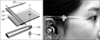 | Fig. 1Major components and assembly of the Outer Canthus Indicator (OCI). Cover (1), indicator (2), ruler (3), base (4), right side arm of the cephalostat (5), and ear rod (6) (from top to bottom in the order of assembly, broken arrow) are indicated. Within the cover and the base (1, 4), the metal ruler and the indicator (2, 3) unit can move up and down (line arrow).
|
 | Fig. 2Procedures for registering and reproducing head posture by the OCI method. To register initial head posture, move the indicator up and down (A-1) until it indicates the outer canthus (A-2). Individual height of the outer canthus from the ear rod plane (OCI value) can be registered by reading the scale on the ruler (A-3). To reproduce initial head posture, adjust the indicator to the individual OCI value (B-1). Then rotate subject's head vertically (B-2) until the outer canthus can be aligned with the indicator (B-3).
|
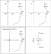 | Fig. 3Landmarks and measurements on the superimposed tracings. True vertical and true horizontal lines crossing at the ear rod point were drawn on each of the photographs taken on T0, T1. Then the tracing papers were superimposed on G, STN, NT, Sn. The angle between the true vertical lines and the distance between the ear rod points were measured on V-ceph 3.5 program (Cybermed, Korea). G, Glabella; STN, soft tissue nasion; NT, nose tip; Sn, subasale; Er, ear rod point; Tv, true vertical line; Th, true horizontal line; Dh, horizontal deviation of Er; Dv, vertical deviation of Er; Ro, angle between Tv0 and Tv1.
|
 | Fig. 4Horizontal and vertical deviations of the ear rod point (same abbreviations as Fig 3; unit, mm).
|
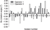
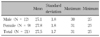
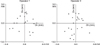
Table 2
Descriptive statistics of the variables
Same abbreviations as Fig 3; SD, standard deviation.
![]()
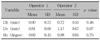
Table 3
Standard errors, maximum and minimum values of the variables

Same abbreviations as Fig 3; SE, standard error; Max, maximum; Min, minimum.
![]()
Table 4
Pearson's correlation coefficients and p values for the horizontal and vertical deviations of ear rod point
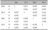
Same abbreviations as Fig 3; R, Pearson's correlation coefficient.
![]()
References
1. Broadbent BH. A new x-ray technique and its application to orthodontia. Angle Orthod. 1931. 1:45–66.
2. Lim SH, Cho JY, Choi GL, Kim KW. Comparison of analysis of the lateral cephalogram and analysis of lateral facial photograph. Korean J Orthod. 2006. 36:74–83.
3. Gottlieb EL, Brazones MM, Malerman AJ, Moskowitz EM, Phipps GS, Sarver DM. Early orthodontic treatment, part 1. J Clin Orthod. 2004. 38:79–90.
4. Sun MK, Uhm GS, Cho JH, Hwang HS. Use of Head Posture Aligner to improve accuracy of frontal cephalograms generated from cone-beam CT scans. Korean J Orthod. 2009. 39:289–299.

5. Jin KH, Hong SJ. The prediction of postsurgical soft tissue profile changes associated with surgical correction of the prognathic mandible by standardized facial photosurgery. Korean J Orthod. 1992. 22:855–868.
6. Hwang HS, Lee JJ, Hwang CH, Choi HH, Lim HJ. Prediction of frontal soft tissue changes after mandibular surgery in facial asymmetry individuals. Korean J Orthod. 2008. 38:252–264.
7. Ackerman JL, Proffit WR. Communication in orthodontic treatment planning: bioethical and informed consent issues. Angle Orthod. 1995. 65:253–261.
8. Takahashi I, Takahashi T, Hamada M, Kawamoto T, Kinoshita Z, Kubo Y, et al. Application of video surgery to orthodontic diagnosis. Int J Adult Orthodon Orthognath Surg. 1989. 4:219–222.
9. Sarver DM, Johnston MW. Video imaging: techniques for superimposition of cephalometric radiography and profile images. Int J Adult Orthodon Orthognath Surg. 1990. 5:241–248.
10. Kazandjian S, Sameshima GT, Champlin T, Sinclair PM. Accuracy of video imaging for predicting the soft tissue profile after mandibular set back surgery. Am J Orthod Dentofacial Orthop. 1999. 115:382–389.
11. Ahlqvist J, Eliasson S, Welander U. The cephalometric projection. Part II. Principles of image distortion in cephalography. Dentomaxillofac Radiol. 1983. 12:101–108.
12. Ahlqvist J, Eliasson S, Welander U. The effect of projection errors on cephalometric length measurements. Eur J Orthod. 1986. 8:141–148.
13. Ahlqvist J, Eliasson S, Welander U. The effect of projection errors on angular measurements in cephalometry. Eur J Orthod. 1988. 10:353–361.
14. Kim KS, Hwang MS, Choi EH, Kim KW, Yoon YJ. Changes of lateral cephalometric values according to the rotation of head. Korean J Orthod. 2000. 30:53–66.
15. Koh EH, Lee KH, Hwang HS. Effects of vertical head rotation on the posteroanterior cephalometric measurements. Korean J Orthod. 2003. 33:73–84.
16. Quintero JC, Trosien A, Hatcher D, Kapila S. Craniofacial imaging in orthodontics: Historical perspective, current status, and future developments. Angle Orthod. 1999. 69:491–506.
17. Lundström F, Lundström A. Natural head position as a basis for cephalometric analysis. Am J Orthod Dentofacial Orthop. 1992. 101:244–247.
18. Peng L, Cooke MS. Fifteen-year reproducibility of natural head posture: A longitudinal study. Am J Orthod Dentofacial Orthop. 1999. 116:82–85.
19. Luyk NH, Whitfield PH, Ward-Booth RP, Williams ED. The reproducibility of the natural head position in lateral cephalometric radiographs. Br J Oral Maxillofac Surg. 1986. 24:357–366.
20. Foster TD, Howat AP, Naish PJ. Variation in cephalometric reference lines. Br J Orthod. 1981. 8:183–187.
21. Solow B, Tallgren A. Head posture and craniofacial morphology. Am J Phys Anthropol. 1976. 44:417–435.
22. Halazonetis D. Estimated natural head position and facial morphology. Am J Orthod Dentofacial Orthop. 2002. 121:364–368.
23. Gu G, Nagata J, Suto M, Anraku Y, Nakamura K, Kuroe K, et al. Hyoid position, pharyngeal airway and head posture in relation to relapse after the mandibular setback in skeletal Class III. Clin Orthod Res. 2000. 3:67–77.
24. Phillips C, Snow MD, Turvey TA, Proffit WR. The effect of orthognathic surgery on head posture. Eur J Orthod. 1991. 13:397–403.
25. Showfety KJ, Vig PS, Matteson S. A simple method for taking natural-head-position cephalograms. Am J Orthod. 1983. 83:495–500.
26. Kim EH, Hwang HS. The validity of head posture aligner in posteroanterior cephalometry. Korean J Orthod. 2000. 30:543–552.
27. Raju NS, Prasad KG, Jayade VP. A modified approach for obtaining cephalograms in the natural head position. J Orthod. 2001. 28:25–28.
28. Chen CM, Lai S, Tseng YC, Lee KT. Simple technique to achieve a natural head position for cephalography. Br J Oral Maxillofac Surg. 2008. 46:677–678.
29. Stefanova N, Stella JP. The predictability of inferior medial canthus as a stable external vertical reference point in maxillary repositioning surgery. Int J Adult Orthodon Orthognath Surg. 2000. 15:305–308.
30. Cooke MS, Wei SH. An improved method for the assessment of the sagittal skeletal pattern and its correlation to previous methods. Eur J Orthod. 1988. 10:122–127.
31. Park EJ, Suhr CH. Study of craniocervical posture and craniofacial morphology in Korean young adults. Korean J Orthod. 1995. 25:129–142.
32. Bister D, Edler RJ, Tom BD, Prevost AT. Natural head posture - considerations of reproducibility. Eur J Orthod. 2002. 24:457–470.
33. Park SH, Yu HS, Kim KD, Lee KJ, Baik HS. A proposal for a new analysis of craniofacial morphology by 3-dimensional computed tomography. Am J Orthod Dentofacial Orthop. 2006. 129:600.e23–600.e34.
 XML Download