

 PDF
PDF ePub
ePub Citation
 Print
Print


Abstract
Objective
The purpose of this study was to assess the changes in height and inclination of the articular eminence during the growth period.
Methods
One hundred and sixty subjects (71 males and 89 females) with a normal skeletal pattern and TMJ function, ranging in age from 5.9 to 19.7 years were divided according to their chronological age into six groups. Lateral individualized corrected TMJ tomograms were taken of all subjects, and the height and inclination of the articular eminence were measured. UNIANOVA was used to compare the differences between the age groups. Mann-Whitney test was used to compare the differences between male and female subjects.
Figures and Tables
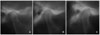 | Fig. 1Lateral individualized corrected TMJ tomogram. A, 7.9 year-old male; B, 12.6 year-old male; C, 18.7 year-old male.
|
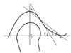 | Fig. 2Measurement of height and inclination of eminence on tomogram. A, Superior point of the fossa; B, inferior point of the articular eminence; Line BC is parallel to the Frankfort horizontal plane; Line AC is perpendicular to the Frankfort horizontal plane and represents the height of the articular eminence; α, the angle between line AB and line BC; β, the angle between the best fit line of the posterior slope of the articular eminence and line BC.
|
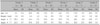
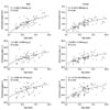 | Fig. 3Scattergram and regression curve for the height and inclination of the articular eminence in male and female subjects. The height and the inclination of the articular eminence was significantly increased with aging (p < 0.001).
|
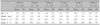
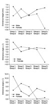 | Fig. 4Growth velocity of the height and inclination of the articular eminence in male and female subjects.
|

References
1. Katsavrias EG, Dibbets JM. The growth of articular eminence height during craniofacial growth period. Cranio. 2001. 19:13–20.

2. Katsavrias EG. Changes in articular eminence inclination during the craniofacial growth period. Angle Orthod. 2002. 72:258–264.
3. Nickel JC, McLachlan KR, Smith DM. Eminence development of the postnatal human temporomandibular joint. J Dent Res. 1988. 67:896–902.
4. Dibbets JM, Dijkman GE. The postnatal development of the temporal part of the human temporomandibular joint. A quantitative study on skulls. Ann Anat. 1997. 179:569–572.
5. Baqaien MA, Al-Salti FM, Muessig D. Changes in condylar path inclination during maximum protrusion between the ages of 6 and 12 years. J Oral Rehabil. 2007. 34:27–33.
6. Ogawa T, Koyano K, Suetsugu T. The influence of anterior guidance and condylar guidance on mandibular protrusive movement. J Oral Rehabil. 1997. 24:303–309.
7. Mimura H, Deguchi T. Morphologic adaptation of temporomandibular joint after chincup therapy. Am J Orthod Dentofacial Orthop. 1996. 110:541–546.
8. Katsavrias EG. The effect of mandibular protrusive (activator) appliances on articular eminence morphology. Angle Orthod. 2003. 73:647–653.
9. Birkebaek L, Melsen B, Terp S. A laminagraphic study of the alterations in the temporo-mandibular joint following activator treatment. Eur J Orthod. 1984. 6:257–266.
10. Eckerdal O, Lundberg M. The structural situation in temporomandibular joints. A comparison between conventional oblique transcranial radiographs, tomograms and histologic sections. Dentomaxillofac Radiol. 1979. 8:42–49.
11. Han JW. Relation of the measuring values in cephalometric radiographs and TMJ tomographs. Korean J Oral Maxillofac Radiol. 2007. 37:19–26.
12. Cohlmia JT, Ghosh J, Sinha PK, Nanda RS, Currier GF. Tomographic assessment of temporomandibular joints in patients with malocclusion. Angle Orthod. 1996. 66:27–35.
13. Hinton RJ. Changes in articular eminence morphology with dental function. Am J Phys Anthropol. 1981. 54:439–455.
14. Ren YF, Isberg A, Westesson PL. Steepness of the articular eminence in the temporomandibular joint. Tomographic comparison between asymptomatic volunteers with normal disk position and patients with disk displacement. Oral Surg Oral Med Oral Pathol Oral Radiol Endod. 1995. 80:258–266.
15. Bland JM, Altman DG. Statistical methods for assessing agreement between two methods of clinical measurement. Int J Nurs Stud. 2010. 47:931–936.
16. Park JG, Kim JC. A study on the angle of articular eminence and the inclination of anterior tooth related to facial types. Korean J Orthod. 1992. 22:869–880.
17. Granados JI. The influence of the loss of teeth and attrition on the articular eminence. J Prosthet Dent. 1979. 42:78–85.
18. Slavicek R. The masticatory organ: functions and dysfunctions. 2002. Klosterneuburg [Austria]: Gamma Med.-wiss. Fortbildungs-AG;136–217.
19. Ingervall B. Relation between height of the articular tubercle of the temporomandibular joint and facial morphology. Angle Orthod. 1974. 44:15–24.
20. Ikai A, Sugisaki M, Young-Sung K, Tanabe H. Morphologic study of the mandibular fossa and the eminence of the temporomandibular joint in relation to the facial structures. Am J Orthod Dentofacial Orthop. 1997. 112:634–638.
21. Akahane Y, Deguchi T, Hunt NP. Morphology of the temporomandibular joint in skeletal class iii symmetrical and asymmetrical cases: a study by cephalometric laminography. J Orthod. 2001. 28:119–128.
22. Katsavrias EG, Halazonetis DJ. Condyle and fossa shape in Class II and Class III skeletal patterns: a morphometric tomographic study. Am J Orthod Dentofacial Orthop. 2005. 128:337–346.
23. Cha BK, Kim CH, Baek SH. Skeletal sagittal and vertical facial types and electromyographic activity of the masticatory muscle. Angle Orthod. 2007. 77:463–470.
24. O'Ryan F, Epker BN. Temporomandibular joint function and morphology: observations on the spectra of normalcy. Oral Surg Oral Med Oral Pathol. 1984. 58:272–279.
25. Sülün T, Cemgil T, Duc JM, Rammelsberg P, Jäger L, Gernet W. Morphology of the mandibular fossa and inclination of the articular eminence in patients with internal derangement and in symptom-free volunteers. Oral Surg Oral Med Oral Pathol Oral Radiol Endod. 2001. 92:98–107.
26. Sato S, Kawamura H, Motegi K, Takahashi K. Morphology of the mandibular fossa and the articular eminence in temporomandibular joints with anterior disk displacement. Int J Oral Maxillofac Surg. 1996. 25:236–238.
27. Atkinson WB, Bates RE Jr. The effects of the angle of the articular eminence on anterior disk displacement. J Prosthet Dent. 1983. 49:554–555.
28. Kurita H, Ohtsuka A, Kobayashi H, Kurashina K. Flattening of the articular eminence correlates with progressive internal derangement of the temporomandibular joint. Dentomaxillofac Radiol. 2000. 29:277–279.
 XML Download