Journal List > Korean J Orthod > v.40(6) > 1043645

Kang, Kim, Park, and Kook: Soft tissue changes associated with ASO/BSSRO and Le Fort I/BSSRO in skeletal Class III malocclusion with upper lip protrusion

Abstract

Objective

The objective of this study was to compare maxillary soft tissue changes and their relative ratios to hard tissue changes after anterior segmental osteotomy (ASO)/bilateral sagittal split ramus osteotomy (BSSRO) and Le Fort I/BSSRO in skeletal Class III malocclusion with upper lip protrusion.

Methods

The study sample comprised the ASO/BSSRO group (n = 14) and the Le Fort I/BSSRO group (n = 15). The Le Fort I/BSSRO group included cases of maxillary posterior impaction only. Lateral cephalograms were taken 2 months before and 6 months after surgery. Linear and angular measurements were performed.

Results

The anterior maxilla moved backward in both groups after surgery, however the amount of change was significantly larger in the ASO/BSSRO group (p < 0.01). The ratios of hard to soft tissue change were 79% (SLS to A point), 80% (LS to A point) in the ASO/BSSRO group, and 15% (SLS to A point), 68% (LS to A point) in the Le fort I/BSSRO group. In addition, there was a 3.23° increase of the occlusal plane in the Le Fort I/BSSRO group.

Conclusions

When two-jaw surgery is indicated in skeletal Class III patients with protrusive lips, ASO may be a treatment of choice for cases with more severe upper lip protrusion, while Le Fort I with posterior impaction may be considered if an increase of occlusal plane angle is required.

Figures and Tables

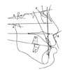
Fig. 1
Reference lines & cephalometric landmarks. 1, Sella (S): the center of the sella turcica; 2, Nasion (N): the most anterior point of the frontonasal suture; 3, Porion11 (Po): the most superior point of contour of external auditory meatus; 4, Orbitale (Or): the lowest point of the lower margin of the bony orbit; 5, Anterior nasal spine (ANS): the tip of the anterior nasal spine of the palatal bone in the hard palate; 6, Posterior nasal spine (PNS): the tip of the posterior nasal spine of the palatal bone in the hard palate; 7, Subspinale (A point): the most posterior point on the anterior contour of the upper alveolar process; 8, Supradentale (SD): the lowermost point on the alveolar portion of the premaxilla; 9, Upper incisor edge (UIE): the incisal edge of the upper central incisor; 10, Supramentale (B point): the most posterior point on the anterior contour of the lower alveolar process; 11, Pronasale (Pn): the most anterior projection of the nose; 12, Columella12 (Cm): the most anterior soft tissue point on the columella of the nose; 13, Subnasale11 (Sn): the point at which the columella merges with the upper cutaneous lip; 14, Superior labial sulcus (SLS): the point of greatest concavity in the midline of the upper lip; 15, Labrale superius (LS): the point indicating the mucocutaneous border of the upper lip; 16, Stomion superius (Stms): the lowermost point on the vermillion of the upper lip.
kjod-40-383-g001
Fig. 2
Horizontal and vertical linear measurements. HRL, Horizontal reference line; VRL, vertical reference line; A, 1, ANS to VRL; 2, PNS to VRL; 3, A point to VRL; 4, supradentale to VRL; 5, upper incisor edge to VRL; 6, pronasale to VRL; 7, columella to VRL; 8, subnasale to VRL; 9, superior labial sulcus to VRL; 10, labrale superius to VRL; B, 1, ANS to HRL; 2, PNS to HRL; 3, A point to HRL; 4, supradentale to HRL; 5, upper incisor edge to HRL; 6, pronasale to HRL; 7, columella to HRL; 8, subnasale to HRL; 9, superior labial sulcus to HRL; 10, labrale superius to HRL.
kjod-40-383-g002
Fig. 3
Angular measurements. 1, SNA; 2, SNB; 3, ANB; 4, FH-occlusal plane; 5, SN-palatal plane; 6, nasolabial angle (columella-subnasale-labrale superius); 7, U1-SN.
kjod-40-383-g003
Fig. 4
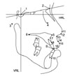
Upper lip length and thickness. HRL, Horizontal reference line; VRL, vertical reference line; A, 1, Subnasale to stomion superius; B, 1, subnasale to subnasale horizontal; 2, superior labial sulcus to superior labial sulcus horizontal; 3, labrale superius to labrale superius horizontal.
kjod-40-383-g004
Table 1
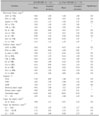
Number, gender, and mean age of sample in anterior segmental osteotomy/bilateral sagittal split ramus osteotomy group (ASO/BSSRO) and Le Fort I osteotomy/bilateral sagittal split ramus osteotomy group (Le Fort I/BSSRO)
kjod-40-383-i001
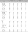
Table 2
Presurgical measurements in anterior segmental osteotomy/bilateral sagittal split ramus osteotomy group (ASO/BSSRO) and Le Fort I osteotomy/bilateral sagittal split ramus osteotomy group (Le Fort I/BSSRO)
kjod-40-383-i002

NS, Not significant; VRL, vertical reference line; HRL, horizontal reference line; SD, supradentale; UIE, upper incisor edge; Pn, pronasale; Cm, columella; Sn, subnasale; SLS, superior labial sulcus; LS, labrale superius; Stms; stomion superius; SnH, subnasale horizontal; SLSH, superior labial sulcus horizontal; LSH, labrale superius horizontal.

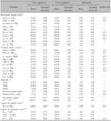
Table 3
Skeletal and soft tissue changes in anterior segmental osteotomy/bilateral sagittal split ramus osteotomy group (n = 14)
kjod-40-383-i003

NS, Not significant; VRL, vertical reference line; HRL, horizontal reference line; SD, supradentale; UIE, upper incisor edge; Pn, pronasale; Cm, columella; Sn, subnasale; SLS, superior labial sulcus; LS, labrale superius; Stms; stomion superius; SnH, subnasale horizontal; SLSH, superior labial sulcus horizontal; LSH, labrale superius horizontal. *p < 0.05; p < 0.01; p < 0.001. Negative values indicate posterior or superior movement.

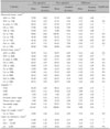
Table 4
Skeletal and soft tissue changes in Le Fort I osteotomy/bilateral sagittal split ramus osteotomy group (n = 15)
kjod-40-383-i004

NS, Not significant; VRL, vertical reference line; HRL, horizontal reference line; SD, supradentale; UIE, upper incisor edge; Pn, pronasale; Cm, columella; Sn, subnasale; SLS, superior labial sulcus; LS, labrale superius; Stms; stomion superius; SnH, subnasale horizontal; SLSH, superior labial sulcus horizontal; LSH, labrale superius horizontal. *p < 0.05; p < 0.01; p < 0.001. Negative values indicate posterior or superior movement.

Table 5
Comparative postsurgical changes in anterior segmental osteotomy/bilateral sagittal split ramus osteotomy group (ASO/BSSRO) and LeFort I osteotomy/bilateral sagittal split ramus osteotomy group (Le Fort I/BSSRO)
kjod-40-383-i005

NS, Not significant; VRL, vertical reference line; HRL, horizontal reference line; SD, supradentale; UIE, upper incisor edge; Pn, pronasale; Cm, columella; Sn, subnasale; SLS, superior labial sulcus; LS, labrale superius; Stms; stomion superius; SnH, subnasale horizontal; SLSH, superior labial sulcus horizontal; LSH, labrale superius horizontal. *p < 0.05; p < 0.01; p < 0.001. Negative values indicate posterior or superior movement.

Table 6
Ratio of horizontal change of soft tissue to hard tissue in anterior segmental osteotomy/bilateral sagittal split ramus osteotomy group (ASO/BSSRO)
kjod-40-383-i006

Pn, Pronasale; VRL, vertical reference line; Sn, subnasale; SLS, superior labial sulcus; LS, labrale superius; A, A point; SD, supradentale; UIE, upper incisor edge. *p < 0.01; p < 0.001.

Table 7
Ratio of horizontal change of soft tissue to hard tissue in LeFort I osteotomy/bilateral sagittal split ramus osteotomy group (Le Fort I/BSSRO)
kjod-40-383-i007

Pn, Pronasale; VRL, vertical reference line; Sn, subnasale; SLS, superior labial sulcus; LS, labrale superius; A, A point; SD, supradentale; UIE, upper incisor edge. *p < 0.01; p < 0.001.

Table 8
Ratio of change of measurements to amount of posterior impaction in LeFort I osteotomy/bilateral sagittal split ramus osteotomy group (Le Fort I/BSSRO)
kjod-40-383-i008

UIE, Upper incisor edge; VRL, vertical reference line; LS, labrale superius; HRL, horizontal reference line. *p < 0.05; p < 0.001. Amount of posterior impaction: ΔPNS to HRL - ΔANS to HRL.

References

1. Cho EJ, Yang WS. Soft tissue changes after double jaw surgery in skeletal Class III malocclusion. Korean J Orthod. 1996. 26:1–16.
2. Choe YK, Suhr CH. Hard and soft tissue changes after orthognathic surgery of mandibular prognathism. Korean J Orthod. 1993. 23:707–724.
3. Rim JS, Choi CM. A cephalometric study on the changes of soft tissue profile (upper lip & nose) following two-jaw surgery. J Korean Assoc Maxillofac Plast Reconstr Surg. 2003. 25:233–237.
4. Han UA, Kim JH, Yoon TH, Park JU, Kook YA. Hard and soft tissue profile changes following anterior subapical osteotomy in bimaxillary dentoalveolar protrusion patients. Korean J Orthod. 2003. 33:475–483.
5. Reyneke JP, Evans WG. Surgical manipulation of the occlusal plane. Int J Adult Orthodon Orthognath Surg. 1990. 5:99–110.
6. Arnett GW, Jelic JS, Kim J, Cummings DR, Beress A, Worley CM Jr, et al. Soft tissue cephalometric analysis: diagnosis and treatment planning of dentofacial deformity. Am J Orthod Dentofacial Orthop. 1999. 116:239–253.
7. Kang SG, Lee YJ, Park YG. A comparative study of soft tissue profile between Korean and Caucasian young adults under NHP. Korean J Orthod. 2003. 33:323–337.
8. Burstone CJ, James RB, Legan H, Murphy GA, Norton LA. Cephalometrics for orthognathic surgery. J Oral Surg. 1978. 36:269–277.
9. Moore JW. Variation of the sella-nasion plane and its effect on SNA and SNB. J Oral Surg. 1976. 34:24–26.
10. Stella JP, Streater MR, Epker BN, Sinn DP. Predictability of upper lip soft tissue changes with maxillary advancement. J Oral Maxillofac Surg. 1989. 47:697–703.
crossref
11. Posnick JC, Fantuzzo JJ, Orchin JD. Deliberate operative rotation of the maxillo-mandibular complex to alter the A-point to B-point relationship for enhanced facial esthetics. J Oral Maxillofac Surg. 2006. 64:1687–1695.
crossref
12. Bell WH, Dann JJ 3rd. Correction of dentofacial deformities by surgery in the anterior part of the jaws. A study of stability and soft-tissue changes. Am J Orthod. 1973. 64:162–187.
crossref
13. Dann JJ 3rd, Fonseca RJ, Bell WH. Soft tissue changes associated with total maxillary advancement: a preliminary study. J Oral Surg. 1976. 34:19–23.
14. Radney LJ, Jacobs JD. Soft-tissue changes associated with surgical total maxillary intrusion. Am J Orthod. 1981. 80:191–212.
crossref
15. Schendel SA, Eisenfeld JH, Bell WH, Epker BN. Superior repositioning of the maxilla: stability and soft tissue osseous relations. Am J Orthod. 1976. 70:663–674.
crossref
16. Chang IH, Lee YJ, Park YG. A comparative study of soft tissue changes with mandibular one jaw surgery and double jaw surgery in Class III malocclusion. Korean J Orthod. 2006. 36:63–73.
17. Enacar A, Taner T, Toroğlu S. Analysis of soft tissue profile changes associated with mandibular setback and double-jaw surgeries. Int J Adult Orthodon Orthognath Surg. 1999. 14:27–35.
18. Jensen AC, Sinclair PM, Wolford LM. Soft tissue changes associated with double jaw surgery. Am J Orthod Dentofacial Orthop. 1992. 101:266–275.
crossref
19. Mansour S, Burstone C, Legan H. An evaluation of soft-tissue changes resulting from Le Fort I maxillary surgery. Am J Orthod. 1983. 84:37–47.
crossref
20. Cha KS, Chung KR, Park JU. Park JU, editor. Diagnosis. Orthognathic surgery. 2003. Seoul: Koon Ja Publishing Inc.;3–43.
21. Kim SY, Kim SG, Lee SH, Kim SH, Chung TY, Ahn TH. Anterior segmental maxillary osteotomy using Cupar's method: preliminary study. J Korean Assoc Maxillofac Plast Reconstr Surg. 2001. 23:422–427.
22. Lew KK, Loh FC, Yeo JF, Loh HS. Profile changes following anterior subapical osteotomy in Chinese adults with bimaxillary protrusion. Int J Adult Orthodon Orthognath Surg. 1989. 4:189–196.
23. Hershey HG, Smith LH. Soft-tissue profile change associated with surgical correction of the prognathic mandible. Am J Orthod. 1974. 65:483–502.
crossref
24. Kajikawa Y. Changes in soft tissue profile after surgical correction of skeletal Class III malocclusion. J Oral Surg. 1979. 37:167–174.
25. Scheideman GB, Legan HL, Bell WH. Soft tissue changes with combined mandibular setback and advancement genioplasty. J Oral Surg. 1981. 39:505–509.
26. Suckiel JM, Kohn MW. Soft-tissue changes related to the surgical management of mandibular prognathism. Am J Orthod. 1978. 73:676–680.
crossref
27. Carlotti AE Jr, Aschaffenburg PH, Schendel SA. Facial changes associated with surgical advancement of the lip and maxilla. J Oral Maxillofac Surg. 1986. 44:593–596.
crossref
28. Ayoub AF, Mostafa YA, el-Mofty S. Soft tissue response to anterior maxillary osteotomy. Int J Adult Orthodon Orthognath Surg. 1991. 6:183–190.
29. Cohn-Stock G. Die chirurgiche Immediatregulierung der Kiefer, speziell die chirurgishce Behandlung der Prognathie. Vjschr Zahnheilk Berlin. 1921. 37:320.
30. Wassmund M. Lehrbuch der Praktischen Chirurgie des Mundes und der Kiefer. 1935. Leipzig: Meusser.
31. Wunderer S. Die prognathie operativen mittels frontal gestiertem maxilla fragment. Ost Z Stomatol. 1962. 59:98–102.
32. Park JU, Baik SH. Classification of Angle Class III malocclusion and its treatment modalities. Int J Adult Orthodon Orthognath Surg. 2001. 16:19–29.
33. Kim YJ, Kim JH. A study on the characteristics of attractive profiles of Korean young women to orthodontists. Korean J Orthod. 2001. 31:479–487.
34. Kim YJ, Nahm DS. Soft tissue cephalometric analysis of aesthetic Korean female. Korean J Orthod. 2002. 32:383–393.
35. Hwang CJ, Lim SA. A study on the postoperative stability of occlusal plane in orthognathic surgery patients depending on the difference of occlusal plane. Korean J Orthod. 1998. 28:237–253.
36. Lew KK. Orthodontic considerations in the treatment of bimaxillary protrusion with anterior subapical osteotomy. Int J Adult Orthodon Orthognath Surg. 1991. 6:113–122.
37. Jeong MH, Choi JH, Kim BH, Kim SG, Nahm DS. Soft tissue changes after double jaw rotation surgery in skeletal Class III malocclusion. J Korean Assoc Oral Maxillofac Surg. 2006. 32:559–565.
TOOLS
Similar articles