

 PDF
PDF ePub
ePub Citation
 Print
Print


Abstract
Objective
This study was designed to assess the diagnostic validity of digital panoramic radiographs compared to cone beam computed tomography (CBCT) in patients with temporomandibular joint disorders.
Methods
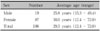
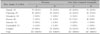
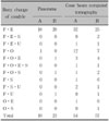
Panoramic radiograph and CBCT were taken from a total of 212 joints from 106 subjects. The joints were examined by two dentists and divided into the following six groups: normal, flattening, osteophyte formation, erosion, sclerosis, and unclassified. The sensitivity and specificity of each observer and inter-observer reliability were statistically analyzed.
Results
The results showed relatively high intra-observer reliability in the diagnosis of both panoramic and CBCT images and the weighted Kappa indices of panoramic and CBCT images were 0.714 and 0.727, respectively. The sensitivities of panoramic images of observer A and B to CBCT images was 82.35% and 84.30%, respectively, while the specificity of observer A and B was 58.06% and 61.54%, respectively. However, guided diagnosis from panoramic and CBCT images were statistically different (p < 0.05).
Conclusions
The present study suggests that the panoramic radiograph could be used as a primary diagnostic device to detect bony changes of temporomandibular joints in clinical orthodontics, because panoramic images showed relatively high sensitivity compared to CBCT images. However, CBCT images may be one of the best choices when a more accurate diagnosis is necessary.
Figures and Tables
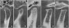 | Fig. 1Different type of condylar shapes in cone beam computed tomography sagittal imaging (A, Normal; B, flattening; C, sclerosis; D, osteophyte; E, erosion).
|


References
1. Graber TM, Vanasdall RL. . Orthodontics: current principles and techniques. 1994. St. Louis: Mosby;41–42.
2. Okeson JP. Management of temporomandibular disorders and occlusion. 2008. St. Louis: Mosby;274–275. 284–285. 322–324.
3. Israel HA, Diamond BE, Saed-Nejad F, Ratcliffe A. Correlation between arthroscopic diagnosis of osteoarthritis and synovitis of the human temporomandibular joint and keratan sulfate levels in the synovial fluid. J Oral Maxillofac Surg. 1997. 55:210–217.

4. Stegenga B, de Bont LG, Boering G. Osteoarthrosis as the cause of craniomandibular pain and dysfunction: a unifying concept. J Oral Maxillofac Surg. 1989. 47:249–256.
5. Kim KA, Koh KJ. Radiographic study of bony changes of the mandibular condyle. Korean J Oral Maxillofac Radiol. 2000. 30:23–32.
6. Goaz PW, White SC. Oral radiology: principles and interpretation. 1994. St. Louis: Mosby;499.
7. An SY, An CH, Choi KS. Efficacy of panoramic radiography as a screening procedure in dental examination compared with clinical evaluation. Korean J Oral Maxillofac Radiol. 2007. 37:83–86.
8. Habets LL, Bezuur JN, Jimenez Lopez V, Hansson TL. The OPG: an aid in TMJ diagnostics. III. A comparison between lateral tomography and dental rotational panoramic radiography (Orthopantomography). J Oral Rehabil. 1989. 16:401–406.
9. Habets LL, Bezuur JN, Naeiji M, Hansson TL. The Orthopantomogram, an aid in diagnosis of temporomandibular joint problems. II. The vertical symmetry. J Oral Rehabil. 1988. 15:465–471.
10. Mongini F. The importance of radiography in the diagnosis of TMJ dysfunctions. A comparative evaluation of transcranial radiographs and serial tomography. J Prosthet Dent. 1981. 45:186–198.
11. Larheim TA, Johannessen S, Tveito L. Abnormalities of the temporomandibular joint in adults with rheumatic disease. A comparison of panoramic, transcranial and transpharyngeal radiography with tomography. Dentomaxillofac Radiol. 1988. 17:109–113.
12. Könönen M. Subjective symptoms from the stomatognathic system in patients with psoriatic arthritis. Acta Odontol Scand. 1986. 44:377–383.
13. Bush FM, Harrington WG, Harkins SW. Interexaminer comparison of bone scintigraphy and panoramic radiography of temporomandibular joints: correlation with signs and symptoms. J Prosthet Dent. 1992. 67:246–251.
14. Ruf S, Pancherz H. Is orthopantomography reliable for TMJ diagnosis? An experimental study on a dry skull. J Orofac Pain. 1995. 9:365–374.
15. Sun MK, Uhm GS, Cho JH, Hwang HS. Use of head posture aligner to improve accuracy of frontal cephalograms generated from cone-beam CT scans. Korean J Orthod. 2009. 39:289–299.
16. Park SB, Park JH, Jung YH, Jo BH, Kim YI. Correlation between menton deviation and dental compensation in facial asymmetry using cone-beam CT. Korean J Orthod. 2009. 39:300–309.
17. Lim MY, Lim SH. Comparison of model analysis measurements among plaster model, laser scan digital model, and cone beam CT image. Korean J Orthod. 2009. 39:6–17.
18. Kang JY, Lim SH, Kim KW. The reliability of the cephalogram generated from cone-beam CT. Korean J Orthod. 2007. 37:391–399.
19. Tsiklakis K, Syriopoulos K, Stamatakis HC. Radiographic examination of the temporomandibular joint using cone beam computed tomography. Dentomaxillofac Radiol. 2004. 33:196–201.
20. Honda K, Larheim TA, Maruhashi K, Matsumoto K, Iwai K. Osseous abnormalities of the mandibular condyle: diagnostic reliability of cone beam computed tomography compared with helical computed tomography based on an autopsy material. Dentomaxillofac Radiol. 2006. 35:152–157.
21. Liang X, Jacobs R, Hassan B, Li L, Pauwels R, Corpas L, et al. A comparative evaluation of Cone Beam Computed Tomography (CBCT) and Multi-Slice CT (MSCT) Part I. On subjective image quality. Eur J Radiol. 2010. 75:265–269.
22. Liang X, Lambrichts I, Sun Y, Denis K, Hassan B, Li L, et al. A comparative evaluation of Cone Beam Computed Tomography (CBCT) and Multi-Slice CT (MSCT). Part II: On 3D model accuracy. Eur J Radiol. 2010. 75:270–274.
23. Loubele M, Jacobs R, Maes F, Denis K, White S, Coudyzer W, et al. Image quality vs radiation dose of four cone beam computed tomography scanners. Dentomaxillofac Radiol. 2008. 37:309–318.
24. Jeon YM, Choi JH, Kim ST, Kwon JS, Ahn HJ. The validity of computed tomography in diagnosis of tempromandibular joint osteoarthritis. Korean J Oral Med. 2008. 33:195–204.
25. Japanese association of TMJ. Temporomandibular joint disorders. 2004. Seoul: DaehanNarae;8–11.
26. Stegenga B, de Bont LG, Boering G, van Willigen JD. Tissue responses to degenerative changes in the temporomandibular joint: a review. J Oral Maxillofac Surg. 1991. 49:1079–1088.
27. Axelsson S. Human and experimental osteoarthrosis of the temporomandibular joint. Morphological and biochemical studies. Swed Dent J Suppl. 1993. 92:1–45.
28. Musgrave MT, Westesson PL, Tallents RH, Manzione JV, Katzberg RW. Improved magnetic resonance imaging of the temporomandibular joint by oblique scanning planes. Oral Surg Oral Med Oral Pathol. 1991. 71:525–528.
29. Lindvall AM, Helkimo E, Hollender L, Carlsson GE. Radiographic examination of the temporomandibular joint. A comparison between radiographic findings and gross and microscopic morphologic observations. Dentomaxillofac Radiol. 1976. 5:24–32.
30. Akerman S, Kopp S, Rohlin M. Macroscopic and microscopic appearance of radiologic findings in temporomandibular joints from elderly individuals. An autopsy study. Int J Oral Maxillofac Surg. 1988. 17:58–63.
31. Cholitgul W, Petersson A, Rohlin M, Tanimoto K, Akerman S. Diagnostic outcome and observer performance in sagittal tomography of the temporomandibular joint. Dentomaxillofac Radiol. 1990. 19:1–6.
32. Hintze H, Wiese M, Wenzel A. Comparison of three radiographic methods for detection of morphological temporomandibular joint changes: panoramic, scanographic and tomographic examination. Dentomaxillofac Radiol. 2009. 38:134–140.
33. Nah KS. Reproducibility of panoramic radiography in patients. Korean J Oral Maxillofac Radiol. 2005. 35:115–119.
34. Crow HC, Parks E, Campbell JH, Stucki DS, Daggy J. The utility of panoramic radiography in temporomandibular joint assessment. Dentomaxillofac Radiol. 2005. 34:91–95.
 XML Download