

 PDF
PDF ePub
ePub Citation
 Print
Print


Abstract
Objective
The aim of this study was to evaluate the occlusal force and contact area and to find its associating factors in Koreans.
Methods
Occlusal force and contact area in maximum intercuspation were measured using the Dental Prescale® system in 651 subjects (15 with normal occlusion, 636 with various malocclusions divided into subgroups according to the skeletal pattern, Angle's molar relationship, age and gender).
Results
Occlusal
force of the normal occlusion group (744.5 ± 262.6 N) was significantly higher than those of the malocclusion group (439.0 ± 229.9 N, p < 0.05). Occlusal force was similar regardless of differences in ANB angle or Angle's molar classification, however the increase in vertical dimension significantly reduced occlusal force (p < 0.05).
Conclusions
Occlusal force was significantly lower in the malocclusion group compared to the normal occlusion group, and in females compared to males, but it was not affected by age, antero-posterior skeletal pattern or molar classification. Although a hyperdivergent facial pattern indicated lower occlusal force compared to a hypodivergent facial pattern, the differences in skeletal pattern were not the primary cause of its decrease, but a secondary result induced by the differences in occlusal contact area according to the facial pattern.
Figures and Tables
Fig. 1
Correlation between occlusal force and contact area (A, normal occlusion group; B, malocclusion group).
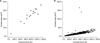

Table 2
Comparison of occlusal force and contact area between normal occlusion and malocclusion groups
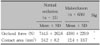
Table 3
Comparison of occlusal force and contact area according to gender (a) and different age groups (b)
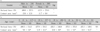
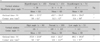
Table 4
Comparison of occlusal force and contact area among different antero-posterior relationships of the maxilla and the mandible


References
1. Throckmorton GS, Finn RA, Bell WH. Biomechanics of differences in lower facial height. Am J Orthod. 1980. 77:410–420.

2. Rios HF, Ma D, Xie Y, Giannobile WV, Bonewald LF, Conway SJ, et al. Periostin is essential for the integrity and function of the periodontal ligament during occlusal loading in mice. J Periodontol. 2008. 79:1480–1490.
3. Proffit WR, Fields HW, Nixon WL. Occlusal forces in normal-and long-faced adults. J Dent Res. 1983. 62:566–570.
4. Ringqvist M. Isometric bite force and its relation to dimensions of the facial skeleton. Acta Odontol Scand. 1973. 31:35–42.
5. Kurusu A, Horiuchi M, Soma K. Relationship between occlusal force and mandibular condyle morphology. Evaluated by limited cone-beam computed tomography. Angle Orthod. 2009. 79:1063–1069.
6. Terespolsky MS, Brin I, Harari D, Steigman S. The effect of functional occlusal forces on orthodontic tooth movement and tissue recovery in rats. Am J Orthod Dentofacial Orthop. 2002. 121:620–628.
7. Keles A, Tokmak EC, Erverdi N, Nanda R. Effect of varying the force direction on maxillary orthopedic protraction. Angle Orthod. 2002. 72:387–396.
8. Melsen B, Bosch C. Different approaches to anchorage: a survey and an evaluation. Angle Orthod. 1997. 67:23–30.
9. Diedrich P. Different orthodontic anchorage systems. A critical examination. Fortschr Kieferorthop. 1993. 54:156–171.
10. Brudevold F. A basic study of the chewing forces of a denture wearer. J Am Dent Assoc. 1951. 43:45–51.
11. Yurkstas A, Curby WA. Force analysis of prosthetic appliances during function. J Prosthet Dent. 1953. 3:82–87.
12. Anderson DJ. Measurement of stress in mastication. II. J Dent Res. 1956. 35:671–673.
13. Linderholm H, Wennström A. Isometric bite force and its relation to general muscle forge and body build. Acta Odontol Scand. 1970. 28:679–689.
14. Fløystrand F, Kleven E, Oilo G. A novel miniature bite force recorder and its clinical application. Acta Odontol Scand. 1982. 40:209–214.
15. Gibbs CH, Mahan PE, Lundeen HC, Brehnan K, Walsh EK, Holbrook WB. Occlusal forces during chewing and swallowing as measured by sound transmission. J Prosthet Dent. 1981. 46:443–449.
16. Harada K, Watanabe M, Ohkura K, Enomoto S. Measure of bite force and occlusal contact area before and after bilateral sagittal ramus osteotomy of the mandible using a new pressure-sensitive device: a preliminary report. J Oral Maxillofac Surg. 2000. 58:370–373.
17. Hirasawa T, Hirano S, Sugita H, Jibiki H, Mori R. Dental application of pressure measuring sheet (author's transl). Shika Rikogaku Zasshi. 1978. 19:298–300.
18. Iwase M, Sugimori M, Kurachi Y, Nagumo M. Changes in bite force and occlusal contacts in patients treated for mandibular prognathism by orthognathic surgery. J Oral Maxillofac Surg. 1998. 56:850–855.
19. Carlsson GE. Bite force and chewing efficiency. Front Oral Physiol. 1974. 1:265–292.
20. Jenkins GN. The physiology of the mouth. 1966. Oxford: Blackwell Scientific Publications.
21. Kim KH, Choy KC, Kim HG, Park KH. Cephalomertic norms of the hard tissues of Korean for orthognathic surgery. J Korean Assoc Oral Maxillofac Surg. 2001. 27:221–230.
22. Bachus KN, DeMarco AL, Judd KT, Horwitz DS, Brodke DS. Measuring contact area, force, and pressure for bioengineering applications: using Fuji Film and TekScan systems. Med Eng Phys. 2006. 28:483–488.
23. Kitafusa Y. Application of "prescale" as an aid to clinical diagnosis in orthodontics. Bull Tokyo Dent Coll. 2004. 45:99–108.
24. Ingervall B, Minder C. Correlation between maximum bite force and facial morphology in children. Angle Orthod. 1997. 67:415–422.
25. Bakke M. Mandibular elevator muscles: physiology, action, and effect of dental occlusion. Scand J Dent Res. 1993. 101:314–331.
26. Helkimo E, Carlsson GE, Helkimo M. Bite force and state of dentition. Acta Odontol Scand. 1977. 35:297–303.
27. Braun S, Bantleon HP, Hnat WP, Freudenthaler JW, Marcotte MR, Johnson BE. A study of bite force, part 1: Relationship to various physical characteristics. Angle Orthod. 1995. 65:367–372.
28. Linke PG, Dette KE, Hoppmann I. Die Entwicklung der Kaukraft bei Kindern in Abhangigkeit von Geschlecht, Alter, Korpergrosse und Gewicht. Arztl Judendk. 1971. 62:338–349.
29. Finn RA. Relationship of vertical maxillary dysplasias, bite force and integrated EMG. Abstracts of conference on craniofacial research. 1978. Ann Arbor, Michigan: University of Michigan Center of Human Growth and Development.
30. Choi WC, Kim TW. Relationship between maximum bite force and facial skeletal pattern. Korean J Orthod. 2003. 33:437–451.
31. Denzinger FW. A study of the correlation of incisal biting force and cephalometric patterns. 1971. Indianapolis, Indiana: Indiana University School of Dentistry.
32. Kwon HK, Yoo JH, Kwon YS, Kim BI. Comparison of bite force with dental prescale and unilateral bite force recorder in healthy subjects. J Korean Acad Prosthodont. 2006. 44:103–111.
 XML Download