

 PDF
PDF ePub
ePub Citation
 Print
Print


Abstract
Objective
The aim of this study was to evaluate the effect of remineralization and inhibition to demineralization after fluoride gel (acidulated phosphate fluoride, APF) or hydroxyapatite (HAp) paste application on interdentally stripped teeth.
Methods
After interdental stripping, 1.23% APF or 5%, 10% HAp paste were applied for 7 days for remineralization. Afterwards, teeth were exposed to lactate carbopol buffer solution for demineralization. Scanning electron microscopy (SEM) and energy dispersive X-ray spectroscopy (EDS) were used to compare change in surface contents and crystal structures after remineralization, and then after demineralization.
Results
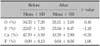
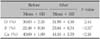
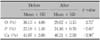
EDS analysis indicated that calcium (p < 0.001) and phosphate (p < 0.01) contents were increased after 10% HAp paste application on stripped enamel, calcium (p < 0.05) and phosphate (p < 0.01) contents were increased after 5% HAp paste application, and fluoride (p < 0.01) contents were increased after 1.23% APF application. SEM image showed that enamel surfaces became smoother and crystal structures became small and compact after APF or HAp application. After demineralization, calcium (p < 0.05) and phosphate (p < 0.05) contents remained increased on the enamel remineralized with 10% HAp paste, and phosphate (p < 0.05) contents remained increased on the enamel remineralized with 5% HAp paste. After demineralization, surfaces looked less destroyed in the enamel remineralized beforehand than those of the control, and small pores between crystal structures, formed by remineralization were remained.
Figures and Tables
Fig. 1
Surface appearance and crystal structure change of stripped enamel after 1.23% acidulated phosphate fluoride (APF) gel application for 7 days (SEM micrograph). A, Stripped enamel (× 250); B, after 1.23% APF gel application on stripped enamel (× 250); C, stripped enamel (× 50 k); D, after 1.23% APF gel application on stripped enamel (× 50 k).
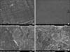
Fig. 2
Surface appearance and crystal structure change of stripped enamel after 5% hydroxyapatite (HAp) paste application for 7 days (SEM micrograph). A, Stripped enamel (× 250); B, after 5% HAp paste application on stripped enamel (× 250); C, stripped enamel (× 50 k); D, after 5% HAp paste application on stripped enamel (× 50 k).
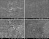
Fig. 3
Surface appearance and crystal structure change of stripped enamel after 10% hydroxyapatite (HAp) paste application for 7 days (SEM micrograph). A, Stripped enamel (× 250); B, after 10% HAp paste application on stripped enamel (× 250); C, stripped enamel (× 50 k); D, after 10% HAp paste application on stripped enamel (× 50 k).
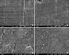
Fig. 4
Surface appearance and crystal structure change after demineralization of stripped enamel treated with 1.23% acidulated phosphate fluoride (APF) gel for 7 days (SEM micrograph). A, After demineralization of stripped enamel (× 250); B, after demineralization of stripped enamel treated with 1.23% APF gel (× 250); C, after demineralization of stripped enamel (× 3 k); D, after demineralization of stripped enamel treated with 1.23% APF gel (× 3 k); E, after demineralization of stripped enamel (× 50 k); F, after demineralization of stripped enamel treated with 1.23% APF gel (× 50 k).
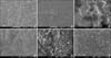
Fig. 5
Surface appearance and crystal structure change after demineralization of stripped enamel treated with 5% hydroxyapatite (HAp) paste for 7 days (SEM micrograph). A, After demineralization of stripped enamel (× 250); B, after demineralization of stripped enamel treated with 5% HAp paste (× 250); C, after demineralization of stripped enamel (× 3 k); D, after demineralization of stripped enamel treated with 5% HAp paste (× 3 k); E, after demineralization of stripped enamel (× 50 k); F, after demineralization of stripped enamel treated with 5% HAp paste (× 50 k).
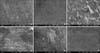
Fig. 6
Surface appearance and crystal structure change after demineralization of stripped enamel treated with 10% hydroxyapatite (HAp) paste for 7 days (SEM micrograph). A, After demineralization of stripped enamel (× 250); B, after demineralization of stripped enamel treated with 10% HAp paste (× 250); C, after demineralization of stripped enamel (× 3 k); D, after demineralization of stripped enamel treated with 10% HAp paste (× 3 k); E, after demineralization of stripped enamel (× 50 k); F, after demineralization of stripped enamel treated with 10% HAp paste (× 50 k).
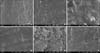
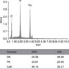
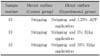
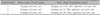
Table 3
Mineral content change (weight %) of stripped enamel after 1.23% acidulated phosphate fluoride (APF) gel application for 7 days
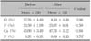
Table 4
Mineral content change (weight %) of stripped enamel after 5% hydroxyapatite paste application for 7 days
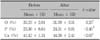
Table 5
Mineral content change (weight %) of stripped enamel after 10% hydroxyapatite paste application for 7 days
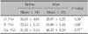
Table 6
Mineral content change (weight %) after demineralization of stripped enamel treated with 1.23% acidulated phosphate fluoride gel (APF) for 7 days
References
1. Bolton WA. Disharmony in tooth size and its relation to the analysis and treatment of malocclusion. Angle Orthod. 1958. 28:113–130.
2. Tuverson DL. Anterior interocclusal relations. Part I. Am J Orthod. 1980. 78:361–370.

3. Sheridan JJ. Air-rotor stripping update. J Clin Orthod. 1987. 21:781–788.
4. Blake M, Bibby K. Retention and stability: a review of the literature. Am J Orthod Dentofacial Orthop. 1998. 114:299–306.
5. Radlanski RJ, Jäger A, Schwestka R, Bertzbach F. Plaque accumulations caused by interdental stripping. Am J Orthod Dentofacial Orthop. 1988. 94:416–420.
6. Radlanski RJ, Jager A, Zimmer B. Morphology of interdentally stripped enamel one year after treatment. J Clin Orthod. 1989. 23:748–750.
7. Twesme DA, Firestone AR, Heaven TJ, Feagin FF, Jacobson A. Air-rotor stripping and enamel demineralization in vitro. Am J Orthod Dentofacial Orthop. 1994. 105:142–152.
8. Joseph VP, Rossouw PE, Basson NJ. Orthodontic microabrasive reproximation. Am J Orthod Dentofacial Orthop. 1992. 102:351–359.
9. Kim KN, Yoon YJ, Kim KW. A study on the enamel surface texture and caries susceptibility in interdentally stripped teeth. Korean J Orthod. 2001. 31:567–578.
10. Row J, Chun YS. A comparative study of roughness of enamel surface to various interdental enamel stripping methods in vitro. Korean J Orthod. 1999. 29:483–490.
11. Brêtas SM, Macari S, Elias AM, Ito IY, Matsumoto MA. Effect of 0.4% stannous fluoride gel on Streptococci mutans in relation to elastomeric rings and steel ligatures in orthodontic patients. Am J Orthod Dentofacial Orthop. 2005. 127:428–433.
12. Chadwick BL, Roy J, Knox J, Treasure ET. The effect of topical fluorides on decalcification in patients with fixed orthodontic appliances: a systematic review. Am J Orthod Dentofacial Orthod. 2005. 128:601–606.
13. Mizrahi E. Enamel demineralization following orthodontic treatment. Am J Orthod. 1982. 82:62–67.
14. Saxton CA. Scanning electron microscope study of the formation of dental plaque. Caries Res. 1973. 7:102–119.
15. Kim MY, Kwon HK, Kim BI. The comparison of remineralizing effect on mouthrinse containing nano sized or micro sized hydroxyapatite. J Korean Acad Oral Health. 2006. 30:325–334.
16. Lee YE, Back HJ, Jeong SH, Choi YH, Song KB. Effect of hydroxyapatite containing dentifrice on the remineralization of early incipient carious lesion. J Korean Acad Oral Health. 2007. 31:305–318.
17. Barone M, Malpassi M. Clinical trial of a 15% supermicronized hydroxyapatite gel for dentin hypersensitivity. G Ital Endod. 1991. 5:43–47.
18. Hüttemann RW, Strübel G, Rzekpa-Glinder V, Dönges H. Investigation of utility and mechanics of use of hydroxyapatite for therapy of hypersensitive tooth root. ZWR. 1989. 98:240–245.
19. Frazier MC, Southard TE, Doster PM. Prevention of enamel demineralization during orthodontic treatment: an in vitro study using pit and fissure sealants. Am J Orthod Dentofacial Orthop. 1996. 110:459–465.
20. Li L. The biochemistry and physiology of metallic fluoride: action, mechanism, and implications. Crit Rev Oral Biol Med. 2003. 14:100–114.
21. Bassin EB, Wypij D, Davis RB, Mittleman MA. Age-specific fluoride exposure in drinking water and osteosarcoma (United States). Cancer Causes Control. 2006. 17:421–428.
22. Silverstone LM. Observations on the dark zone in early enamel caries and artificial caries-like lesions. Caries Res. 1967. 1:260–274.
23. Margolis HC, Murphy BJ, Moreno EC. Development of caries-like lesions in partially saturated lactate buffers. Caries Res. 1985. 19:36–45.
24. Clarkson BH, Krell D, Wefel JS, Crall J, Feagin FF. In vitro caries-like lesion production by Streptococcus mutans and Actinomyces viscosus using sucrose and starch. J Dent Res. 1987. 66:795–798.
25. Boyle EL, Higham SM, Edgar WM. The production of subsurface artificial caries lesions on third molar teeth. Caries Res. 1998. 32:154–158.
26. Arends J, Schuthof J, Jongebloed WG. Lesion depth and microhardness indentations on artificial white spot lesions. Caries Res. 1980. 14:190–195.
27. Silverstone LM. Structure of carious enamel, including the early lesion. Oral Sci Rev. 1973. 3:100–160.
28. Herkstroter FM, Noordmans J, ten Bosch JJ. Wavelength-independent microradiography: its use to measure mineral changes in curved and thick samples. Caries Res. 1990. 24:399.
29. ten Bosch JJ, van der Mei HC, Borsboom PC. Optical monitor of in vitro caries. A comparison with chemical and microradiographic determination of mineral loss in early lesions. Caries Res. 1984. 18:540–547.
30. Kim SR, Hong SJ, Roh BD, Lee CY, Kum KY. The remineralizing effects of early enamel carious lesion by supersaturated buffer solution under pH cycling model. J Korean Acad Conserv Dent. 2001. 26:341–349.
31. Hicks J, Garcia-Godoy F, Flaitz C. Biological factors in dental caries: role of remineralization and fluoride in the dynamic process of demineralization and remineralization (part 3). J Clin Pediatr Dent. 2004. 28:203–214.
32. Lee KH. Effect of fluoride and calcium on enamel remineralization in vitro. J Korean Acad Pediatr Dent. 2004. 31:624–629.
33. Murray JJ, Rugg-Gunn AJ. Fluorides in caries prevention. 1982. 2nd ed. Bristol: Jone Wright & Sons;100–121.
34. Alexander SA, Ripa LW. Effects of self-applied topical fluoride preparations in orthodontic patients. Angle Orthod. 2000. 70:424–430.
35. Rogers GA, Wagner MJ. Protection of stripped enamel surfaces with topical fluoride applications. Am J Orthod. 1969. 56:551–559.
36. Yamazaki H, Litman A, Margolis HC. Effect of fluoride on artificial caries lesion progression and repair in human enamel: regulation of mineral deposition and dissolution under in vivo-like conditions. Arch Oral Biol. 2007. 52:110–120.
 XML Download