Abstract
Objective:
The aim of this study was to validate the Periotest values for the prediction of orthodontic mini-implants’ stability.
Methods:
Sixty orthodontic mini-implants (7.0 mm x Ø1.45 mm; ACR, Biomaterials Korea, Seoul, Korea) were inserted into the buccal alveolar bone of 5 twelve month-old beagle dogs. Insertion torque (IT) and Periotest values (PTV) were measured at the installation procedure, and removal torque (RT) and PTV were recorded after 12 weeks of orthodontic loading. To correlate PTV with variables, the cortical bone thickness (mm) and bone mineral density (BMD) within the cortical bone and total bone area were calculated with the help of CT scanning.
Results:
The BMD and cortical bone thickness in mandibular alveolus were significantly higher than those of the maxilla (p < 0.05). The PTV values ranged from −3.2 to 4.8 for 12 weeks of loading showing clinically stable mini-implants. PTV at insertion was significantly correlated with IT (−0.51), bone density (−0.48), cortical bone thickness (−0.42) (p < 0.05) in the mandible, but showed no correlation in the maxilla. PTV before removal was significantly correlated with RT (−0.66) (p < 0.01) in the mandible.
Go to : 
REFERENCES
1.Huja SS., Litsky AS., Beck FM., Johnson KA., Larsen PE. Pull-out strength of monocortical screws placed in the maxillae and mandibles of dogs. Am J Orthod Dentofacial Orthop. 2005. 127:307–13.

2.Huja SS., Katona TR., Burr DB., Garetto LP., Roberts WE. Microdamage adjacent to endosseous implants. Bone. 1999. 25:217–22.

3.Roberts WE., Simmons KE., Garetto LP., DeCastro RA. Bone physiology and metabolism in dental implantology: risk factors for osteoporosis and other metabolic bone diseases. Implant Dent. 1992. 1:11–21.
4.Chen Y., Shin HI., Kyung HM. Biomechanical and histological comparison of self-drilling and self-tapping orthodontic micro-implants in dogs. Am J Orthod Dentofacial Orthop. 2008. 133:44–50.

5.Brown GA., McCarthy T., Bourgeault CA., Callahan DJ. Mechanical performance of standard and cannulated 4.0-mm cancellous bone screws. J Orthop Res. 2000. 18:307–12.

6.Heidemann W., Gerlach KL., Gröbel KH., Köllner HG. Influence of different pilot hole sizes on torque measurements and pullout analysis of osteosynthesis screws. J Craniomax-illofac Surg. 1998. 26:50–5.

7.Miyawaki S., Koyama I., Inoue M., Mishima K., Sugahara T., Takano-Yamamoto T. Factors associated with the stability of titanium screws placed in the posterior region for orthodontic anchorage. Am J Orthod Dentofacial Orthop. 2003. 124:373–8.

8.Schulte W., d'Hoedt B., Lukas D., Muhlbradt L., Scholz F., Bretschi J, et al. Periotest—a new measurement process for periodontal function. Zahnarztl Mitt. 1983. 73:1229–40.
9.van Steenberghe D., Quirynen M. Reproducibility and detection threshold of peri-implant diagnostics. Adv Dent Res. 1993. 7:191–5.

10.Friberg B., Sennerby L., Meredith N., Lekholm U. A comparison between cutting torque and resonance frequency measurements of maxillary implants. A 20-month clinical study. Int J Oral Maxillofac Surg. 1999. 28:297–303.

11.Alsaadi G., Quirynen M., Michiels K., Jacobs R., van Steen-berghe D. A biomechanical assessment of the relation between the oral implant stability at insertion and subjective bone quality assessment. J Clin Periodontol. 2007. 34:359–66.

12.Schulte W., Lukas D. Periotest to monitor osseointegration and to check the occlusion in oral implantology. J Oral Implantol. 1993. 19:23–32.
13.Schulte W., Lukas D. The Periotest method. Int Dent J. 1992. 42:433–40.
14.Kim JW., Chang YI. Effects of drilling process in stability of micro-implants used for the orthodontic anchorage. Korean J Orthod. 2002. 32:107–15.
15.Park SH., Kim SH., Ryu JH., Kang YG., Chung KR., Kook YA. Bone-implant contact and mobility of surface-treated orthodontic micro-implants in dogs. Korean J Orthod. 2008. 38:416–26.

16.Meredith N., Alleyne D., Cawley P. Quantitative determination of the stability of the implant-tissue interface using resonance frequency analysis. Clin Oral Implants Res. 1996. 7:261–7.

17.Tricio J., Laohapand P., van Steenberghe D., Quirynen M., Naert I. Mechanical state assessment of the implant-bone continuum: a better understanding of the Periotest method. Int J Oral Maxillofac Implants. 1995. 10:43–9.
18.Truhlar RS., Morris HF., Ochi S. Stability of the bone-implant complex. Results of longitudinal testing to 60 months with the periotest device on endosseous dental implants. Ann Periodontol. 2000. 5:42–55.

19.Truhlar RS., Lauciello F., Morris HF., Ochi S. The influence of bone quality on periotest values of endosseous dental implants at stage II surgery. J Oral Maxillofac Surg. 1997. 55(12 Suppl 5):55–61.

20.Kim JW., Ahn SJ., Chang YI. Histomorphometric and mechanical analyses of the drill-free screw as orthodontic anchorage. Am J Orthod Dentofacial Orthop. 2005. 128:190–4.

21.Chavez H., Ortman LF., DeFranco RL., Medige J. Assessment of oral implant mobility. J Prosthet Dent. 1993. 70:421–6.

22.OlivéJ. Aparicio C. Periotest method as a measure of osseoin-tegrated oral implant stability. Int J Oral Maxillofac Implants. 1990. 5:390–400.
23.Salonen MA., Raustia AM., Kainulainen V., Oikarinen KS. Factors related to periotest values in endosseal implants: a 9-year follow-up. J Clin Periodontol. 1997. 24:272–7.

24.Tricio J., van Steenberghe D., Rosenberg D., Duchateau L. Implant stability related to insertion torque force and bone density: an in vitro study. J Prosthet Dent. 1995. 74:608–12.

25.van Steenberghe D., Tricio J., Naert I., Nys M. Damping characteristics of bone-to-implant interfaces. A clinical study with the periotest device. Clin Oral Implants Res. 1995. 6:31–9.
26.Bränemark R., Öhrnell LO., Nilsson P., Thomsen P. Biomechanical characterization of osseointegration during healing: an experimental in vivo study in the rat. Biomaterials. 1997. 18:969–78.

27.Deguchi T., Nasu M., Murakami K., Yabuuchi T., Kamioka H., Takano-Yamamoto T. Quantitative evaluation of cortical bone thickness with computed tomographic scanning for orthodontic implants. Am J Orthod Dentofacial Orthop. 2006. 129:721. .e7-12.

28.Carr AB., Papazoglou E., Larsen PE. The relationship of Periotest values, biomaterial, and torque to failure in adult baboons. Int J Prosthodont. 1995. 8:15–20.
29.Aparicio C., Orozco P. Use of 5-mm-diameter implants: periotest values related to a clinical and radiographic evaluation. Clin Oral Implants Res. 1998. 9:398–406.

30.Haas R., Bernhart T., Dörtbudak O., Mailath G. Experimental study of the damping behaviour of IMZ implants. J Oral Rehabil. 1999. 26:19–24.

31.Huang HM., Chiu CL., Yeh CY., Lee SY. Factors influencing the resonance frequency of dental implants. J Oral Maxillofac Surg. 2003. 61:1184–8.

32.Al-Nawas B., Wagner W., Grötz KA. Insertion torque and resonance frequency analysis of dental implant systems in an animal model with loaded implants. Int J Oral Maxillofac Implants. 2006. 21:726–32.
33.Nkenke E., Hahn M., Weinzierl K., Radespiel-Tröger M., Neukam FW., Engelke K. Implant stability and histomor-phometry: a correlation study in human cadavers using stepped cylinder implants. Clin Oral Implants Res. 2003. 14:601–9.

34.Schliephake H., Sewing A., Aref A. Resonance frequency measurements of implant stability in the dog mandible: experimental comparison with histomorphometric data. Int J Oral Maxillofac Surg. 2006. 35:941–6.

Go to : 
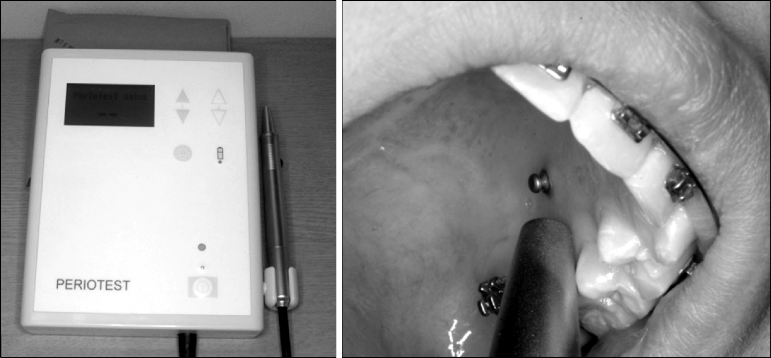
 | Fig 1.Periotest (Siemens, Bensheim, Germany) and clinical application for the mobility test of mini-implants. |
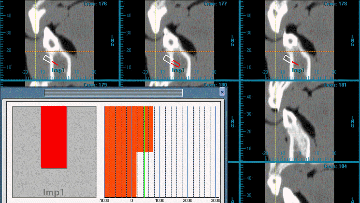
 | Fig 2.Bone mineral density calibration procedure. The scanned images were reconstructed to a transaxial image for each occlusal plane of the maxilla and mandible, and the average bone mineral density inside of cylindrical area (red box; diameter, 2 mm; depth, 5 mm) was measured by Hounsfield unit using V-implant program (CyberMed, Seoul, Korea). |
Table 1.
Range of periotest value (PTV), insertion torque, and removal torque for loading periods of maxilla and mandible
Table 2.
Comparison of bone mineral density (BMD) and cortical bone thickness of implantation site
Table 3.
Correlation of periotest value (PTV) with each variable at implantation of mini-implant
| Position | Variables | Insertiontorque (Ncm) | BMDcortical (mgHA/cm3) | BMDtotal (mgHA/cm3) | Corticalbone thickness(mm) | |
|---|---|---|---|---|---|---|
| PTV | Maxilla | Correlationcoefficient | −0.32 | −0.07 | −0.29 | −0.11 |
| p value | 0.14 | 0.75 | 0.18 | 0.63 | ||
| Mandible | Correlationcoefficient | −0.51∗ | −0.12 | −0.48∗ | −0.42∗ | |
| p value | 0.01 | 0.55 | 0.021 | 0.047 | ||
Table 4.
Correlation of periotest value (PTV) with each variable at removal of mini-implant
| Position | Variables | Removaltorque (Ncm) | BMDcortical (mgHA/cm3) | BMDtotal (mgHA/cm3) | Corticalbone thickness(mm) |
|---|---|---|---|---|---|
| PTV Maxilla | Correlationcoefficient | −0.27 | −0.22 | 0.24 | 0.23 |
| p value | 0.084 | 0.16 | 0.12 | 0.15 | |
| Mandible | Correlationcoefficient | −0.66∗ | −0.09 | −0.11 | 0.078 |
| p value | 0.00 | 0.53 | 0.46 | 0.62 |


 PDF
PDF ePub
ePub Citation
 Print
Print


 XML Download