

 PDF
PDF ePub
ePub Citation
 Print
Print


Abstract
Objective
The aim of this study was to evaluate the volumes and areas of the upper airways in children with Class II malocclusion, using three dimensional cone-beam computed tomography (CBCT) and to compare the volumetric and cross-sectional measurements and cephalometric variables to investigate possible relationships between the upper airway and facial morphology.
Methods
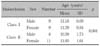
CBCT scans were obtained from 37 subjects (17 boys and 20 girls; average age, 11.02 years). The upper airway volumes and areas were measured, and compared with cephalometric variables.
Results
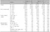
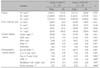
The area of the PNS-posterior plane (SPP) was significantly smaller in the Class II malocclusion group (p < 0.05). Also, the volumetric and cross-sectional measurements were lower in Class II than in Class I malocclusion groups, although the differences were not significant between the two groups (p > 0.05). The Class II malocclusion group showed significantly smaller values of PFH, mandibular body length, pog to N perp and showed larger values of FMA, ANB, and facial convexity than the Class I malocclusion group. The volume of the upper airway in front of PNS point (WN) showed negative correlation with ANB (p < 0.05).
Figures and Tables
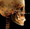 | Fig. 1The three-dimensional image was reoriented, using the FH plane as its horizontal reference plane. The FH plane was constructed from the right and left porions (Po(R), Po(L)) and the right orbitale (Or(R)).
|
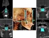 | Fig. 2Reference planes and the cross-sectional area measurements of the upper airway. Reference planes: A, ANS plane, the coronal plane passing through the anterior nasal spine (ANS); B, PNS plane, the coronal plane passing through the posterior nasal spine (PNS); C, PNS-posterior plane, the coronal plane passing through the point on the anterior pharyngeal wall extending from PNS (PNS-posterior); D, PNS-axial plane, the axial plane perpendicular to the coronal plane from PNS. The cross-sectional area measurements of these planes: the ANS plane area (SA), the PNS plane area (SP), the PNS-posterior plane area (SPP), the PNS-axial plane area (SPA).
|
 | Fig. 3Upper airway volumetric measuremets. To isolate the space of the airway, the threshold value was set at a range of -1,024 to -300 Hounsfield units. The airway was sculpted to be isolated and divided into two parts by the PNS plane; the volume of the upper airway in front of PNS point (WN), the volume of the upper airway just behind of PNS point (WP). Volumetric measurements were carried out using InVivoDental software (Anatomage Inc., San Jose, CA, USA).
|
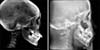 | Fig. 4Two-dimensional cephalometric images were derived from the three dimensional CT scans by creating an orthogonal projection with parallel rays.
|
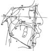 | Fig. 5Cephalometric measurements used in this study. 1, A to N perp; 2, Pog to N perp; 3, facial convexity; 4, mandibular body length; 5, ANB; 6, gonial angle; 7, anterior facial height; 8, posterior facial height; 9, FMA; 10, Ba-SE-FMN.
|
References
1. Ceylan I, Oktay H. A study on the pharyngeal size in different skeletal patterns. Am J Orthod Dentofacial Orthop. 1995. 108:69–75.

2. Holmberg H, Linder-Aronson S. Cephalometric radiographs as a means of evaluating the capacity of the nasal and nasopharyngeal airway. Am J Orthod. 1979. 76:479–490.
3. Joseph AA, Elbaum J, Cisneros GJ, Eisig SB. A cephalometric comparative study of the soft tissue airway dimensions in persons with hyperdivergent and normodivergent facial patterns. J Oral Maxillofac Surg. 1998. 56:135–139.
4. Ricketts RM. Respiratory obstruction syndrome. Am J Orthod. 1968. 54:495–507.
5. Lopatiene K, Babarskas A. Malocclusion and upper airway obstruction. Medicina (Kaunas). 2002. 38:277–283.
6. Angle E. Treatment of malocclusion of the teeth. 1907. 7th ed. Philadelphia: SS White Manufacturing Company;45.
7. Linder-Aronson S. Adenoids. Their effect on mode of breathing and nasal airflow and their relationship to characteristics of the facial skeleton and the denition. A biometric, rhino-manometric and cephalometro-radiographic study on children with and without adenoids. Acta Otolaryngol Suppl. 1970. 265:1–132.
8. Moore A. Observations on mouth breathing. Bull N Z Soc Periodontol. 1972. 33:9–11.
9. Hwang CJ, Ryu YK. A longitudinal study of nasopharynx and adenoid growth of Korean children. Korean J Orthod. 1985. 15:93–104.
10. Lee YS, Kim JC. A cephalometric study on the airway size according to the types of the malocclusion. Korean J Orthod. 1995. 25:19–29.
11. Lee YS, Baik HS, Lee KJ, Yu HS. The structural change in the hyoid bone and upper airway after orthognathic surgery for skeletal class III anterior open bite patients using 3-dimensional computed tomography. Korean J Orthod. 2009. 39:72–82.
12. Alves PV, Zhao L, O'Gara M, Patel PK, Bolognese AM. Three-dimensional cephalometric study of upper airway space in skeletal class II and III healthy patients. J Craniofac Surg. 2008. 19:1497–1507.
13. Chang HS, Baik HS. A proposal of landmarks for craniofacial analysis using three-dimensional CT imaging. Korean J Orthod. 2002. 32:313–325.
14. Iwasaki T, Hayasaki H, Takemoto Y, Kanomi R, Yamasaki Y. Oropharyngeal airway in children with Class III malocclusion evaluated by cone-beam computed tomography. Am J Orthod Dentofacial Orthop. 2009. 136:318.e1–318.e9.
15. Kim YI, Kim SS, Son WS, Park SB. Pharyngeal airway analysis of different craniofacial morphology using cone-beam computed tomography (CBCT). Korean J Orthod. 2009. 39:136–145.
16. Dahlberg G. Statistical methods for medical and biological students. 1940. London: G. Allen & Unwin Ltd;1–140.
17. Lagravère MO, Carey J, Toogood RW, Major PW. Three-dimensional accuracy of measurements made with software on cone-beam computed tomography images. Am J Orthod Dentofacial Orthop. 2008. 134:112–116.
18. Major MP, Flores-Mir C, Major PW. Assessment of lateral cephalometric diagnosis of adenoid hypertrophy and posterior upper airway obstruction: a systematic review. Am J Orthod Dentofacial Orthop. 2006. 130:700–708.
19. Abramson ZR, Susarla S, Tagoni JR, Kaban L. Three-dimensional computed tomographic analysis of airway anatomy. J Oral Maxillofac Surg. 2010. 68:363–371.
20. Fouke JM, Strohl KP. Effect of position and lung volume on upper airway geometry. J Appl Physiol. 1987. 63:375–380.
21. Pae EK, Lowe AA, Sasaki K, Price C, Tsuchiya M, Fleetham JA. A cephalometric and electromyographic study of upper airway structures in the upright and supine positions. Am J Orthod Dentofacial Orthop. 1994. 106:52–59.
22. Proffit WR. Proffit WR, Fields HW, Sarver DM, editors. Concepts of growth and development. Contemporary orthodontics. 2007. 4th ed. St Louis: Mosby;27–29.
23. Hwang YI, Lee KH, Lee KJ, Kim SC, Cho HJ, Cheon SH, et al. Effect of airway and tongue in facial morphology of prepubertal Class I, II children. Korean J Orthod. 2008. 38:74–82.
24. McNamara JA. Influence of respiratory pattern on craniofacial growth. Angle Orthod. 1981. 51:269–300.
25. Subtelny JD, Baker HK. The significance of adenoid tissue in velopharyngeal function. Plast Reconstr Surg. 1956. 17:235–250.
26. Mergen DC, Jacobs RM. The size of nasopharynx associated with normal occlusion and Class II malocclusion. Angle Orthod. 1970. 40:342–346.
27. Kim YJ, Bok GS, Lee KH, Hwang YI, Park YH. The relationship between upper airway width and facial growth changes in orthodontic treatment of growing children. Korean J Orthod. 2009. 39:168–176.
28. Behlfelt K, Linder-Aronson S, McWilliam J, Neander P, Laage-Hellman J. Cranio-facial morphology in children with and without enlarged tonsils. Eur J Orthod. 1990. 12:233–243.
29. Solow B, Siersbaek-Nielsen S, Greve E. Airway adequacy, head posture, and craniofacial morphology. Am J Orthod. 1984. 86:214–223.
30. Sosa FA, Graber TM, Muller TP. Postpharyngeal lymphoid tissue in Angle Class I and Class II malocclusions. Am J Orthod. 1982. 81:299–309.
 XML Download