

 PDF
PDF ePub
ePub Citation
 Print
Print


Abstract
Objective
The purpose of this study was to evaluate the possibility of using a digital model and cone beam computed tomograph (CBCT) image for model analysis.
Methods
Model analyses of CBCT images, plaster models, and digital models of 20 orthodontic patients with a permanent dentition with no proximal metal restorations, were compared.
Results
The average differences of tooth size measurements were 0.01 to 0.20 mm, and the average difference of arch length discrepancy measurements were 0.41 mm in the maxilla and 0.82 mm in the mandible. The difference in Bolton discrepancy measurements was 0.17 mm for the anterior region and 0.44 mm overall but with no statistically significant difference. When comparing CBCT images with plaster models, the average differences in tooth size measurements were -0.22 to 0.01 mm, and the average differences in arch length discrepancy measurements were 0.43 mm in the maxilla and 0.32 mm in the mandible. Difference in Bolton discrepancy measurements were 0.35 mm in the anterior region and 1.25 mm overall. CBCT images showed significantly smaller overall Bolton discrepancy measurements.
Figures and Tables
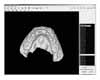 | Fig 1Measurements of tooth size and dental arch width of a laser scan digital model. 3DXer ver 3.5 was used.
|
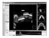 | Fig 2Volume rendering model of CBCT data constructed using Accurex ver 1.0. 3D zoom mode was applied and displayed in the main window for measurement of lower left lateral incisor width. The thresholding ranges of teeth 3 preset that were used in this study are shown in the fine tuning tab on the bottom of the program window.
|
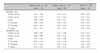
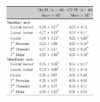
Table 1
Absolute differences of repeated measurements of tooth sizes of plaster model, laser scan digital model and CBCT image (unit, mm)
![]()
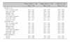
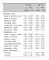
Table 2
Absolute differences of repeated measurements of arch length discrepancies, arch width, Bolton discrepancies of plaster model, laser scan digital model and CBCT image (unit, mm)
![]()
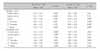
Table 3
Absolute differences of tooth size measurements between laser scan digital model and plaster model, and between CBCT image and plaster model (unit, mm)
![]()
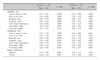
Table 4
Differences of tooth size measurements between laser scan digital model and plaster model, and between CBCT image and plaster model (unit, mm)
![]()
Table 5
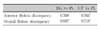
Absolute differences of arch length discrepancy, arch width, and Bolton discrepancy measurements between laser scan digital model and plaster model, and between CBCT image and plaster model (unit, mm)
![]()
References
1. OrthoCAD. USA: Available at:
http://www.orthocad.com.
2. Emodels. USA: Available at:
http://www.dentalemodels.com.
3. Zilberman O, Huggare JA, Parikakis KA. Evaluation of the validity of tooth size and arch width measurements using conventional and three-dimensional virtual orthodontic models. Angle Orthod. 2003. 73:301–306.
4. Kuroda T, Motohashi N, Tominaga R, Iwata K. Three-dimensional dental cast analyzing system using laser scanning. Am J Orthod Dentofacial Orthop. 1996. 110:365–369.

5. Sohmura T, Kojima T, Wakabayashi K, Takahashi J. Use of an ultrahigh-speed laser scanner for constructing three-dimensional shapes of dentition and occlusion. J Prosthet Dent. 2000. 84:345–352.
6. Stevens DR, Flores-Mir C, Nebbe B, Raboud DW, Heo G, Major PW. Validity, reliability, and reproducibility of plaster vs digital study models: comparison of peer assessment rating and Bolton analysis and their constituent measurements. Am J Orthod Dentofacial Orthop. 2006. 129:794–803.
7. Garino F, Garino GB. Comparison of dental arch measurements between stone and digital casts. World J Orthod. 2002. 3:250–254.
8. Baik HS, Lee HJ, Jeon JM. A proposal of soft tissue landmarks for craniofacial analysis using three-dimensional laser scan imaging. Korean J Orthod. 2006. 36:1–13.
9. Baik HS, Jeon JM, Lee HJ. A study of facial soft tissue of Korean adults with normal occlusion using a three-dimensional laser scanner. Korean J Orthod. 2006. 36:14–29.
10. Ko SD, Cha KS. A Study on the labial & buccal surface contour in Korean permanent teeth using three-dimensional laser scanning. Korean J Orthod. 2002. 32:275–291.
11. Chang YJ, Kim TW, Yoo KH. The effect of variations in the vertical position of the bracket on the crown inclination. Korean J Orthod. 2002. 32:401–411.
12. You HW. The optimal scanning pose generating algorithm for 3D dental model scanner [thesis]. 2000. Seoul: Myongji University.
13. Park JW. The study on the measurement error of the 3D-digital model made by laser scan method [thesis]. 2003. Seoul: Seoul National University.
14. Han JH. Comparison of measurements in three-dimensional digital model and dental plaster model [thesis]. 2004. Seoul: Yonsei University.
15. Park SI. The comparison between manual and 3D-digital measurement in dental cast measurements according to the degree of crowding [thesis]. 2006. Seoul: Korea University.
16. Holberg C, Steinhäuser S, Geis P, Rudzki-Janson I. Conebeam computed tomography in orthodontics: benefits and limitations. J Orofac Orthop. 2005. 66:434–444.
17. Mischkowski RA, Pulsfort R, Ritter L, Neugebauer J, Brochhagen HG, Keeve E, et al. Geometric accuracy of a newly developed cone-beam device for maxillofacial imaging. Oral Surg Oral Med Oral Pathol Oral Radiol Endod. 2007. 104:551–559.
18. Chae JH, Song JW, Cha JY, Choi JS, Park YC. Labial and buccal surface contours of Korean normal occlusion in a three-dimensional digital model. Korean J Orthod. 2008. 38:95–103.
19. Misch KA, Yi ES, Sarment DP. Accuracy of cone beam computed tomography for periodontal defect measurements. J Periodontol. 2006. 77:1261–1266.
20. Pinsky HM, Dyda S, Pinsky RW, Misch KA, Sarment DP. Accuracy of three-dimensional measurements using cone-beam CT. Dentomaxillofac Radiol. 2006. 35:410–416.
21. Ludlow JB, Laster WS, See M, Bailey LJ, Hershey HG. Accuracy of measurements of mandibular anatomy in cone beam computed tomography images. Oral Surg Oral Med Oral Pathol Oral Radiol Endod. 2007. 103:534–542.
22. Chang J, Yenice KM, Narayana A, Gutin PH. Accuracy and feasibility of cone-beam computed tomography for stereotactic radiosurgery setup. Med Phys. 2007. 34:2077–2084.
23. Bolton WA. Disharmony in tooth size and its relation to the analysis and treatment of malocclusion. Angle Orthod. 1958. 28:113–130.
24. Lascala CA, Panella J, Marques MM. Analysis of the accuracy of linear measurements obtained by cone beam computed tomography (CBCT-NewTom). Dentomaxillofac Radiol. 2004. 33:291–294.
25. Santoro M, Galkin S, Teredesai M, Nicolay OF, Cangialosi TJ. Comparison of measurements made on digital and plaster models. Am J Orthod Dentofacial Orthop. 2003. 124:101–105.
26. Quimby ML, Vig KW, Rashid RG, Firestone AR. The accuracy and reliability of measurements made on computer-based digital models. Angle Orthod. 2004. 74:298–303.
27. Lee JH, Paik KS, Chang MS, Lee SP. An evaluation of validity of measurements using digital caliper and three-dimensional virtual dental models. Korean J Anat. 2004. 37:209–218.
28. Bolton WA. The clinical application of tooth-size analysis. Am J Orthod. 1962. 48:504–529.
29. Tomassetti JJ, Taloumis LJ, Denny JM, Fischer JR Jr. A comparison of 3 computerized Bolton tooth-size analyses with a commonly used method. Angle Orthod. 2001. 71:351–357.
30. Lagravère MO, Fang Y, Carey J, Toogood RW, Packota GV, Major PW. Density conversion factor determined using a cone-beam computed tomography unit NewTom QR-DVT 9000. Dentomaxillofac Radiol. 2006. 35:407–409.
31. Sim EJ, Hwang HS, Moon JD. A study on the error of tooth size measurements. Korean J Orthod. 1999. 29:491–501.
32. Anatomage. USA: Available at:
http://www.anatomage.com.
33. OrthoProofUSA. USA: Available at:
http://www.orthoproofusa.com.
 XML Download