Abstract
This paper describes the case of a 50-year-old female with a Class II malocclusion who presented with severe bimaxillary protrusion and generalized alveolar bone loss due to adult periodontitis. The treatment plan consisted of extracting both upper and lower first premolars and periodontal treatment. Anterior segmental osteotomy (ASO) of the mandible and upper anterior segment retraction using compression osteogenesis after peri-segmental corticotomy (Speedy orthodontics) was performed. Correct overbite and overjet, facial balance, and improvement of lip protrusion were obtained. However, a slight root resorption tendency was observed on the lower anterior dentition. The active treatment period was 9 months and the results were stable for 27 months after debonding. This new type of treatment mechanics can be an effective alternative to orthognathic surgery. (Korean J Orthod 2009;39(1):54-65)
Go to : 
REFERENCES
1.Melsen B. Limitations in adult orthodontics. In: Melsen B editor. Current controversies in orthodontics. Chicago: Quintessence Publishing;1991.
2.Handelman CS. The anterior alveolus: its importance in limiting orthodontic treatment and its influence on the occurrence of iatrogenic sequelae. Angle Orthod. 1996. 66:95–109.
3.Proffit WR., White RP Jr. Who needs surgical-orthodontic treatment? Int J Adult Orthodon Orthognath Surg. 1990. 5:81–9.
4.Miyajima K., Nagahara K., Lizuka T. Orthodontic treatment for a patient after menopause. Angle Orthod. 1996. 66:173–8.
6.Bell WH., Jacobs JD., Legan HL. Treatment of Class II deep bite by orthodontic and surgical means. Am J Orthod. 1984. 85:1–20.

7.Bojrab DG., Dumas JE., Lahrman DE. JCO/interviews Dr. David G. Bojrab, Dr. James E. Dumas, Dr. Don E. Lahrman on surgical-orthodontics. J Clin Orthod. 1977. 11:330–42.
8.Laigan DT., Hey JH., West HA. Aseptic necrosis following maxillary osteotomies: report of 36 cases. J Oral Maxillofac Surg. 1990. 48:142–56.
9.Köle H. Surgical operations on the alveolar ridge to correct occlusal abnormalities. Oral Surg Oral Med Oral Pathol. 1959. 12:515–29.

10.Bell WH. Surgical-orthodontic treatment of interincisal diaste-mas. Am J Orthod. 1970. 57:158–63.

11.Anholm JM., Crites DA., Hoff R., Rathbun WE. Corticotomy-fa-ciliated orthodontics. CDA J. 1986. 14:7–11.
12.Düker J. Experimental animal research into segmental alveolar movement after corticotomy. J Maxillofac Surg. 1975. 3:81–4.

13.Gantes B., Rathbun E., Anholm M. Effects on the periodontium following corticotomy-facilitated orthodontics. Case reports. J Periodontol. 1990. 61:234–8.

14.Park WK., Kim SS., Park SB., Son WS., Kim YD., Jun ES, et al. The effect of cortical punching on the expression of OPG, RANK, and RANKL in the periodontal tissue during tooth movement in rats. Korean J Orthod. 2008. 38:159–74.

15.Wilcko WM., Wilcko T., Bouquot JE., Ferguson DJ. Rapid orthodontics with alveolar reshaping: two cases reports of decrowding. Int J Periodontics Restorative Dent. 2001. 21:9–19.
16.Chung KR. Text book of speedy orthodontics. Seoul: Jeesung;2001.
17.Chung KR., Kim SH., Kook YA. Speedy surgical orthodontic treatment with skeletal anchorage in adults. In: Bell WH, Guerrero CA editors. Distraction osteogenesis of the facial bones. Hamilton: BC Deckers;2007. p. 167–86.
18.Chung KR., Oh MY., Ko SJ. Corticotomy-assisted orthodontics. J Clin Orthod. 2001. 35:331–9.
19.Suya H. Corticotomy in orthodontics. In: Hosl E, Baldauf A editors. Mechanical and biological basics in orthodontic therapy. Heidelberg: Huthig Buch Verlag;1991.
20.Kanno T., Mitsugi M., Furuki Y., Kozato S., Ayasaka N., Mori H. Corticotomy and compression osteogenesis in the posterior maxilla for treating severe anterior open bite. Int J Oral Maxillofac Surg. 2007. 36:354–7.

21.Kim S., Park Y., Chung K. Severe anterior open bite malocclusion with multiple odontoma treated by C-lingual retractor and horseshoe mechanics. Angle Orthod. 2003. 73:206–12.
22.Kim SH., Park YG., Chung K. Severe Class II anterior deep bite malocclusion treated with a C-lingual retractor. Angle Orthod. 2004. 74:280–5.
23.Chung KR., Kim YS., Linton JL., Lee YJ. The miniplate with tube for skeletal anchorage. J Clin Orthod. 2002. 36:407–12.
24.Chung KR., Kim SH., Kook YA., Mo SS., Jung JA. Class II malocclusion treated by combining a lingual retractor and a palatal plate. Am J Orthod Dentofacial Orthop. 2008. 133:112–23.

25.Kawakami T., Nishimoto M., Matsuda Y., Deguchi T., Eda S. Histologic suture changes following retraction of the maxillary anterior bone segment after corticotomy. Endod Dent Trauma-tol. 1996. 12:38–43.
26.Ericsson I., Thilander B., Lindhe J., Okamoto H. The effect of orthodontic tilting movements on the periodontal tissues of in-fected and non-infected dentitions in dogs. J Clin Periodontol. 1977. 4:278–93.

27.Ǻrtun J., Urbye KS. The effect of orthodontic treatment on periodontal bone support in patients with advanced loss of marginal periodontium. Am J Orthod Dentofacial Orthop. 1988. 93:143–8.
28.Yoshikawa Y. Effects of corticotomy on maxillary retraction induced by orthopedic force. J Matsumoto Dent Coll Soc. 1987. 13:292–320.
29.Fukunaga T., Kurodaa S., Kurosaka H., Takano-Yamamoto T. Skeletal anchorage for orthodontic correction of maxillary protrusion with adult periodontitis. Angle Orthod. 2006. 76:148–55.
30.Lee HK., Chung KR. The vertical location of the center of resistance for maxillary six anterior teeth during retraction using three dimensional finite element analysis. Korean J Orthod. 2001. 31:425–38.
Go to : 
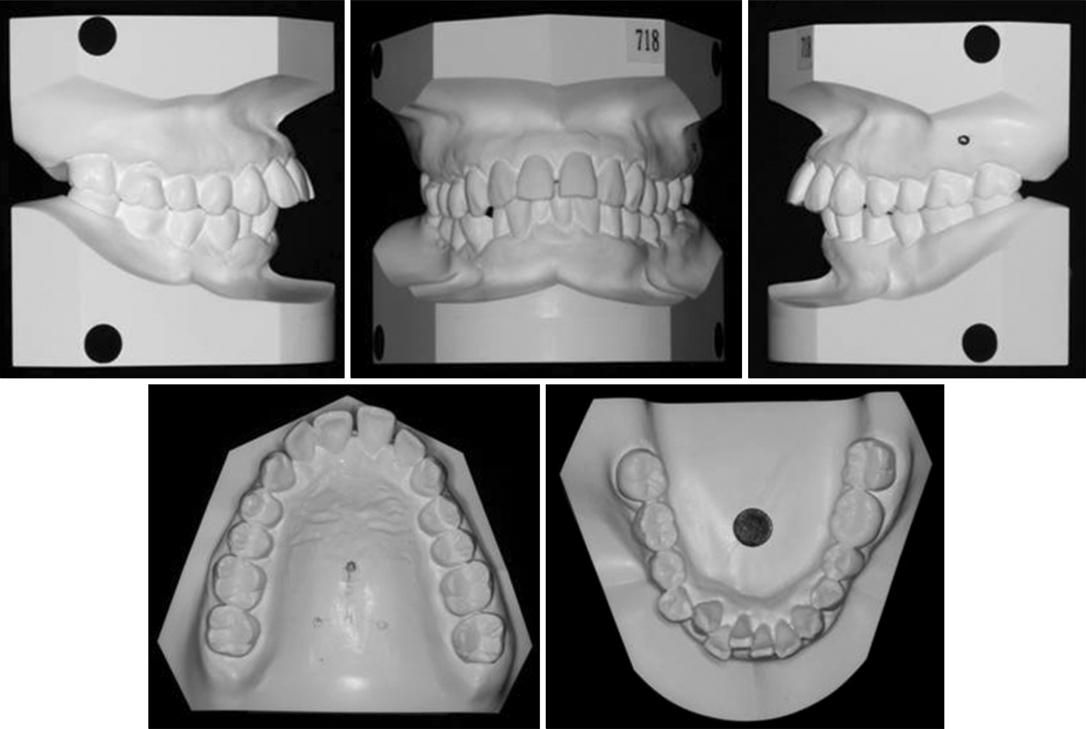
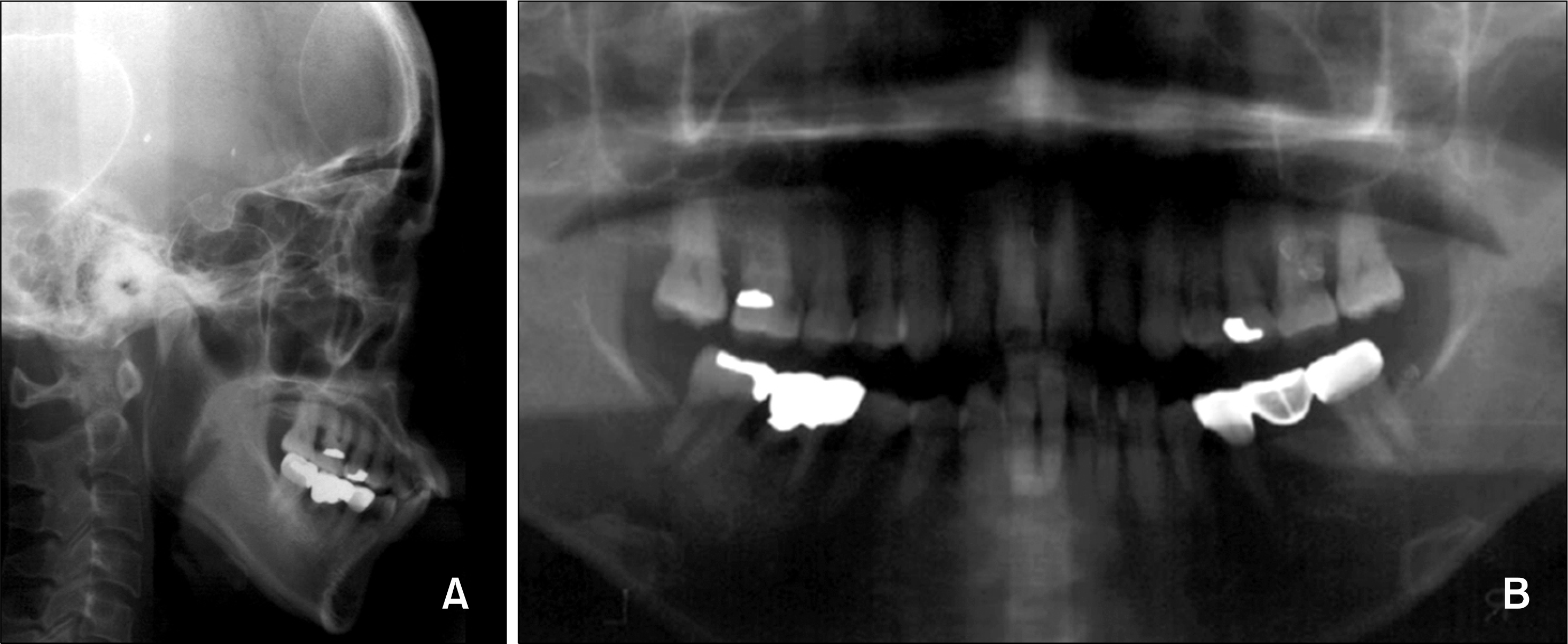
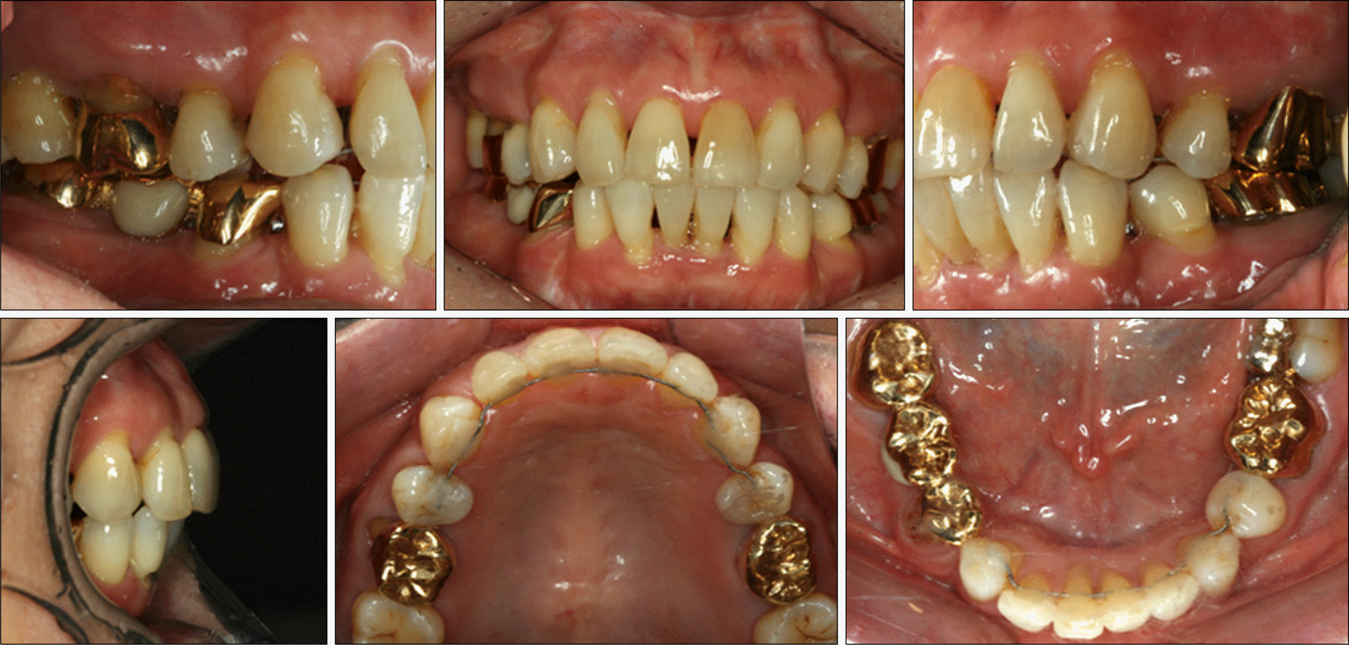
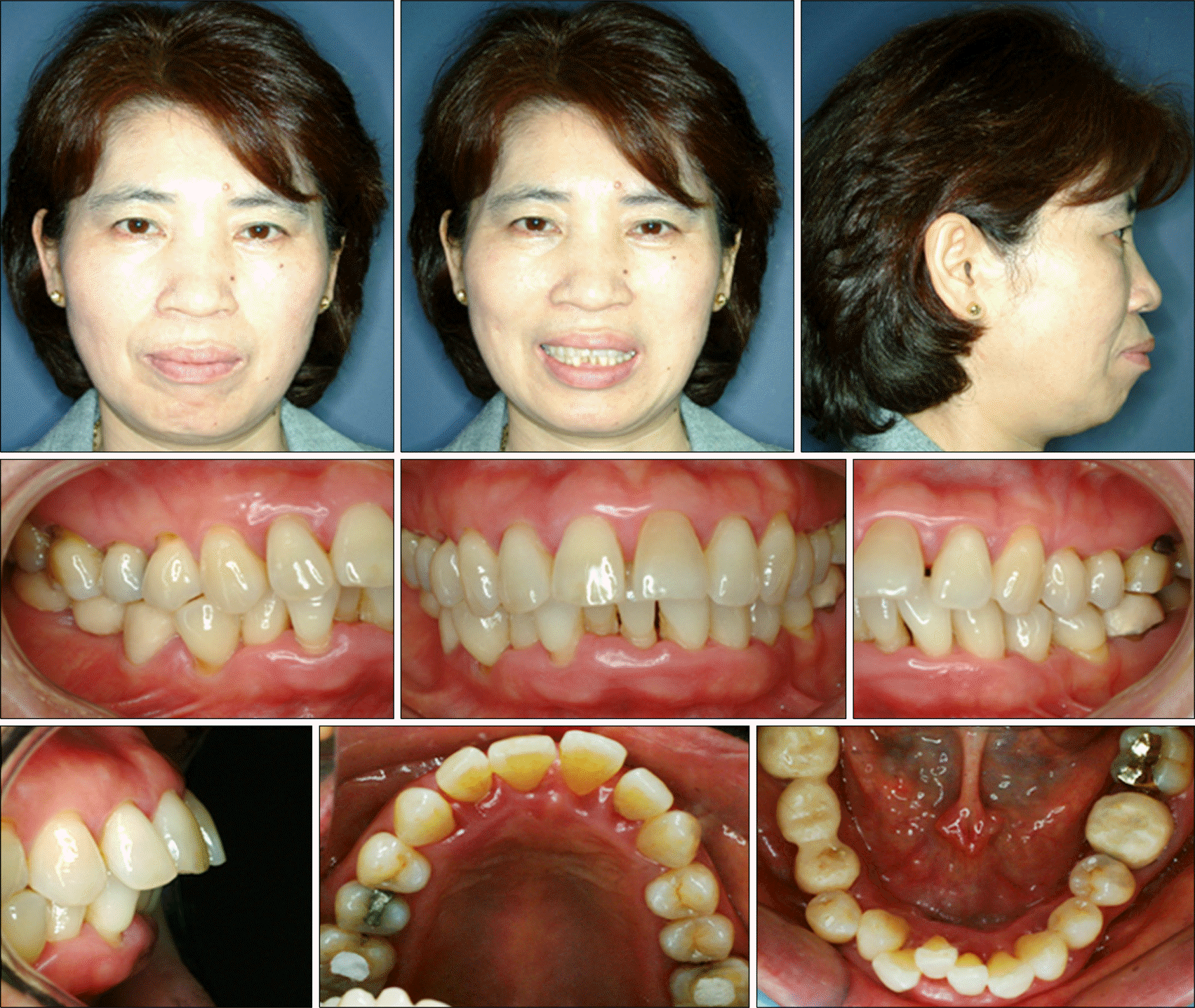 | Fig 1.Facial and intraoral photographs before treatment show a very convex profile with significant mentalis muscle strain and reveal a Class II canine and Class I molar relationship. |
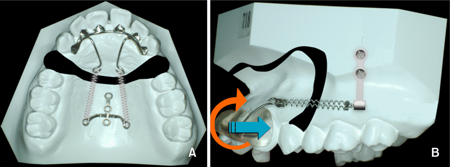 | Fig 4.Schematic illustration of the anterior segment retraction method after perisegmental corticotomy. A, Titanium C palatal plate, drill free screws and C lingual retractor combined lingual retraction; B, labial retractor and C tube combined retraction mechanics. |
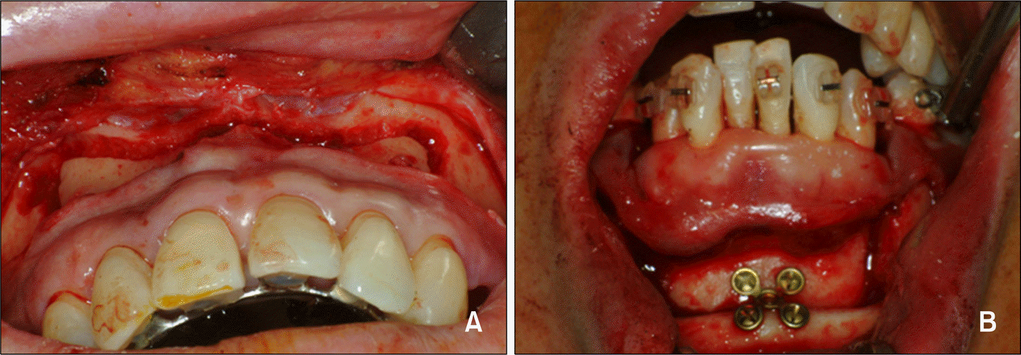 | Fig 5.Oral view during speedy orthodontics surgery. A, Oral view after buccal perisegmental corticotomy; B, anterior segmental osteotomy (ASO) on the lower anterior segment. |
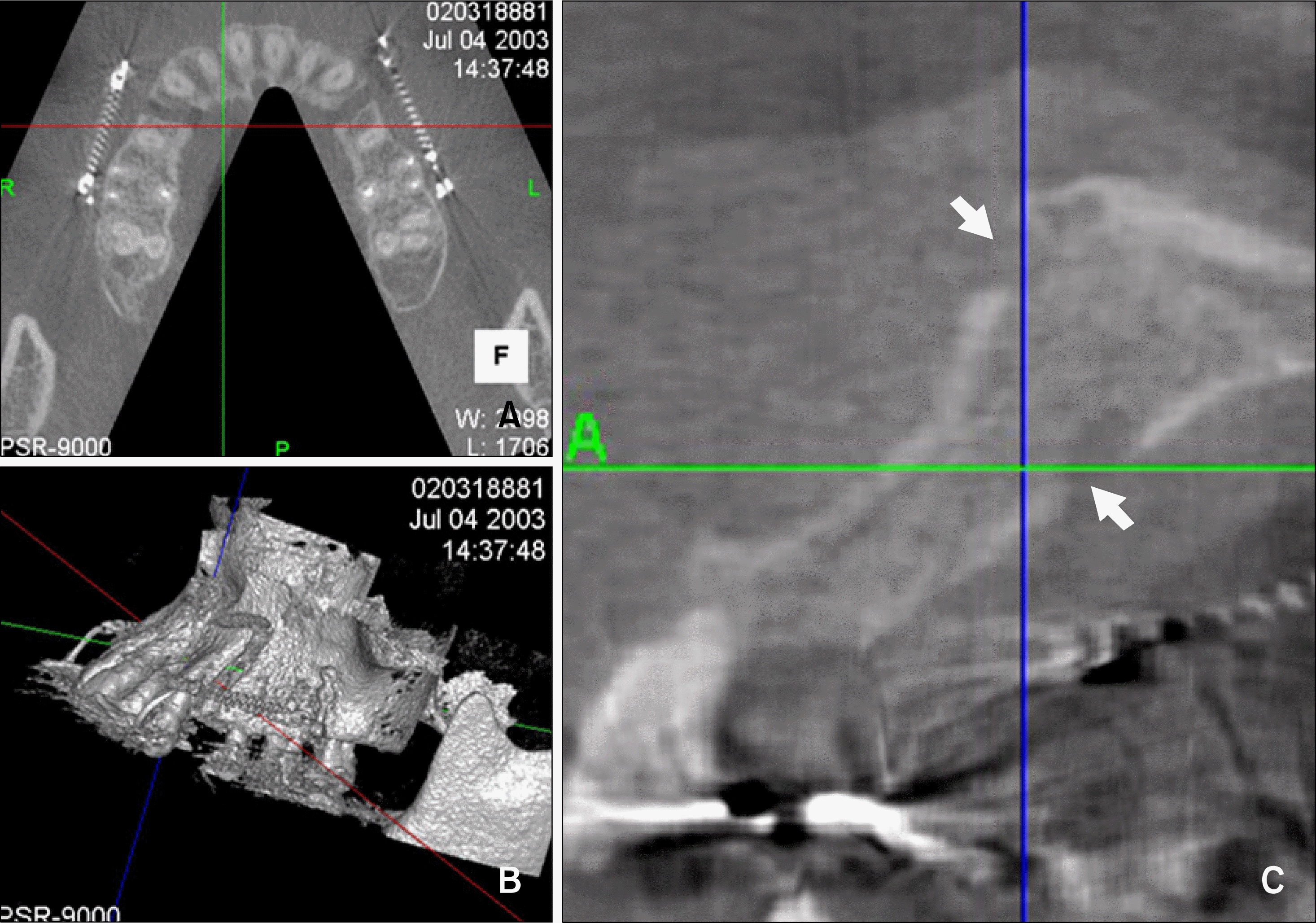 | Fig 6.Cone beam CT view (PSR-9000N, Asahi Roentgen, Kyoto, Japan) after perisegmental corticomy. A, Transaxial view; B, 3 dimensional reconstruction view shows the labial perisegmental corticotomized area; C, arrows in sagittal view show depth of corticotomy. |
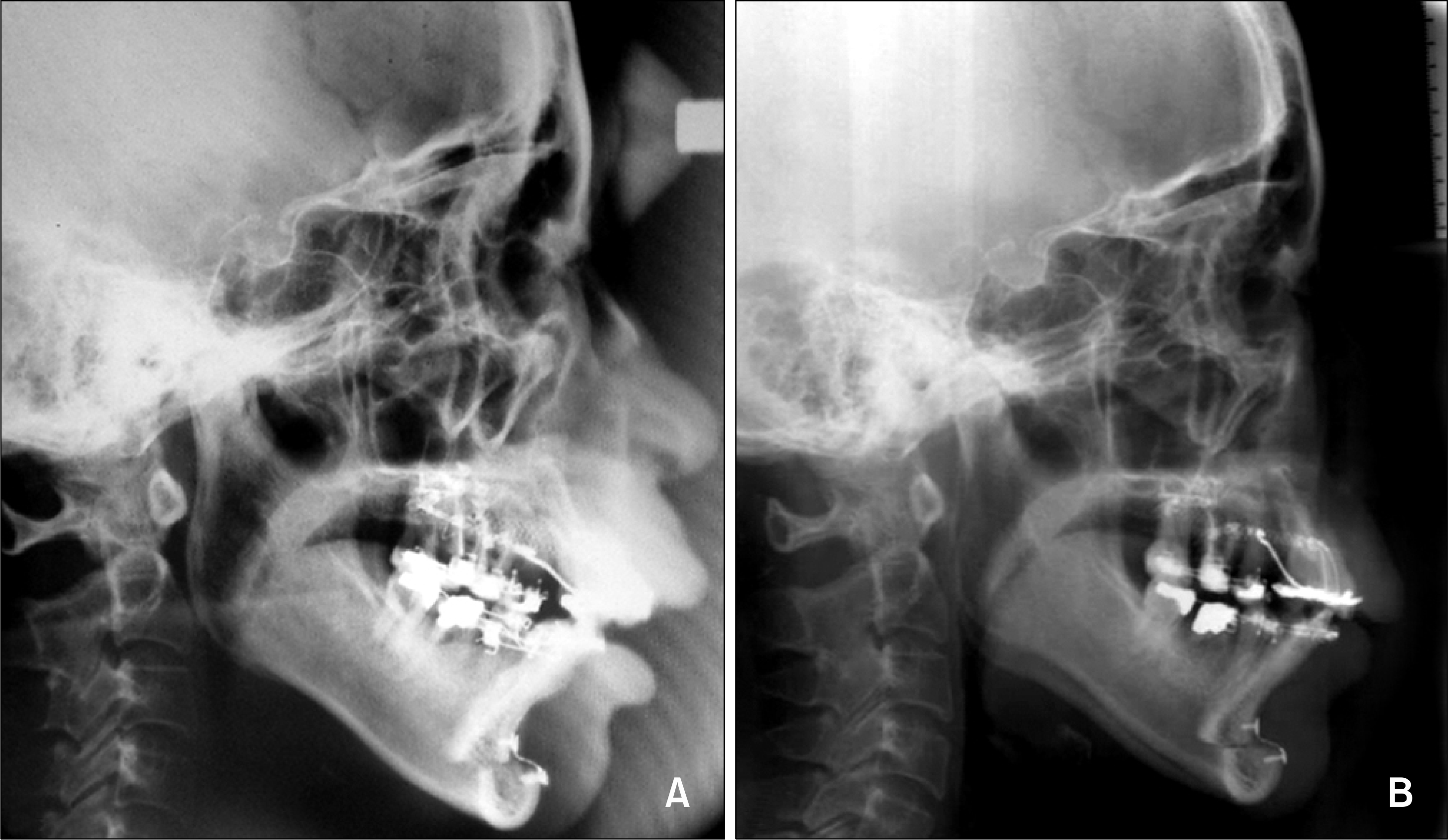 | Fig 7.Progress on lateral cephalograms. A, 1 week after immediate upper retraction; B, 7 weeks after retraction. |
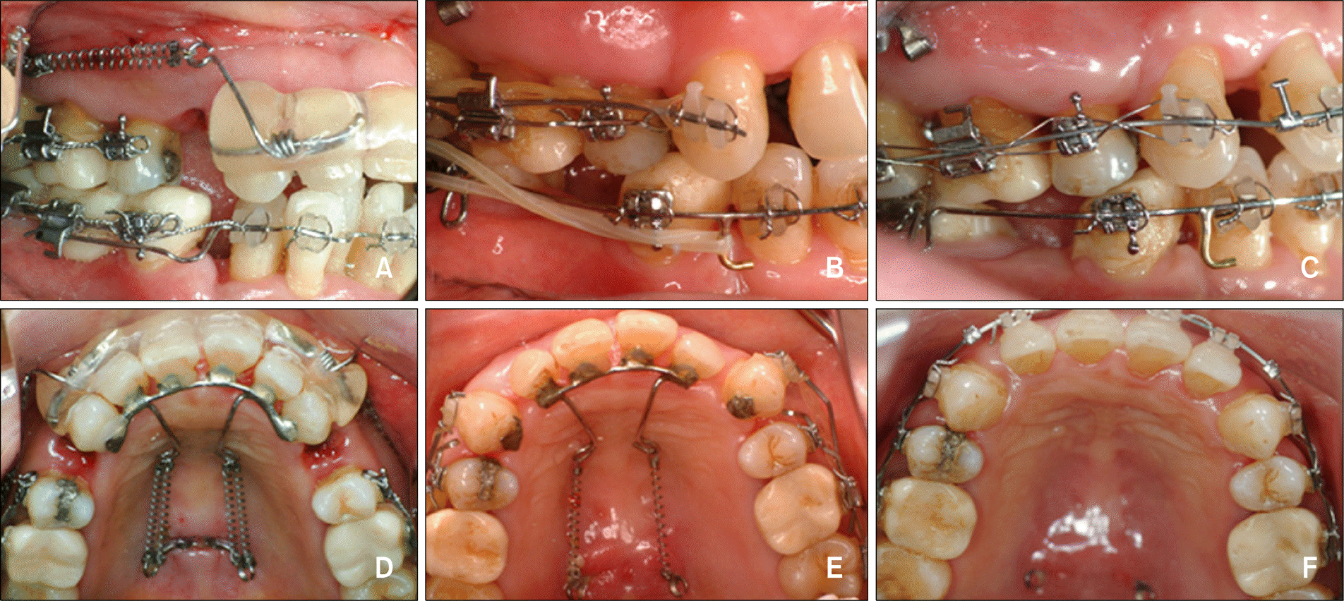 | Fig 8.Progress in oral views. A and D, 1 week after retraction; B and E, 5 months after retraction; C and F, 6 months after treatment. Fixed appliances were applied for conventional orthodontic treatment. |
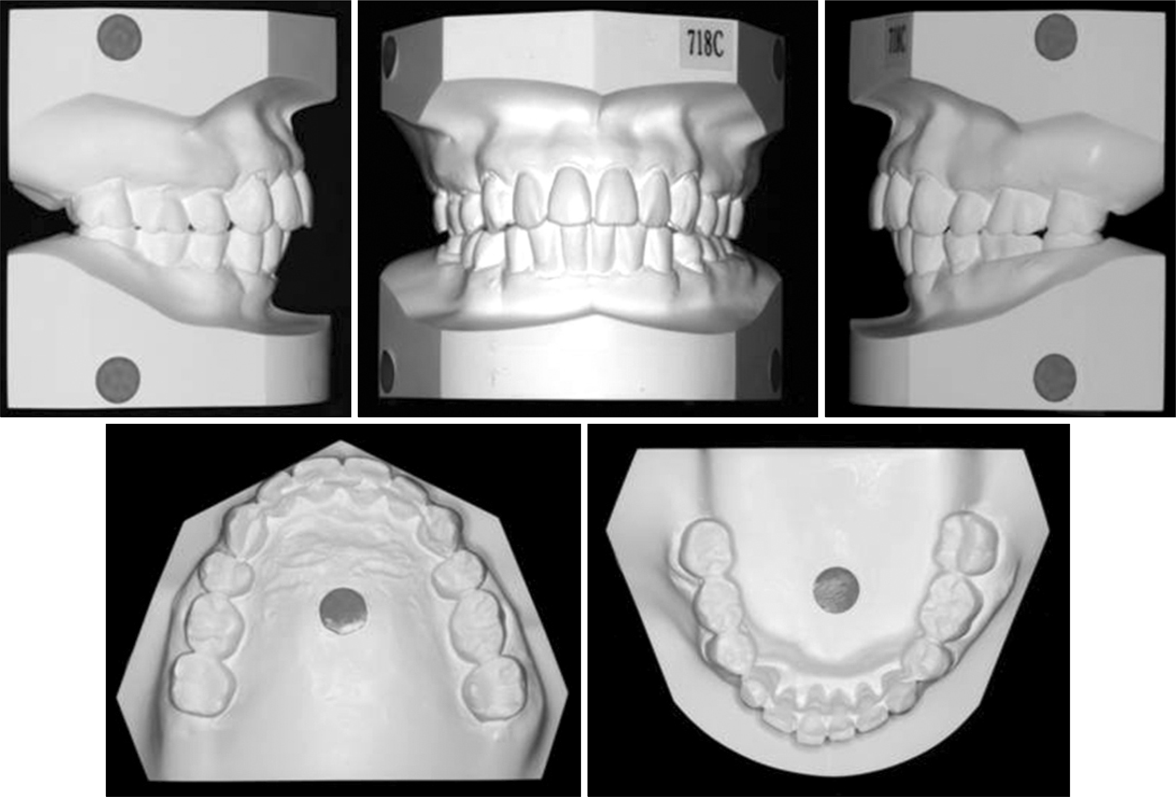
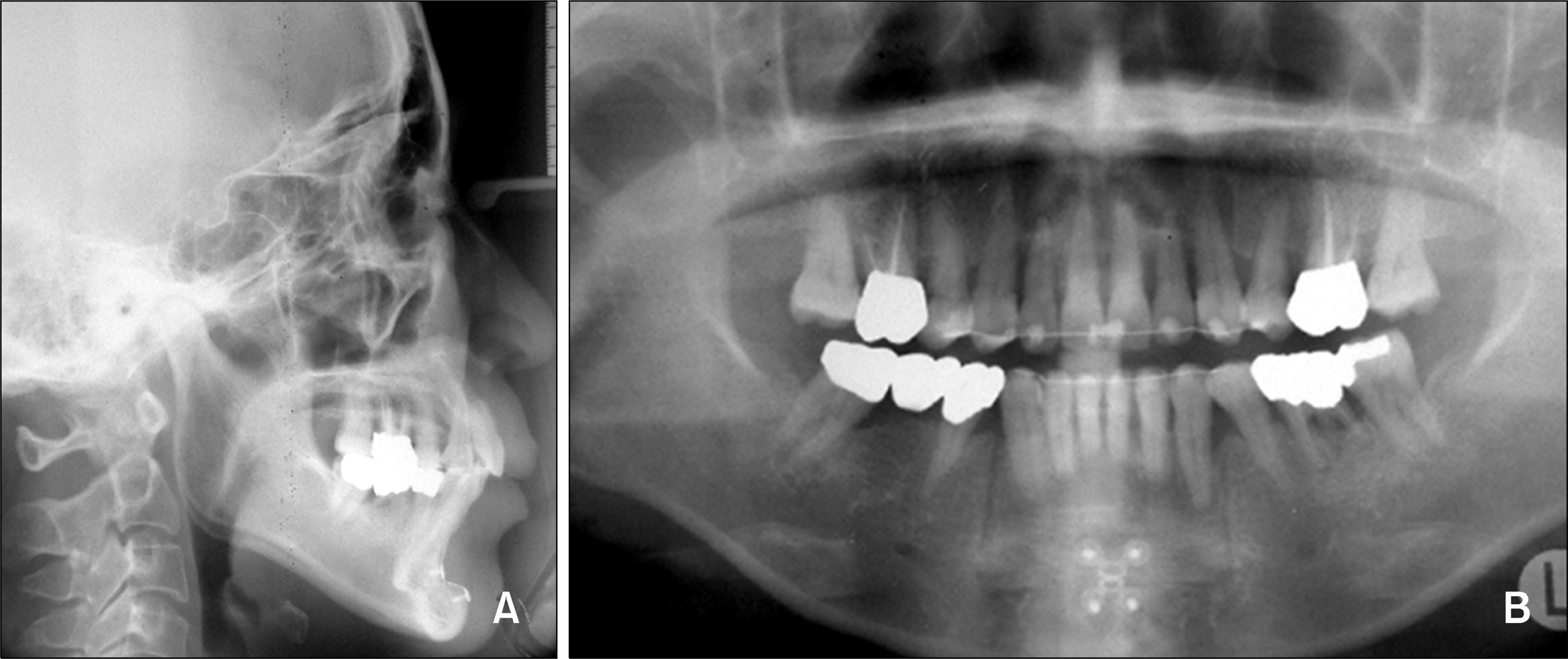
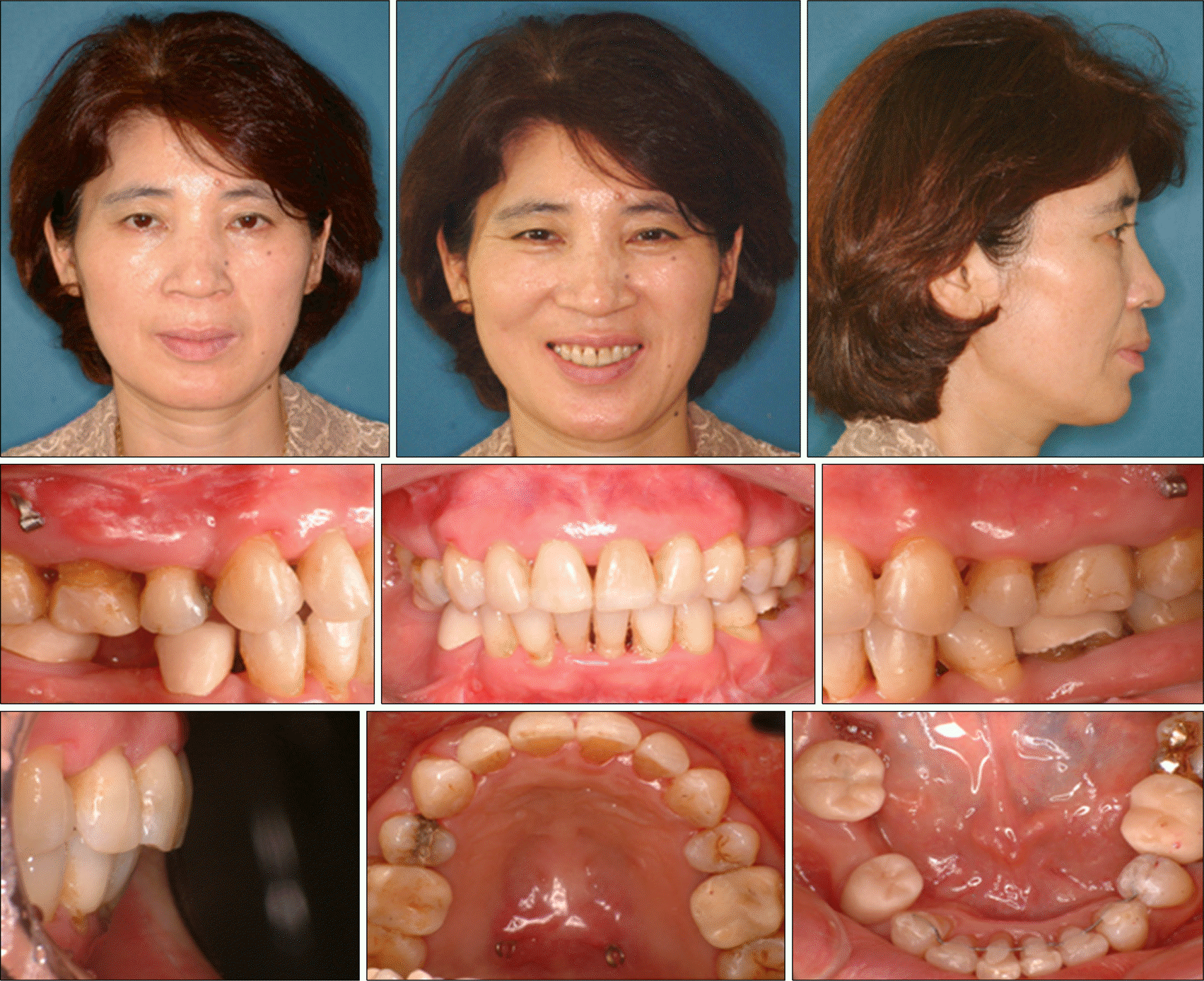 | Fig 9.Facial and intraoral photographs after treatment show good overjet, overbite, facial balance, and a reduction of hypermentalis activity. |
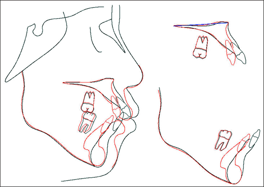 | Fig 12.Superimpositions of lateral cephalograms: pretreatment (black line) to post-treatment (red line). |
Table 1.
Cephalometric measurements pre- and post treatment


 PDF
PDF ePub
ePub Citation
 Print
Print


 XML Download