

 PDF
PDF ePub
ePub Citation
 Print
Print


Abstract
Objective
The purpose of this study was to compare the difference in three dimensional tooth movement using three different wire sizes (0.018 × 0.025-in, 0.016 × 0.022-in, 0.016-in) on a NiTi scissors-bite corrector.
Methods
Computed tomography (CT) images of the experimental model before and after tooth movement were taken and reconstructed into three dimensional models for superimposition. The direction and the amount of tooth movement were measured and analyzed statistically.
Results
The lingual and intrusive movements of the crown of the maxillary second molar were increased as the size of the NiTi wire increased. The roots of the maxillary second molars moved buccally except for the 0.016-in group. The intrusive movement of the roots of the maxillary second molars was increased as the size of the NiTi wire increased. Due to the use of orthodontic mini-implants, anchorage loss was under 0.2 mm on average.
Figures and Tables
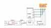
Fig 2
Dragon Helix and NiTi scissors-bite corrector. A, B, Dragon Helix; C, D, NiTi scissors-bite corrector. The second molar was aligned in line of occlusion and overintruded.
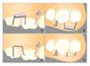

Fig 4
Process of the experiments. A, Before tooth movement; B, after tooth movement; C, forming stl files from dicom files using V-works; D, superimposition and measuring of tooth movement.
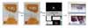
Fig 5
Superimposition of experimental models (0.018 × 0.025 in). Blue color shows before tooth movement, and red color, after tooth movement. Red circle was one of the reference markers for superimposition, X axis is the bucco-lingual direction, Y axis is the occluso-gingival direction and Z axis is the mesio-distal direction. A, Buccal aspect; B, distal aspect.
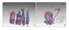
Fig 6
Schematic drawing of superimposition. A, B, 0.018 × 0.025 in group; C, D, 0.016 × 0.022 in group; E, F, 0.016 in group A,C,E in occlusal view; B, D, F in distal view. Green teeth show initial position and purple teeth show final position.
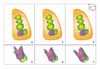
Fig 7
Comparison of mesiolingual cusp displacements of the second molar between 3 groups. X axis, Bucco-lingual direction; Y axis, occluso-gingival direction. *, significantly different as p < 0.05 between groups.

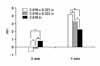
Fig 8
Comparison of palatal root displacements of the second molar between 3 groups. *, significantly different as p < 0.05 between groups.

References
1. Ogihara K, Nakahara R, Koyanagi S, Suda M. Treatment of a Brodie bite by lower lateral expansion: a case report and fourth year follow-up. J Clin Pediatr Dent. 1998. 23:17–21.
2. Heikinheimo K, Salmi K, Myllärniemi S. Identification of cases requiring orthodontic treatment. A longitudinal study. Swed Dent J Suppl. 1982. 15:71–77.
3. Thilander B, Wahlund S, Lennartsson B. The effect of early interceptive treatment in children with posterior cross-bite. Eur J Orthod. 1984. 6:25–34.

4. Pirttiniemi P, Kantomaa T. Relation of glenoid fossa morphology to mandibulofacial asymmetry, studied in dry human Lapp skulls. Acta Odontol Scand. 1992. 50:235–243.
5. Kucher G, Weiland FJ. Goal-oriented positioning of upper second molars using the palatal intrusion technique. Am J Orthod Dentofacial Orthop. 1996. 110:466–468.
6. Enacar A, Pehlivanoglu M, Akcan CA. Molar intrusion with a palatal arch. J Clin Orthod. 2003. 37:557–559.
7. Nakamura Y, Murata K, Ogino T, Sekiya T, Hirashita A. Intrusion of overerupted upper second molars with a modified lingual arch. J Clin Orthod. 2004. 38:622–626.
8. Legan HL. Orthodontic planning and biomechanics for transverse distraction ostegenesis. Semin Orthod. 2001. 7:160–168.
9. Baeten LR. Canine retraction: a photoelastic study. Am J Orthod. 1975. 67:11–23.
10. Burstone CJ, Pryputniewicz RJ. Holographic determination of centers of rotation produced by orthodontic forces. Am J Orthod. 1980. 77:396–409.
11. Caputo AA, Chaconas SJ, Hayashi RK. Photoelastic visualization of orthodontic forces during canine retraction. Am J Orthod. 1974. 65:250–259.
12. Moss ML, Skalak R, Patel H, Sen K, Moss-Salentijn L, Shinozuka M, et al. Finite element method modeling of craniofacial growth. Am J Orthod. 1985. 87:453–472.
13. Weijs WA, de Jongh HJ. Strain in mandibular alveolar bone during mastication in the rabbit. Arch Oral Biol. 1977. 22:667–675.
14. Ogura M, Yamagata K, Kubota S, Kim JH, Kuroe K, Ito G. Comparison of tooth movements using Friction-Free and preadjusted edgewise bracket systems. J Clin Orthod. 1996. 30:325–330.
15. Drescher D, Bourauel C, Thier M. Application of the orthodontic measurement and simulation system (OMSS) in orthodontics. Eur J Orthod. 1991. 13:169–178.
16. Rhee JN, Chun YS, Row J. A comparison between friction and frictionless mechanics with a new typodont simulation system. Am J Orthod Dentofacial Orthop. 2001. 119:292–299.
17. Cha BK, Lee JY, Bae SH, Park DI. Preliminary study of future orthodontic model analysis: the orthodontic application of 3-dimensional reverse engineering technologies. J Korean Dent Assoc. 2002. 40:107–107.
18. Yun SW, Lim WH, Chong DR, Chun YS. Scissors-bite correction on second molar with a dragon helix appliance. Am J Orthod Dentofacial Orthop. 2007. 132:842–847.
19. Chun YS, Row J, Suh MS, Park IK. An experimental study on the dynamic tooth moving effects of two precision lingual archs(Pla) for correction of posterior scissor bite by the calorific machine. Korean J Orthod. 1998. 28:29–41.
20. Proffit WR, Fields HW, Sarver DM. Contemporary orthodontics:. 2007. St.Louis: Mosby;340.
21. Melsen B, Agerbaek N, Eriksen J, Terp S. New attachment through periodontal treatment and orthodontic intrusion. Am J Orthod Dentofacial Orthop. 1988. 94:104–116.
 XML Download